![]()
![]()
![]()
Use LEFT and RIGHT arrow keys to navigate between flashcards;
Use UP and DOWN arrow keys to flip the card;
H to show hint;
A reads text to speech;
24 Cards in this Set
- Front
- Back
- 3rd side (hint)
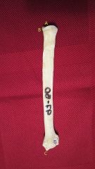
![Name the bone (include view [medial/lateral] and orientation [left/right])](https://images.cram.com/images/upload-flashcards/43/21/79/6432179_m.jpg)
Name the bone (include view [medial/lateral] and orientation [left/right]) |
Scapula (Right), Lateral View |
|
|
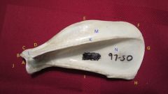
A. B C D |
A: Glenoid Cavity B: Supraglenoid Tubercle C: Coracoid Process D: Scapular Notch |
A: articulates with ventral angle; concave surface B: proximal to A C: small prominence on craniomedial aspect of B D: concavity that joins E and J |
|

E F G H I J |
E: Cranial Border F: Cranial Angle G: Dorsal Border H: Caudal Angle I: Caudal Border J: Ventral Angle |
E: thin and rounded side, thickens dorsally towards F F: corner that connects E & G G: shortest, relatively straight side H: corner that connects G & I I: thick and straight, roughened proximally and distally J: articulates with the humerus and includes A |
|

K L M N |
K: Spine L: Acromion M: Supraspinous Fossa N: Infraspinous Fossa |
K: divides the current view of the bone into two L: distally-extending process of K M: region cranial to K N: region caudal to K |
|

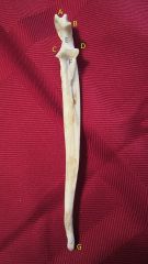
Name the bone view |
Medial View |
|
|
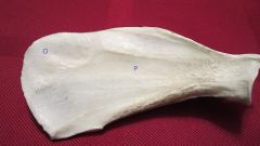
O P |
O: Serrated Face P: Subscapular Fossa |
O: dorsocranial portion, roughened P: smooth, bears three lines that converge ventrally |
|
![Name the bone (include view [cranial/caudal] and orientation [left/right])](https://images.cram.com/images/upload-flashcards/43/40/48/6434048_m.jpg)
Name the bone (include view [cranial/caudal] and orientation [left/right]) |
L Humerus, Cranial View |
|
|

A. B C |
A: Greater Tubercle B: Lesser Tubercle C: Intertubecular Groove
|
A: large rounded ridge craniolateral to N B: smaller ridge on medial aspect of N C: area between A & B |
|

D E F G |
D: Tricipital Line E: Deltoid Tuberosity F: Brachial Groove G: Crest of Greater Tubercle |
D: ridge running distally from the junction of A and N E: distal pointed end of D F: smooth service starting distal to N on lateral aspect, curving craniodistally around bone G: Poorly defined ridge extending distally from the cranial end of A |
|
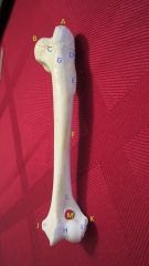
H I J K |
H: Trochlea I: Capitulum J: Medial Epicondyle K: Lateral Epicondyle |
H: larger division of articular surface of humeral condyle I: smaller division of articular surface J: larger side portion of condyle, squared-off caudal border K: smaller side portion, rounded caudal border |
|

Name the bone view |
Caudal View |
|
|
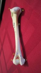
L M N O |
L: Radial Fossa M: Supratrochlear Foramen N: Head O: Tuberosity for Teres Major
|
L: smaller depression on cranial surface M: opening between L & P N: smooth, rounded articular surface of caudomedial aspect O: roughened area on medial side at junction of proximal and middle thirds |
|

P Q R |
P: Olecranon Fossa Q: Lateral Supracondylar Crest R: Medial Supracondylar Crest |
P: depression on caudal surface, sits between Q & R Q: starts at distal third and runs distally along caudal surface to end on K R: starts at distal third and runs distally along caudal surface to end on J |
|
![Name the bone (include view [cranial/caudal] and orientation [Left/Right])](https://images.cram.com/images/upload-flashcards/43/42/55/6434255_m.jpg)
Name the bone (include view [cranial/caudal] and orientation [Left/Right]) |
Radius (Right), Caudal View |
|
|
A. B C D |
A: Foeva Capitis B: Articular Circumference C: Styloid Process D: Ulnar Notch |
A: Articular surface, oval in shape, concave B: narrow band on caudal surface, contacts radial notch of ulna C: pointed projection on medial side D: smooth surface on caudolater border |
|
![Name the bone (include view [cranial/caudal] and orientation [Left/Right])](https://images.cram.com/images/upload-flashcards/43/42/64/6434264_m.jpg)
Name the bone (include view [cranial/caudal] and orientation [Left/Right]) |
Ulna (Left), Cranial View |
|
|
A. B C D |
A: Tuber Olecrani B: Anconeal Process C: Medial Coronoid Process D: Lateral Coronoid Process |
A: Proximal portion of olecranon, grooved on cranial surface, thickens caudally B: Sharp, pointed end proximal to extent of notch C: larger distal portion of E D: smaller distal portion of E |
|

E F G |
E: Trochlear Notch F: Radial Notch G: Styloid Process |
E: Proximal portion of olecranon F: Curved surface between C & D G: Pointed distal extremity |
|
![Name the bone (include view [dorsal/palmar] and orientation [Left/Right])](https://images.cram.com/images/upload-flashcards/43/42/91/6434291_m.jpg)
Name the bone (include view [dorsal/palmar] and orientation [Left/Right]) |
Forepaw (Left), Dorsal View |
|
|

A. B C D |
A: Intermedioradial Carpal Bone B: Ulnar Carpal Bone C: Accessory Carpal Bone D: Distal Carpal Bones |
A: most medial of proximal row B: extends distally almost to E C: located palmar D: get progressively larger, articulate with one E except for the most lateral, which articulates with two |
|

E F G H |
E: Metacarpal Bones F: Proximal Phalanges G: Middle Phalanges H: Distal Phalanges |
E: numbered from medial to lateral; 1 is smallest, 2&5 middle, 3&4 longest F: have proximal base and distal head G: have proximal base and distal head H: has proximal base, I & J |
|
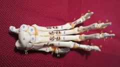
I J K L M |
I: Ungual Crest J: Ungual Process K: Metacarpophalangeal Joints L: Proximal Interphalangeal Joints M: Distal Interphalangeal Joints |
I: proximally surrounds J J: pointed portion of H K: connects E & F L: connects F & G M: connects G & H |
|
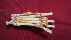
Name the bone view |
Palmar View |
|
|

N O |
N: Proximal Sesamoid Bones O: Dorsal Sesamoid Bone |
N: two on the flexor surface, embedded in the muscle tissue O: small, embedded in muscle tissue |