Reading...
![]()
Play button
![]()
Play button
![]()
Use LEFT and RIGHT arrow keys to navigate between flashcards;
Use UP and DOWN arrow keys to flip the card;
H to show hint;
A reads text to speech;
89 Cards in this Set
- Front
- Back
|
1. List the structures that make up the integumentary system:
|
1. the skin,
2. the accessory structures, 3. the hair, 4. the nails and 5. the glands. |
|
|
2. What type of tissue is the skin made up of?
|
1. epidermis &
2. dermis. *The hypodermis is not part of the layers of the skin but is the foundation for the skin to build on. The epidermis is made up of 'squamous epithelium' that sits on top of the dermis. The dermis is a 'connective tissue' layer that sits on top of the hypodermis and forms the support structures of the skin. The hypodermis is made up of 'loose connective tissue' with collagen fibres, elastin fibres, adipose cells, fibroblasts and macrophages. |
|
|
3. List & describe the five functions of skin:
|
1. Protection (Immunity)
2. Sensation (Touch) 3. Vitamin D production 4. Excretion (Sweat) 5. Temperature regulation |
|
|
4. Describe the foundation layer that underlies the 2 layers of the skin:
|
1. The hypodermis is :
2. also called the subcutaneous layer. 3. It is not part of the layers of the skin but is the foundation for the skin to build on. 4. The hypodermis attaches the dermis to the underlying bone and muscle 5. supplies the skin with blood vessels and nerves. 6. made up of loose connective tissue with collagen fibres, elastin fibres, adipose cells, fibroblasts and macrophages. 7. The adipose tissue (fat) insulates the body, helping to conserve heat and protect the underlying organs from mechanical shock. 8. Over half the body's fat is contained in the hypodermis, 9. the distribution of this fat is responsible for the contours of the female body. 10. As people age, fat and elastic fibres within the subcutaneous layer decreases causing wrinkles. |
|
|
5. Describe the structure & function of the dermis:
|
1. don't (DERMIS:)
2. recycle (Reticular) 3. paper (Papillary) 4. diapers (Dermal) |
|
|
5. Describe the structure & function of the dermis:
|
1. The dermis sits between the hypodermis and epidermis.
2. is the thickest part of the two layers of skin. 3. thicker on soles of feet and palms of hands, thinner on scrotum and eyelids. 4. typically is thinner in females than males. 5. The dermis is connective tissue and consists of collagen and elastic fibres in between a few fat cells and blood vessels and many nerves, hair follicles and glands. 6. The dermis is divided into two distinct connective tissue layers: * The superficial papillary layer. * The deep reticular layer. |
|
|
6. How are finger prints formed?
|
'The superficial papillary layer' contains
'fine finger-like projections' called 'papillae'. The patterns in these papillae are what produces your distinct finger prints. |
|
|
7. Describe the structure & function of the epidermis:
|
1. made up of keratinised stratified squamous epithelium.
2. thinner than the dermis layer. 3. contains no blood vessels. 4. supplied with oxygen from the capillaries in the papillary layer of the dermis. 5. four layers of the epidermis which combined are 0.1 mm thick. 6. The palms of the hands and soles of the feet have an extra epidermal layer. |
|
|
8. List & briefly describe the five layers of the epidermis:
|
every ( epidermis )
1. cute (corneum) 2. lady (lucidum) 3. gets (granulosum) 4. sexy (spinosum) 5. boys/guys (basale/germinativum) |
|
|
8. List & briefly describe the five layers of the epidermis:
|
5. Stratum basale or stratum germinativum - deepest layer, made up of a single layer cuboidal or columnar cells known as keratinocytes, enables the skin to repair itself.
4. Stratum spinosum - sits above the stratum basale (#5) and consists of 8 - 10 layers of multi-sided keratinocytes, gives the skin strength and flexibility. 3. Stratum granulosum - made up of 2 - 5 layers of elongated diamond shape cells, gives the skin its water-repellent properties. 2. Stratum lucidum - only present in the thick skin of the fingertips, palms and soles of the feet. It is a thin clear zone, consisting of 3 - 5 layers of keratinocytes that are full of keratin. 1. Stratum corneum - most superficial layer of the epidermis containing 25 - 30 layers of squamous cells that are completely filled with keratin. The cells in this layer flake off by the millions, in a process known as desquamation, while millions of epithelial cells are reproduced daily to replace the ones that are shed. |
|
|
9. Which layer of the stratum is only found in thick skin?
|
Layer #2. Stratum lucidum - only present in the thick skin of the fingertips, palms and soles of the feet.
|
|
|
10. Where does skin cell growth occur?
|
Layer #5. Stratum basale or stratum germinativum - enables the skin to repair itself.
|
|
|
11. Describe the movement of & changes in keratinocytes as they move from the basale layer to the corneum layer:
|
5. The keratinocytes in (#5) the stratum basale are stem cells that are continuously dividing and reproducing more keratinocytes, enabling the skin to repair itself.
* As new cells are produced, they push the cells above them towards the surface. 4. Keratinisation of the cells begins in (#4) the stratum spinosum as keratin fibres appear. These keratin fibres gives the skin strength and flexibility. 3. The cytoplasm of the cells in (#3) the stratum granulosum fill with keratin and the nucleus and organelles fragment and die. The cells look grainy, as more protein granules are produced within the cell. The cells start to release their lipid contents into the spaces between the cells which gives the skin its water-repellent properties. 2. The stratum lucidium is a thin clear zone, consisting of 3 - 5 layers of keratinocytes that are full of keratin. 1. The stratum corneum is the most superficial layer of the epidermis containing 25 - 30 layers of squamous cells that are completely filled with keratin. The cells in this layer flake off by the millions, in a process known as desquamation, while millions of epithelial cells are reproduced daily to replace the ones that are shed. * It can take 40-56 days for the cells of the stratum basale to make their way to the top layer of the epidermis. (a month +1/3rd - 2 months) |
|
|
12. What gives skin its waterproof properties?
|
3. The cytoplasm of the cells in (#3) the stratum granulosum fill with keratin and the nucleus and organelles fragment and die. The cells look grainy, as more protein granules are produced within the cell. *The cells start to release their lipid contents into the spaces between the cells which gives the skin its water-repellent properties.
|
|
|
13. What is the name of the process when dead skin cells flake off?
|
Desquamation.
|
|
|
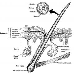
14. Describe the structure of hair?
|
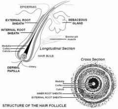
*Hair is composed of a keratin similar to that in the epidermis,
* is divided into the shaft (found above the skin) *and the root (found below the skin). *Shaft *Follicle *External root sheath *Internal root sheath *Medulla (inner) *Cortex *Cuticle * Derma Papilla (Base/Bulb) |
|
|
15. What is the function of hair?
|
The hair has a 'protective function'.
for example, eyelashes prevent things falling in the eyes and hair on the head protects from sunburn in summer and heat loss in winter. |
|
|
16. What is the purpose of the arrector pili muscle & where is it found?
|
* These muscles contract in response to fear or cold to pull the hair follicle into a vertical position and make the hair stand up straight.
* attached to the papillary layer of the dermis and the hair follicles. *The skin around the shaft of the hair also gets pulled up straight causing goose bumps to form. |
|
|
17. List the 4 types of glands found in the skin, where they are found & what are their secretions
|
1. Sudoriferous glands = Sweat Glands:
* Eccrine glands - secrete water to the 'total body surface', eccrine glands open by a duct directly onto the skin surface. * Apocrine glands - apocrine glands usually develop in association with hair follicles and open into them. They secrete a fatty sweat into the gland tubule. Emotional stress causes the tubule wall to contract, expelling the fatty secretion to the skin, where local bacteria break it down into odorous fatty acids, in the 'underarm' and in 'genital regions'. 2. Sebaceous glands secrete an oily substance known as 'sebum' into the 'hair follicle'. found in the 'face' and 'scalp', the 'lips', 'tip of the penis' and 'labia minora'. 3. Ceruminous glands are only found in the 'ear canal'. They are a type of modified sweat gland which produces 'cerumen' (ear wax). 4. Mammary glands are modified sweat glands located in the breasts that produce milk. |
|
|
18. What are finger nails composed of?
|
*composed of 'stratum corneum'.
They are tightly packed with keratin that becomes hard and plate-like and protect the ends of the fingers and toes. (Corn-Hard-Nail) |
|
|
19. How do stretch marks occur?
|
Stretch marks, or striae, are due to tears in the 'reticular layer' of the 'dermis' caused by extreme stretching.
|
|
|
20. What contributes to skin colour?
|
* Melanocytes are found in the stratum basale and produce melanin. This melanin is a pigment that produces the colour of the skin, hair and eyes, is produced in greater amounts after exposure to UV light.
* circulating blood, * the thickness of stratum corneum. |
|
|
21. How is vitamin D synthesized by the body?
|
* skin is exposed to ultraviolet radiation,
* a precursor molecule in the skin is activated to form vitamin D, * This molecule is modified by the liver and kidneys to produce an active form of vitamin D. |
|
|
22. List the changes that occur in the skin due to aging?
|
As people age, the amount of fat and elastic fibres within the subcutaneous (hypodermis {bottom}) layer decreases causing wrinkles.
|
|
|
23. What is the function of the skeletal system?
|
1. Support
2. Protection 3. Movement 4. Blood cell production (Bone Marrow) 5. Storage (Fat, Calcium, Potassium) |
|
|
24. Name the different bones in the body:
|
1: Compact bone,
2: Cancellous (Spongy) bone. |
|
|
25. Discuss briefly the functions of bone:
|
* Bone provides a framework for the body.
* Supports soft tissues. * Provides points of attachment for many skeletal muscles. * Encloses organs to protect them. * Bone marrow found in the cavities of bones produce blood cells & platelets. * Fat, Ca++ (Calcium) & PO4ˉ (Potassium) are stored in bone from the blood stream. |
|
|
26. Describe the difference between compact bone & cancellous bone:
|
Compact Bone:
* Forms the external layer of all bones. * Provides protection & support. * Helps bones resist weight bearing stress. * Contains more bone matrix than spaces. * Matrix is tightly packed & arranged in circles (osteon) around a central canal which contain blood vessels & nerves. * Osteocytes found in spaces of the matrix. Cancellous bone: * Also called spongy bone as it resembles a sponge. * Contains more spaces than bone matrix. * Bone matrix is made up of interconnecting beams of bone called trabeculae. * Osteocytes exist within the trabeculae. * Osteoblasts & osteoclasts exist on the walls of the trabeculae. * Blood vessels & bone marrow fill the canals between the trabeculae. * Found where strength but lightness is required. |
|
|
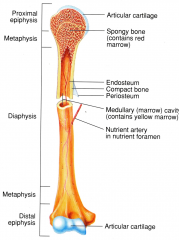
27. Draw a diagram of a long bone and label the following components:
* Diaphysis, * epiphysis, * metaphysis, * medullary cavity, * periosteum, * endosteum, * articular cartilage |
|
|
|
28. What are an osteoblast, osteocyte & osteoclast?
|
* Osteoblasts: produced from stem cells.
* Osteocytes: are mature osteoblasts. * Osteoclasts: made in the red bone marrow. They have a ruffled border which has an acid environment causing decalcification of bone . |
|
|
29. What are the functions of these cells?
|
* Osteoblasts: are the bone forming cells that create the matrix of bone & mineralises bone.
* Osteocytes: they maintain the matrix. * Osteoclasts: are responsible for the breakdown of bone (resorption). They have a ruffled border which has an acid environment causing decalcification of bone . |
|
|
30. Briefly describe the five different types of bone:
|
1. Long:Length is > than width & slightly curved for strength.
2. Short: Cube shaped nearly equal in length & width. 3. Flat: Generally thin & composed of 2 parallel plates of compact bone with spongy bone in between. 4. Irregular: Complex shaped & do not fit into any other group. 5. Sesamoid: Small bones embedded in tendons where considerable pressure develops. Eg, thumb, great toe, patella. (6. Sutural: Small bones located within the cranial sutures or joints.) |
|
|
31. What are the functions of the vertebral column?
|
* Protects the spinal cord.
* Supports the head & trunk. * Ribs & muscles of the back attach to the vertebral column. |
|
|
32. Name the five regions in the vertebrae & how many vertebrae are in each of those regions.
|
C = Cervical = 7.
T = Thoracic = 12. L = Lumbar = 5. S = Sacrum = 5 fused into 1, C = Coccyx = 4 fused into 1, |
|
|
33. What are the functions of the atlas and axis?
|
* C1 (atlas) supports the head.
It allows nodding of the head (nodding for yes). * C2 (axis) acts as a pivot on which the atlas and head rotate. It allow side to side rotation (turning head from side to side for no). |
|
|
34. How many true ribs are there?
|
7 pairs.
|
|
|
35. What is a false rib?
|
3 pairs 8, 9, and 10 attach to each other and then to the cartilage of the 7th rib.
|
|
|
36. Differentiate between the bones in the appendicular and the bones in the axial skeleton.
|
Appendicular:
* Upper and lower limbs. * Pelvic girdle. * Pectoral girdles. Axial: * Skull. * Vertebrae. * Thorax (ribs & sternum). |
|
|
37. What are the identified subdivisions of the sternum?
|
* manubrium,
* body & * xiphoid process. * Sternal angle is the junction of the manubrium & body. * Suprasternal notch is the depression in the anterior surface of the manubrium. |
|
|
38. Identify the bones & suture lines of the skull:
|
8 cranial =
1 Frontal bone . 2 parietals. 2 temporals. 1 Occipital. 1 Ethmoid. 1 Sphenoid . 14 facial bones. 2 nasal. 2 maxilla. 2 zygomatic arch. 2 lacrimal. 2 palatine . 2 inferior nasal concha. 1 Mandible . 1 Vomer. Coronal: suture unites the frontal & 2 parietal bones. Sagittal: suture unites the 2 parietal bones. Lambdoid: suture unites the parietal & occipital bones. Squamous: suture unites the parietal & temporal bones. C S L S |
|
|
39. List the three different types of joints classified according to structure. Give an example of each.
|
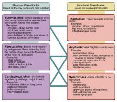
Fibrous - Suture Joints in skull bones.
Cartilaginous - epiphyseal plates of growing bones & Vertebra joints. Synovial - Knee, Hip. |
|
|
40. List the different types of joints classified according to function. Give an example of each.
|

Synarthrotic - Joints with little or no movement = Suture Joints in skull bones.
Amphiarthrotic - Slightly moveable joints = intervertebral disks (spine). Diarthrotic - Freely moveable synovial joints = Knee, Hip. |
|
|
41. What is the function of bursae?
|
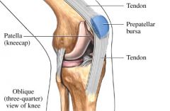
A closed sac or envelope lined with synovial membrane and containing synovial fluid, usually found or formed in areas subject to friction (over an exposed or prominent body part or where a tendon passes over a bone).
|
|
|
42. Describe the functions of muscle.
|
8 functions of muscle:
1: Body movement, 2: Maintain body posture, 3: Produces heat (75% of body heat), 4: Ventilation (breathing), 5: Communication (speech), 6: Maintain blood pressure, 7: Movement of organs, 8: Blood flow (the heart!). |
|
|
43. What are the characteristics of muscle tissue?
|
1: Contractility.
2: Excitability. 3: Extensibility. 4: Elasticity. |
|
|
44. Differentiate between the three types of muscle tissue and give an example of each in the human body.
|
1: Skeletal,
2: Cardiac, 3: Smooth. |
|
|
45. What is the cell membrane of the muscle cell called?
|
EnPEpF
1: Endomysium: surrounds individual muscle cells (fibres) 2: Perimysium: surrounds fasciculus bundles of 10-100 muscle cells. 3: Epimysium: surrounds the whole muscle & is also called the 'fascia'. |
|
|
47. Briefly describe a muscle cell fibre:
|
a ‘Fibre’.
Long cylindrical cells 1 - 4mm long. Developed from myoblasts. Their cytoplasm (sarcoplasm) is filled with protein myofibrils which gives muscle its striped appearance. Each muscle cell has many nuclei. They lay just inside the plasma membrane (sarcolemma). Myofibrils extend from one end of the muscle fibre to the other. |
|
|
48. Describe a fascicle:
|
Several muscle cells fibres then form into a group which is held together by the perimysium. (another connective tissue layer).
This group of muscle cells is called a muscle fascicle. |
|
|
49. Describe the fascia:
|
Epimysium: surrounds the whole muscle & is also called the 'fascia'.
The fascia forms a sheet under the skin & surrounds individual muscles or groups of muscles. Continuous with the connective tissue of tendons & bones. |
|
|
50. What is the contractile unit of muscle?
|
A sarcomere, or contractile unit.
|
|
|
51. What is the role of muscles in maintaining body temperature?
|
Muscle contractions produce heat and as much as 70% of body heat is produced by energy produced in muscle tissue.
When the internal heat of the body reaches too low a level thermoreceptors in the skin relay a message to the hypothalamus in the brain. In response to this signal, the skeletal muscles contract and relax in an involuntary manner (shivering) increasing muscle activity to generate heat. In turn, muscles are also responsive to exterior heat - cold air increases muscle tone, and hot conditions have a relaxing effect on muscles. |
|
|
52. Describe the function of a ligament.
|
Ligaments connect two or more bones together and help stabilize joints.
|
|
|
53. Describe the function of a tendon.
|
Tendons attach muscle to bone. Tendons vary in size and are somewhat elastic.
|
|
|
54. Describe the function of cartilage.
|
Cartilage provides support within structures.
|
|
|
55. Differentiate between the layers of the pericardium?
|
The heart is enclosed in a protective sac called the 'pericardium'
The pericardium consists of two parts: Layer of fibrous pericardium, Layer of serous pericardium. |
|
|
56. State the structure and function of the fibrous pericardium.
|
* Thick superficial (outer) layer.
* Is a tough, dense irregular connective tissue. * Holds the heart in position in the chest. * It prevents over stretching of the heart. * Anchors heart to diaphragm and sternum. * Provides protection from injury and infection. |
|
|
57. State the structure and function of the serous pericardium.
|
* Thin delicate inner layer of pericardium:
- Consists of 2 layers: 1. Parietal layer. * Internal layer. *Attached to inside of fibrous pericardium. 2. Visceral layer. * External layer. * Also called the epicardium. * Attached to heart muscle. |
|
|
58. What is the other name for the visceral pericardium?
|
Also called the epicardium.
|
|
|
59. Briefly describe the three layers of the heart wall.
|
EpMEn
1. Epicardium. 2. Myocardium. 3. Endocardium. |
|
|
60. List the chambers of the heart.
|
The heart has four chambers.
- Two upper (superior) chambers: 'atria'. - Two lower (inferior) chambers: 'ventricles'. |
|
|
61. Which blood vessels enter the heart and which blood vessels leave the heart?
|
All 'veins' of the heart 'enter' the heart.
|
|
|
62. How does blood return to the heart from the coronary circulation?
|
.
|
|
|
63. Name the semi-lunar valves and state their location.
|
.
|
|
|
64. Name the atrioventricular valves & state their location.
|
.
|
|
|
65. What is the function of heart valves?
|
.
|
|
|
66. What are chordea tendinae?
|
.
|
|
|
67. List the components of the electrical conduction system.
|
.
|
|
|
68. Why is the sinoatrial node (SA Node) called the pace maker of the heart?
|
.
|
|
|
69. Describe blood flow through the heart, linking the opening and closing of valves, contraction of the chambers and the electrical conduction.
|
.
|
|
|
70. What is stroke volume?
|
.
|
|
|
71. What is an ECG?
|
.
|
|
|
72. What does the P wave represent?
|
.
|
|
|
73. What does the QRS complex represent?
|
.
|
|
|
74. What does the T wave represent?
|
.
|
|
|
75. Define repolarisation and depolarisation.
|
.
|
|
|
76. Describe the phases of the cardiac cycle.
|
.
|
|
|
77. What causes heart sounds?
|
.
|
|
|
78. When does S1 occur?
|
.
|
|
|
79. When does S2 occur?
|
.
|
|
|
80. Where the cardiovascular centre located and what is its function?
|
.
|
|
|
81. What is the role of chemoreceptors, baroreceptors and proprio-receptors in regulating the heart?
|
.
|
|
|
82. What factors excluding illness affect heart rate?
|
.
|
|
|
83. Draw a diagram of the heart including the vessels entering and leaving the heart, the chambers, the valves, the conduction system and the wall dividing the right and left side.
|
.
|
|
|
84. Describe the differences between structure & function of arteries & veins.
|
.
|
|
|
85. Draw a simple diagram of the direction of blood flow from the left ventricle through the systemic circulation and back to the right atrium.
|
.
|
|
|
86. What mechanisms assist the return of venous blood to the heart?
|
.
|
|
|
87. What is the function of the endothelium?
|
.
|
|
|
88. Differentiate between the systemic and pulmonary circulation.
|
.
|