![]()
![]()
![]()
Use LEFT and RIGHT arrow keys to navigate between flashcards;
Use UP and DOWN arrow keys to flip the card;
H to show hint;
A reads text to speech;
300 Cards in this Set
- Front
- Back
|
What specific type of information is carried by the Dorsal Columns? |
--Touch --Proprioception --2-pt. discrimination |
|
|
What specific type of information is carried by the Anterior Corticospinal Tract? |
15% of voluntary motor fibers |
|
|
What specific type of information is carried by the Lateral Corticospinal Tract? |
85% of voluntary motor fibers |
|
|
What specific type of information is carried by the Lateral Spinothalamic Tract? |
--Pain --Temperature |
|
|
Which pathways have their level of decussation in the medulla? |
--Dorsal Columns --Lateral Corticospinal Tract |
|
|
What is the level of decussation for the Anterior Corticospinal Tract? |
Level of exit |
|
|
(Sympathetic Division) Preganglionic Nueron |
--Myelinated
--1st of 2 motor neurons --Cell bodies in lateral grey horns |
|
|
(Sympathetic Division) Postganglionic Neurons |
--Unmyelinated --2nd of 2 motor neurons --Cell bodies in trunk |
|
|
Grey Horns of Spinal Cord |
(Sympathetic Division) --Lateral grey horn --T1-L2 --Cell bodies in lateral grey horn |
|
|
Dorsal Root of Spinal Cord |
(Sympathetic Division) --Sensory neuron axon |
|
|
Ventral Root of Spinal Cord |
(Sympathetic Division) Pathway for ANS & somatic motor neurons |
|
|
Dorsal Rami |
(Sympathetic Division) Nerve supply for posterior of body |
|
|
Ventral Rami |
(Sympathetic Division)
Nerve supply for anterior of body |
|
|
White ramus communicans |
(Sympathetic Division) --T1-L2 --Myelinated --Preganglionic nuerons |
|
|
Grey ramus communicans |
(Sympathetic Division) --Unmyelinated --Postganglionic neurons --Connects back w/ spinal nerves |
|
|
Paravertebral Ganglion |
(Sympathetic Division) Chain of ganglia running alongside length of spinal cord |
|
|
Superior Cervical Ganglion |
#86 (Sympathetic Division) Nerves supplying head & heart |
|
|
Middle Cervical Ganglion |
#88 (Sympathetic Division) Nerves supplying heart |
|
|
Inferior Cervical Ganglion |
#89 (Sympathetic Division) Nerves supplying heart |
|
|
Name 3 Prevertebral Ganglia. Where are they located? |
(Sympathetic Division) --Celiac ganglion --Superior mesenteric ganglion --Inferior mesenteric ganglion Location: below diaphragm |
|
|
(Parasympathetic Division) Preganglionic Nueron |
From brain stem or sacral neurons to terminal ganglia |
|
|
(Parasympathetic Division) Postganglionic Nueron |
From terminal ganglia to effector |
|
|
Cranial Nerve III--Oculomotor Nerve |
(Parasympathetic Division) Serves Ciliary Ganglion |
|
|
Cranial Nerve VII--Facial Nerve |
(Parasympathetic Division) Serves Pterygopalatine Ganglion |
|
|
Cranial Nerve IX--Glossopharyngeal Nerve |
(Parasympathetic Division) Serves Submandibular Ganglion |
|
|
Cranial Nerve X--Vagus Nerve |
(Parasympathetic Division) Serves Otic Ganglion |
|
|
What is the function of Spinal Nerves 2-4? |
(Parasympathetic Division) Help to serve Pelvic Splanchnic Nerves |
|
|
What do the Pelvic Splanchnic Nerves innervate? |
(Parasympathetic Division) --Bladder --Intestines --Genitals |
|
|
(Parasympathetic Division) What are 4 of the Terminal Ganglia? |
--Ciliary Ganglion --Pterygopalatine Ganglion --Submandibular Ganglion --Otic Ganglion |
|
|
Function of Ciliary Ganglion |
Goes to eye; constricts pupil |
|
|
Function of Pterygopalatine Ganglion |
Goes to lacrimal gland & mucus membrane of nose & palate |
|
|
Function of Submandibular Ganglion |
Goes to submandibular & sublingual glands |
|
|
Function of Otic Ganglion |
Goes to parotid gland (salivation) |
|
|
What is the CNS exit of the Sympathetic Division? |
T1 - L2 Thoracolumbar |
|
|
What is the CNS exit of the Parasympathetic Division? |
S2 - S4 Cranial Nerves III, IIX, IX, & X Craniosacral |
|
|
Where are preganglionic neuronal cell bodies of the Sympathetic Division located? |
Lateral grey horn |
|
|
Where are preganglionic neuronal cell bodies of the Parasympathetic Division located? |
--Brainstem (Cranial Nerves III, IIX, IX, X) --S2-S4 |
|
|
Compare preganglionic neuron length in Sympathetic Division vs. Parasympathetic Division |
SD: SHORT PSD: LONG |
|
|
Compare postganglionic neuron length in Sympathetic Division vs. Parasympathetic Division |
SD: LONG PSD: SHORT |
|
|
Where are preganglionic axons located in the Sympathetic Division? |
White Ramus |
|
|
Where are preganglionic axons located in the Parasympathetic Division? |
Along somatic nerves |
|
|
What neurotransmitter is released from preganglionic neurons? |
Acetylcholine (ACh) |
|
|
Where are postganglionic neuronal cell bodies of the Sympathetic Division located? |
Paravertebral & Prevertebral |
|
|
Where are postganglionic neuronal cell bodies of the Parasympathetic Division located? |
Terminal Ganglia --Ciliary --Pterygopalatine --Submandibular --Otic |
|
|
Where are Sympathetic postganglionic axons located? |
Grey ramus
|
|
|
Where are Parasympathetic postganglionic axons located? |
Adjacent to or on target organs |
|
|
What neurotransmitter is released from postganglionic neurons? |
SD: Norepinephrine PSD: Acetylcholine (ACh) |
|
|
What organs are only innervated by the Sympathetic Division (not dual innervated)? |
--Sweat Glands --Arrector Pilli --Kidneys --Adrenal Glands --Spleen --Blood Vessels |
|
|
What are the 4 E's? |
Main effects of the Sympathetic Division: --Emergency --Exercise --Excitement --Embarrassment |
|
|
What is SLUDD? |
Main effects of the Parasympathetic Division: --Salivation --Lacrimation --Urination --Digestion --Defecation |
|
|
Axon of Sympathetic Preganglionic Neuron may... |
--Synapse in 1st ganglion it enters --Pass up/down chain to another ganglion --Pass through chain to prevertebral ganglion --Pass through chain to adrenal gland |
|
|
Characteristics of the Fibrous Tunic (of eye)? |
--Outtermost layer of eye --Protects & shapes eye --Location of muscle attachment |
|
|
What is the specific layer of the Fibrous Tunic called? |
Sclera |
|
|
What is the conjunctiva? |
Mucus membrane |
|
|
What are the 3 main portions of the conjunctiva? |
--Palpebral conjunctiva --Bulbar conjunctiva --Conjunctival sac |
|
|
Location of the Palpebral Conjunctiva? |
Inner eyelid
|
|
|
Location of the Bulbar Conjunctiva? |
Against sclera |
|
|
Location of Conjunctival Sac |
Where Palpebral & Bulbar Conjunctivas meet |
|
|
What are the characteristics of the Cornea? |
--Transparent covering to iris --Refracts 75% of incoming light |
|
|
What are the 3 main layers of the cornea? |
--Corneal Epithelium (NKSSET) --Corneal Stroma (DICT) --Corneal Endothelium (SSET) |
|
General Layer? |
Fibrous Tunic |
|

Specific Layer? |
Sclera |
|
|
Sclera |
(AKA - "Fibrous Tunic") Dense CT |
|

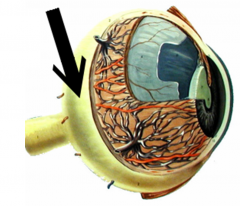
General layer? |
Vascular Tunic |
|
Specific layer? |
Choroid |
|
General layer? |
Nervous Tunic |
|

Specific layer? |
Retina |
|
Structure? |
Palpebral Conjunctiva |
|
Structure? |
Bulbar Conjunctiva |
|
Structure? |
Conjunctival Sac |
|
Structure? |
Cornea |
|
|
What is the outer surface of the cornea called? |
Corneal Epithelium |
|
|
What is the middle layer of cornea called? |
Corneal Stroma |
|
|
What is the innermost surface of the cornea called? |
Corneal Endothelium |
|
|
What kind of tissue comprises Corneal Epithelium? |
Non-keratinized stratified squamous epithelial tissue (NKSSET) |
|
|
What kind of tissue comprises Corneal Stroma? |
Dense irregular CT (DICT) |
|
|
What kind of tissue comprises Corneal Endothelium? |
Simple squamous epithelial tissue (SSET) |
|
|
What is LASIK? |
Precise, controlled removal of corneal tissue by a special laser to reshape the cornea, thereby changing its focusing power |
|
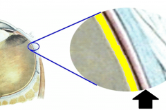
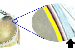
What is this layer? |
Corneal Epithelium |
|
What is this layer? |
Corneal Stroma |
|
What is this layer? |
Corneal Endothelium |
|
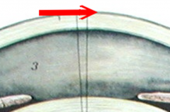
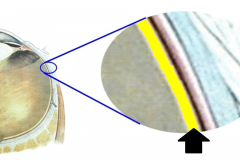
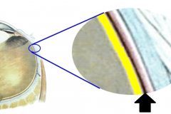
What general layer is this? What specific layer is this? |
General layer: Fibrous Tunic Specific layer: Sclera |
|
What structure is this? |
Sclera |
|
|
What structure reflects light & gives animals night vision? |
Tapetum (NOT in humans) |
|
|
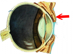
What is the anterior portion of the vascular tunic called? |
Ciliary Bodies |
|
|
What structures comprise the Ciliary Bodies? |
--Ciliary muscle --Ciliary process |
|
|
What is the ciliary muscle? |
Circular band of smooth muscle that adjusts the shape of the lens |
|
|
What is the function of ciliary processes? |
Secrete aqueous humor |
|
What is this structure? |
Ciliary Muscle |
|
What are these structures? |
Ciliary processes |
|
|
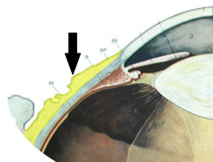
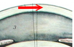
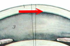
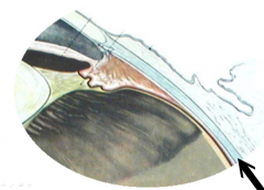
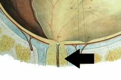
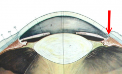
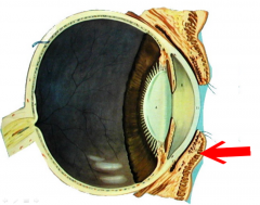
What is the junction between the retina & ciliary bodies called? |
Ora Serrata |
|
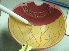
What is this structure? |
Ora Serrata |
|
|
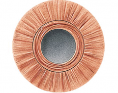
What is the "flat donut" shaped structure that helps regulate light entering the eye? |
Iris |
|
|
What is the hole in the eye that light passes through? |
Pupil |
|
What is this structure? |
Iris |
|
What is this structure? |
Iris |
|
What is this structure? |
Iris |
|
What is this structure? |
Pupil |
|
|
What is the posterior 3/4 of the eyeball that contains vision receptors called? |
Retina |
|
|
What are the 4 main structures of the Retina? |
--Fovea centralis --Macula lutea --Optic disc --Optic nerve |
|

What is this structure? |
Fovea Centralis |
|
What is this structure? |
Macula Lutea |
|
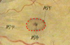
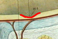
What is this structure (outlined in red dashed line)? |
Macula Lutea |
|
What is this structure? |
Fovea Centralis |
|
|
Characteristics of Fovea Centralis? |
--High concentration of cones --Highest visual acuity |
|
|
What is the small indentation containing fovia centralis called? |
Macula lutea |
|
|
What is the Optic Disc? |
--Blind spot --Point of Optic Nerve exit |
|
|
What does the Optic Nerve consist of? |
Axons of ganglion cells |
|
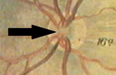
What is this structure? |
Optic Disc |
|
What is this structure? |
Optic Disc |
|
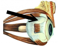
What is this structure? |
Optic Nerve (CN II) |
|
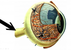
What is this structure? |
Optic Nerve (CN II) |
|
|
What is the Pigmented Epithelium? |
Layer that contains melanin & absorbs stray light |
|
What layer is this? |
Retina |
|
What layer is this? |
Pigmented Epithelium |
|
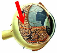
What layer is this? |
Choroid |
|
What layer is this? |
Sclera |
|
|
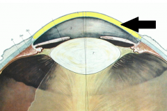
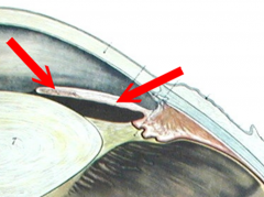
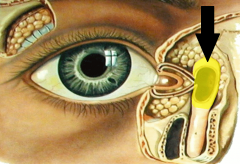
What is the Anterior Cavity? |
Space between lens & cornea |
|
|
What does the Anterior Cavity contain? |
Aqueous Humor |
|
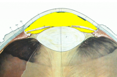
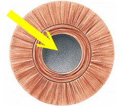
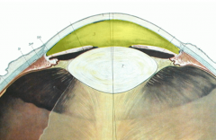
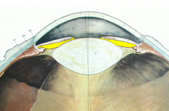
What is the structure highlighted in yellow? |
Anterior Cavity |
|
What is the structure highlighted in yellow? |
Anterior Chamber (of Anterior Cavity) |
|
What is the structure highlighted in yellow? |
Posterior Chamber (of Anterior Cavity) |
|
|
What is produced by the ciliary processes? |
Aqueous Humor |
|
|
What is the function of aqueous humor? |
Nourish lens & cornea |
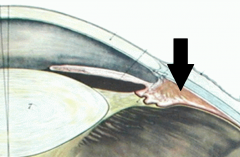
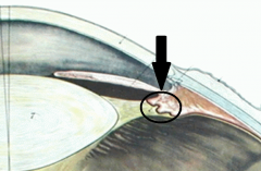
|
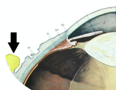
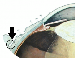
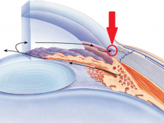
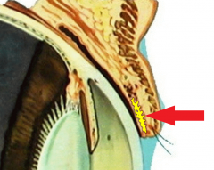
What is this structure? |
Canal of Schlemm |
|
What is this structure? |
Canal of Schlemm |
|
What is this structure? |
Canal of Schlemm |
|
|
What is the function of the Canal of Schlemm? |
Absorbs aqueous humor & returns it to blood |
|
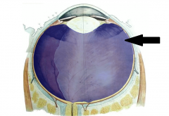
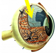
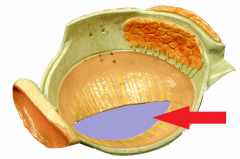
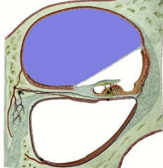
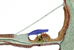
What is this structure (highlighted in blue)? |
Vitreous Chamber |
|
|
What is contained in the Vitreous Chamber? |
Vitreous Humor |
|
|
What are characteristics of Vitreous Humor? |
--Jelly-like --Doesn't regenerate --Contains phagocytes |
|
|
What is the function of Vitreous Humor? |
Holds retina in place |
|
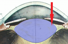
What is this structure (highlighted in blue)? |
Lens |
|
|
What are the components of the lens? |
--Lens Capsule --Suspensory Ligaments |
|
|
What material is the lens comprised of? |
Crystallins protein |
|
|
What is the function of the lens? |
Refracts 25% of light |
|
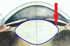
What is this structure (highlighted in blue)? |
Lens Capsule |
|
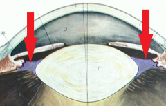
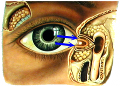
What are these structures (highlighted in blue)? |
Suspensory Ligaments |
|
|
What is the CT that surrounds crystallins proteins? |
Lens Capsule |
|
|
What is the function of the Suspensory Ligaments? |
--Connects Lens to Ciliary Muscles (muscle contraction decreases tension) |
|
What is this structure?
|
Upper Palpebrae |
|
What is this structure? |
Lower Palpebrae |
|
|
What is the function of the upper & lower palebrae? |
--Protect Eye --Spread Tears |
|
What is this structure (highlighted in yellow)? |
Tarsal Glands |
|
|
What is the function of Tarsal Glands? |
Produce fluid to keep eyelids from sticking |
|
|
What is the function of Ciliary Glands (on eyelash follicles)? |
Waterproofing |
|
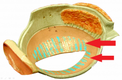
What are the structures (highlighted in turquoise)? |
Tarsal Glands |
|
What is the structure (highlighted in blue)? |
Palpebral Fissure |
|
|
What are the corners of the eye called? |
Medial & Lateral Commissures |
|
What is this structure? |
Caruncle |
|
|
What is the Caruncle comprised of? |
Sebaceous & Sudoriferous Glands
|
|
|
What is the function of the Caruncle? |
Make eye boogers ;) |
|
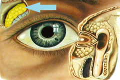
What is this structure (highlighted in yellow)? |
Lacrimal Gland |
|
|
What is the function of the Lacrimal Gland? |
Produces tears |
|
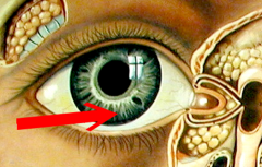
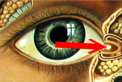
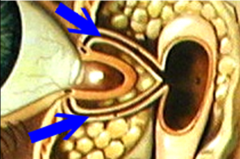
What are these structures (at tips of pointers)? |
Lacrimal Punctum |
|
|
What is the function of the Lacrimal Punctum? |
Opening that tears drain into |
|
What are the structures shown? |
Lacrimal Canals |
|
What is the structure shown (highlighted in yellow)? |
Lacrimal Sac |
|

What is the structure shown (highlighted in yellow)? |
Nasolacrimal Duct |
|
|
What are the structures of the Lacrimal Apparatus? |
--Lacrimal Gland --Lacrimal Punctum --Lacrimal Canal --Lacrimal Sac --Nasolacrimal Duct |
|
|
Trace the path tears follow from eye to nose |
Created in Lacrimal Gland Drain into Lacrimal Punctum Which drain into Lacrimal Canal Which drains into the Lacrimal Sac Which drains into the Nasolacrimal Duct |
|
What is this structure? |
Nasolacrimal Duct |
|
|
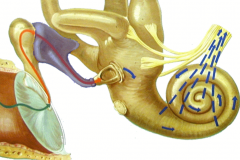
What are the main structures of the External Ear? |
--Pinna --External Auditory Meatus --Ceruminous Glands |
|
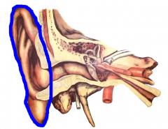
What is this structure (outlined in blue)? |
Pinna |
|
|
What is the upper part of the External Ear called? |
Helix |
|
|
What is the lower part of the External Ear called? |
Lobule |
|
|
What is the Pinna comprised of? |
Elastic cartilage (covered in skin) |
|
What are the structures shown (at tip of pointer)? |
Ceruminous Glands |
|
|
What is the function of Ceruminous Glands? |
Produce earwax to trap debris |
|
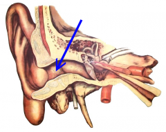
What is the structure shown (outlined in blue)? |
External Auditory Meatus |
|
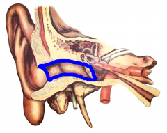
What is the area shown (outlined in blue)? |
Middle Ear |
|
What is the structure shown (outlined in blue)? |
Tympanic Membrane |
|
|
What is the function of the Tympanic Membrane? |
Vibrates in response to sound |
|
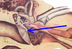
What is the structure shown (at the tip of pointer)? |
Umbo |
|
|
What is the Umbo? |
Attachment point for malleus |
|
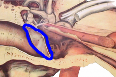
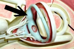
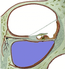
What is the space shown (outlined in blue)? |
Tympanic Cavity |
|
|
What is the area surrounding the Ossicles? |
Tympanic Cavity |
|
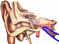
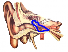
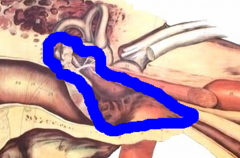
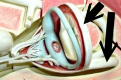
What is the structure shown (canal outlined in blue)? |
Eustachian Tube |
|
|
What is the function of the Eustachian Tube? |
Help equalize pressure between the external environment & middle ear |
|
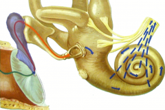
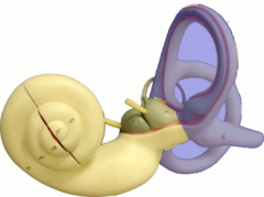
What is the area shown? |
Inner Ear |
|
|
What are the 3 Ossicles? |
--Malleus --Incus --Stapes |
|
What is the structure shown (highlighted in blue)? |
Malleus |
|
|
What does the Malleus articulate with? |
--Umbo --Incus |
|
What is the structure shown (highlighted in blue)? |
Incus |
|
|
What does the Incus articulate with? |
--Malleus --Stapes |
|
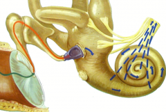
What is the structure shown (highlighted in blue)? |
Stapes |
|
|
What does the Stapes articulate with? |
--Incus --Oval Window |
|
|
What is the function of Ossicles? |
Transmit sound waves
|
|
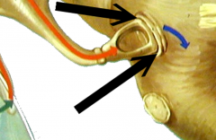
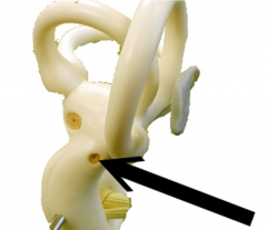
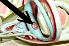
What is the structure shown? |
Oval Window |
|
|
Where does the Stapes articulate with the Cochlea? |
Oval Window |
|
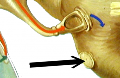
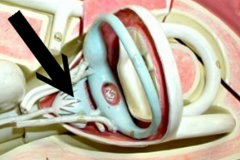
What is the structure shown? |
Round Window |
|
|
What is the function of the Round Window |
Where sound is diffused out of the Cochlea |
|
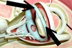
What is the structure shown (at tip of pointer)? |
Round Window |
|
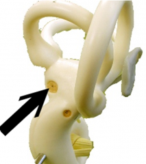
What is the structure shown (at tip of pointer)? |
Oval Window |
|
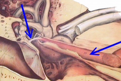
What is the structure shown (at tip of pointers)? |
Tensor Tympani |
|
|
What is the function of the Tensor Tympani muscle? |
Decrease vibration by increasing tension to prevent damage to Ossicles |
|
|
Where does the Tensor Tympani muscle insert? |
(Handle of) Malleus |
|
|
What is the function of the Stapedius muscle? |
Decrease vibration by increasing tension to prevent damage to Oval Window |
|
|
Where does the Stapedius muscle insert? |
(End of) Incus |
|
|
Where does the Stapedius muscle articulate? |
Stapes |
|
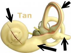
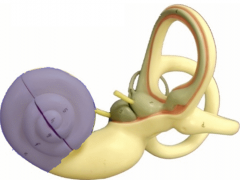
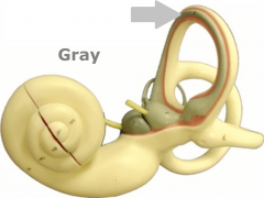
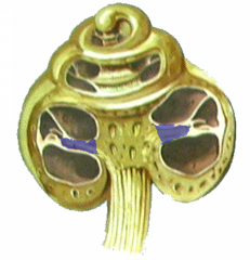
What is the structure shown (in tan)? |
Osseous Labyrinth |
|
|
What is the Osseous Labyrinth filled with? |
Perilymph |
|
|
What is the function of the Osseous Labyrinth? |
Receptors for hearing & balance |
|
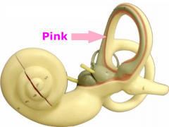
What is the cavity shown (in pink) filled with? |
Perilymph |
|

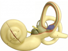
What is the structure shown (in grey)? |
Membranous Labyrinth |
|
|
What surrounds the Vestibule, Cochlea, & Semicircular Canals? |
Osseous Labyrinth |
|
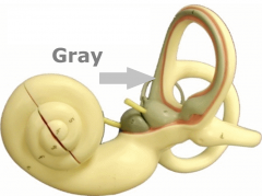
What is the cavity shown (in grey) filled with? |
Endolymph |
|
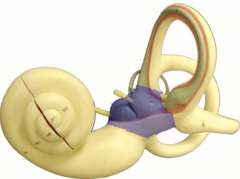
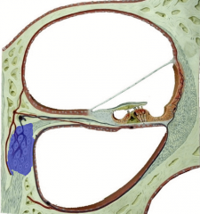
What is the structure shown (highlighted in blue)? |
Vestibule |
|
|
What is the function of the Vestibule? |
Receptors for equilibrium (static) |
|
What are the structures shown (highlighted in blue)? |
Semicircular Canals |
|
|
What is the function of the Semicircular Canals? |
Receptors for equilibrium (dynamic) |
|
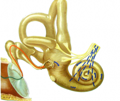
What is the structure shown (highlighted in blue)? |
Cochlea |
|
|
What is the function of the Cochlea? |
Receptors for hearing |
|
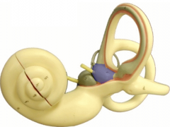
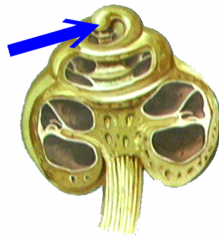
What is the structure shown (highlighted in blue)? |
Utricle ("little bag") |
|
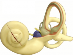
What is the structure shown (highlighted in blue)? |
Saccule ("little sac") |
|
|
What is the function of the Utricle & Saccule? |
Static equilibrium |
|

What is the structure shown (at tip of pointer)? |
Utricle |
|
What is the structure shown (at tip of pointer)? |
Saccule |
|
What is the structure shown (at tip of pointer)? |
Macula |
|
What are the canals shown (shown in grey)? |
Semicircular Ducts |
|
|
What is the function of Semicircular Ducts? |
Dynamic Equilibrium |
|
What is the structure shown (highlighted in blue)? |
Ampulla |
|
|
What is the function of the Ampula? |
Dynamic equilibrium |
|
|
What is the end of the Semicircular Duct called? |
Ampulla |
|
What are the structures shown (at tips of pointers)? |
Semicircular Canals |
|
What are the structures shown (at tips of pointers)? |
Semicircular Ducts |
|
What is the structure shown (at tip of pointer)? |
Ampulla |
|
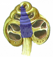
What is the structure shown (highlighted in blue)? |
Modiolus |
|
What is the structure shown (highlighted in bue)? |
Osseous Spiral Lamina |
|
|
What is the function of the Osseous Spiral Lamina? |
Anchors Basilar Membrane |
|
What is the structure shown (highlighted in blue)? |
Osseous Spiral Lamina |
|
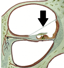
What is the structure shown (at tip of pointer)? |
Vestibular Membrane |
|
|
What is the function of the Vestibular Membrane? |
Separates Scala Vestibuli from Scala Media |
|
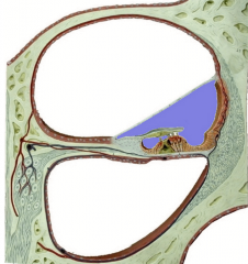
What is the structure shown (highlighted in blue)? |
Scala Media |
|

What is contained in the structure shown (highlighted in blue)? |
Endolymph |
|
|
What is the function of the Scala Media? |
Receptors for hearing |
|
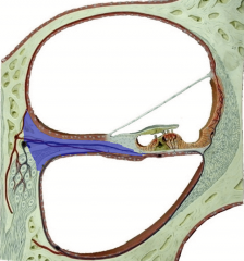
What is the structure shown (highlighted in blue)? |
Scala Vestibuli |
|

What is contained in the structure shown (highlighted in blue)? |
Perilymph |
|
|
What structure starts at the Oval Window? |
Scala Vestibuli |
|
|
What structure ends at the Round Window? |
Scala Tympani |
|
What structure is shown (highlighted in blue)? |
Scala Tympani |
|

What is contained in the structure shown (highlighted in blue)? |
Perilymph |
|
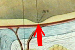
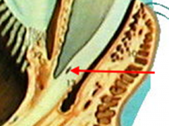
What structure is shown (at tip of pointer)? |
Helicotrema |
|
|
Where does the Scala Tympani meet the Scala Vestibuli? |
Helicotrema |
|
What is the structure shown (highlighted in blue)? |
Spiral Ganglion |
|
|
Where in the Coclea are the cell bodies of Bipolar Neurons located? |
Spiral Ganglion |
|
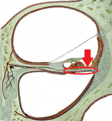
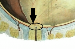
What is the structure shown (circled in red)? |
Basilar Membrane |
|
|
What is the function of the Basilar Membrane? |
Receptors for hearing |
|
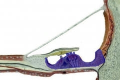
What is the structure shown (highlighted in blue)? |
Spiral Organ of Corti |
|
|
What are the 3 main structures of the Organ of Corti? |
--Hair Cells --Stereocilia (the "hairs") --Tectorial Membrane |
|
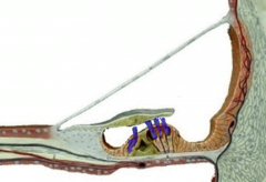
What are the structures shown (highlighted in blue)? |
Hair Cells (of Organ of Corti) |
|
|
What is the function of the Hair Cells in the Organ of Corti? |
Hearing receptors |
|
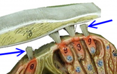
What are the structures shown (at tips of pointers)? |
Hairs (of Organ of Corti) |
|
What is the structure shown (highlighted in blue)? |
Tectorial Membrane |
|
|
What is the function of the Tectorial Membrane? |
Covers hair cells above Spiral Organ of Corti |
|

What are the structures shown (at tips of red pointers) collectively called? |
Photoreceptors |
|
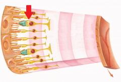
What is the structure shown (at tip of pointer)? |
Cone Cells (Photoreceptor) |
|
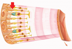
What is the structure shown (at tip of pointer)? |
Rod Cells (Photoreceptor) |
|
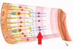
What type of cells are shown (at tip of pointer)? |
Bipolar Cells (1st Order Neurons) |
|

What type of cells are shown (at tip of pointer)? |
Ganglion Cells (2nd Order Neurons) |
|
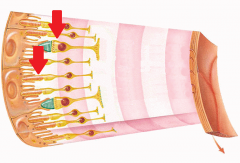
What type of cells are shown (at tips of pointers)? |
Photoreceptors (Receptor Cells) |
|

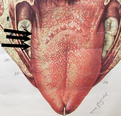
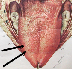
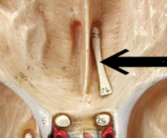
What are the structures shown (at tips of pointers)? |
Vallate Papillae |
|
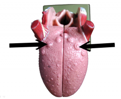
What is the function of the structures shown (at tips of pointers)? |
Vallate Papillae house 100-300 taste buds
|
|
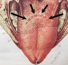
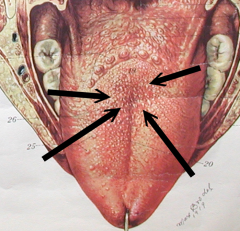
What are the structures shown (at tips of pointers)? |
Vallate Papillae |
|
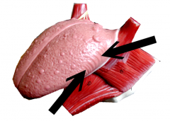
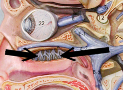
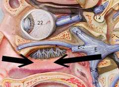
What are the structures shown (at tips of pointers)? |
Foliate Papillae |
|
|
What do Foliate Papillae NOT have now that they did in childhood? |
Taste Buds |
|
What are the structures shown (at tips of pointers)? |
Foliate Papillae |
|
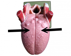
What are the structures shown (at tips of pointers)? |
Filiform Papillae |
|
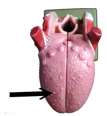
What are the structures shown (at tips of pointers)? |
Filiform Papillae |
|
|
What are the 4 types of Papillae of the Tongue? |
--Vallate Papillae --Foliate Papillae --Filiform Papillae --Fungiform Papillae |
|
What are the structures shown (at tips of pointers)? |
Fungiform Papillae |
|
|
What is the function of Filiform Papillae? |
Increase surface area for friction
(aids w/ bolus formation) |
|
|
What is the function of Fungiform Papillae? |
House 5 taste buds per papillae |
|
|
Which of the Papillae of the tongue do NOT house taste buds? |
Filiform Papillae |
|
|
What is the function of Taste Buds? |
House chemoreceptors (to taste food) |
|
What are the structures shown (at tips of pointers)? |
Fungiform Papillae |
|
What type of Papillae (of the Tongue) are shown? |
Fungiform Papillae |
|
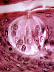
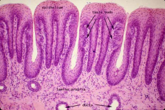
What is the large round structure shown called? |
Taste Bud |
|
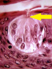
What is the structure shown (at tip of pointer)? |
Taste Pore |
|
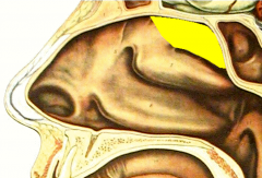
What is the structure shown (highlighted in yellow)? |
Superior Nasal Concha
|
|
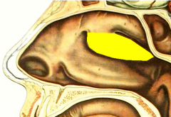
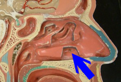
What is the structure shown (highlighted in yellow)? |
Middle Nasal Concha |
|
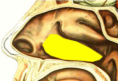
What is the structure shown (highlighted in yellow)? |
Inferior Nasal Concha |
|
|
What is the function of the Nasal Concha? |
--Warm & humidify air --Move air to smell receptors |
|
What is the structure shown (at tip of pointer)? |
Olfactory Bulb |
|
|
What is the function of the Olfactory Bulb? |
Pathway for signals |
|
What is the structure shown (at tip of pointer)? |
Olfactory Tract |
|
|
What is the function of the Olfactory Tract? |
Pathway for signals |
|
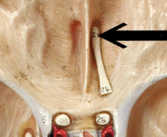
What are the structures shown (at tips of pointers)? |
Olfactory Nerves |
|
|
What is the function of Olfactory Nerves? |
Pathway for signals |
|
|
Where do Olfactory Dendrites enter into the Nasal Cavity? |
Cribriform Plate (of skull) |
|
What cells would be present at tips of pointers? |
Olfactory epithelium (Receptor Cells) |
|
|
What are the functions of Olfactory Epithelium? |
--House smell receptors --Produce Mucus (in Bowman's Glands) --Support |
|
|
What is an Emmetropic Eye? |
A normal Eye |
|
|
What is a Myopic Eye? |
A nearsighted eye |
|
|
What lens is used to correct for a Myopic Eye? |
Concave |
|
|
What is a Hyperopic Eye? |
A farsighted eye |
|
|
What lens is used to correct for a Hyperopic Eye? |
Convex |
|
|
What is the condition of becoming farsighted with age? |
Presbyopia |
|
|
What is an Astigmatic Eye? |
An eye with an irregularly shaped cornea or lens |
|
|
What type of lens is used to correct for an Astigmatic Eye? |
Toric ("donut" shaped) |