![]()
![]()
![]()
Use LEFT and RIGHT arrow keys to navigate between flashcards;
Use UP and DOWN arrow keys to flip the card;
H to show hint;
A reads text to speech;
132 Cards in this Set
- Front
- Back
- 3rd side (hint)
|
1. vision 2. taste 3. smell 4. hearing 5. equilibrium |
5 special senses |
|
|
|
special sensory receptors |
distinct receptor cells that are highly localized, and housed in complex organs (eyes and ears) or structures (taste buds, olfactory epithelium) |
|
|
|
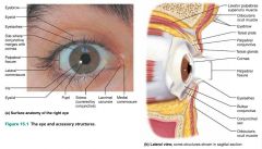
1. eyebrows 2. eyelids 3. conjunctiva 4. lacrimal apparatus 5. extrinsic eye muscles |
5 accessory structures of the eye |

|
|
|
eyebrows |
short, coarse hairs that help protect the eyes from sunlight and perspiration |

|
|
|
eyelids (palpebrae) |
protect the anterior portion of the eyes |

|
|
|
palpebral fissure |
the slit separating the upper and lower eyelids |
|
|
|
commisures |
medial and lateral angles of the eye |

|
|
|
lacrimal caruncle |
the fleshy elevation of the medial commissure that contains sebaceous and sweat glands; whitish oily secretion ("Sandman's eye sand") |

|
|
|
eyelashes |
richly innervated hairs projecting from the free margin of each eyelid; extremely sensitive to touch |

|
|
|
tarsal glands |
glands that secrete an oily secretion that lubricates the eyelid and eye |

|
|
|
conjunctiva |
transparent mucous membrane lining the anterior eye (except the cornea) and inner eyelids |

|
|
|
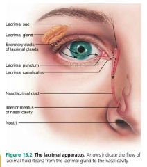
lacrimal apparatus |
consists of the lacrimal gland and the ducts that drain lacrimal secretions into the nasal cavity |

|
|
|
lacrimal gland |
orbital gland that secretes tears; superior and lateral to the eye |

|
|
|
lacrimal secretion (tears) |
secretion that cleans, protects, and lubricates the eyes |

|
|
|
lysozyme |
lacrimal fluid enzyme that destroys bacteria |
|
|
|
lacrimal canal |
drain for lacrimal secretions, run from the lacrimal sac into the nasolacrimal duct (nasal cavity) |
|
|
|
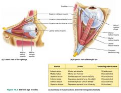
extrinsic eye muscles |
six straplike muscles that control eyeball movement |
|
|
|
1. lateral rectus 2. medial rectus 3. superior rectus 4. inferior rectus 5. inferior oblique 6. superior oblique |
6 extrinsic eye muscles |

|
|
|
1. oculomotor (III) 2. trochlear (IV) 3. abducens (VI) |
3 cranial nerves that innervate the extrinsic eye muscles |
|
|
|
diplopia |
double vision |
|
|
|
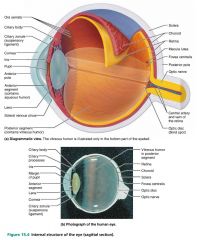
1. fibrous layer 2. vascular layer 3. inner layer |
3 layers of the eyeball |
|
|
|
fibrous layer (tunic) |
outermost layer of the eyeball; dense avascular connective tissue |

|
|
|
1. sclera 2. cornea |
2 regions of the fibrous layer of the eyeball |

|
|
|
sclera |
white part of the eye; continuous with the dura mater of the brain |

|
|
|
cornea |
transparent, anterior portion of the fibrous layer of the eye; consists of collagen fibers, refracts light |

|
|
|
1. sensitive 2. regenerates quickly 3. avascular |
3 important characteristics of the cornea |
|
|
|
vascular layer (tunic) |
pigmented middle layer of the eyeball |

|
|
|
1. choroid 2. ciliary body 3. iris |
3 regions of the vascular layer of the eye |

|
|
|
choroid |
highly vascular, dark brown membrane forming the posterior 5/6 of the vascular tunic; absorbs light (preventing it from scattering) |

|
|
|
ciliary body |
ring of tissue in the vascular layer of the eye that encircles the lens |

|
|
|
ciliary muscles |
smooth muscle controlling the shape of the lens |
|
|
|
ciliary processes |
posterior folds of the ciliary body that secrete aqueous humor |

|
|
|
ciliary zonules (suspensory ligaments) |
ligaments that connect the ciliary body to the lens, suspending it |

|
|
|
iris |
visible colored portion of the eye that controls pupil size |

|
|
|
pupil |
round central opening of the eye that controls the amount of light entering; ANS controlled |

|
|
|
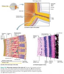
retina (sensory layer) |
innermost layer of the eyeball |

|
|
|
1. pigmented layer 2. neural layer |
2 layers of the retina |

|
|
|
pigmented layer |
outer layer of the retina that absorbs light and prevents it from scattering; abuts the choroid |

|
|
|
neural layer |
inner layer of the retina that transduces (converts) and transmits the visual signal |

|
|
|
1. photoreceptors 2. bipolar cells 3. ganglion cells |
3 types of neurons forming the neural layer of the retina |

|
|
|
photoreceptors |
outer neuron cells of the neural layer of the retina; contains rods and cones |

|
|
|
1. rods 2. cones |
2 types of photoreceptors in the eye |

|
|
|
rods |
photoreceptors for dim light (black and white) |

|
|
|
cones |
photoreceptors for bright light and high-resolution color vision |

|
|
|
bipolar cells |
middle cells of the neural layer of the retina; transmit signals from photoreceptors to ganglion cells |
|
|
|
ganglion cells |
innermost cells of the neural layer of the retina; generate action potentials through their axons (the optic nerve) |

|
|
|
optic disc |
where the optic nerve leaves the eye, 20% off center; blind spot because it has no photoreceptors |

|
|
|
macula lutea |
posterior center of the eye; high concentration of cones (acute vision) |

|
|
|
fovea centralis |
center of the macula lutea; highest concentration of cones |

|
|
|
vitreous humor |
clear viscous gel that maintains the shape of the eye; fills the posterior cavity (behind the lens) |

|
|
|
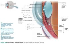
aqueous humor |
clear fluid that provides nutrients and O2 to the lens and cornea; fills the anterior cavity (around and in front of the lens) |

|
|
|
canal of Schlemm (scleral venous sinus) |
canal encircling the eye that returns aqueous humor to the blood |
|
|
|
glaucoma |
caused by blocked drainage of aqueous humor that compresses the retina and optic nerve |
|
|
|
lens |
flexible structure that changes shape to focus light on the retina; avascular |

|
|
|
crystallins |
tightly packed, transparent proteins that form the body of the lens |
|
|
|
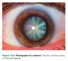
cataract |
clouding of the lens caused by hardening or thickening of the lens (crystallins clumping together) |
|
|
|
visible light |
part of the electromagnetic spectrum that eyes respond to; small particles called photons |
|
|
|
refraction |
the bending of light as it passes through different mediums |
|
|
|
- cornea - aqueous humor - lens - vitreous humor - retina - photoreceptors |
trace the path of light as it enters the eye (6 areas) |

|
|
|
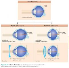
distant vision |
vision of parallel light; the cornea refracts the light |
|
|
|
close vision |
vision of diverging light; the lens must also refract the light, in addition to the cornea |
|
|
|
myopia (nearsightedness) |
when distant objects focus in front of the retina, rather than on it; eyeball is too long |

|
|
|
hyperopia (farsightedness) |
when objects focus behind the retina; eyeball is too short |
|
|
|
astigmatism |
blurry images caused by unequal curvature in different parts of the cornea or lens |
|
|
|
retinal |
a light-absorbing molecule that changes shape when exposed to light; comes from vitamin A |
|
|
|
opsin |
protein attached to retinal; 1 type for rods, 3 types for cones |
|
|
|
rhodopsin |
the photopigment of rods; breaks down in light, increases with dark |
|
|
|
light adaptation |
light completely bleaches rhodopsin, so cones take over; moving from darkness to light |
|
|
|
dark adaptation |
rod's rhodopsin accumulates in the dark, and cones cannot function; moving from light to darkness |
|
|
|
optic nerve (CN II) |
nerve formed by axons from the ganglion cells of the eye |
|
|
|
optic chiasma |
the point where fibers from the medial eye cross over to the opposite side; the right visual field heads to the left optic tract and left visual cortex, and vice versa |
|
|
|
optic tract |
the bundle of fibers carrying information from the visual field to the thalamus |
|
|
|
visual cortex |
responsible for conscious perception of a visual image; located in the occipital lobe |
|
|
|
superior colliculi |
area of the midbrain responsible for visual reflexes (head and eye movements) |
|
|
|
stereoscopic vision (binocular vision) |
the same image seen from different angles allows for depth perception |
|
|
|
thalamus |
area of the brain that receives input from both eyes; detects movement and depth perception |
|
|
|
primary visual cortex |
area of the brain that forms a topographical map of the retina |
|
|
|
visual association area |
area of the brain that interprets dynamic images, form, color, depth, and motion |
|
|
|
chemoreceptors |
receptors that respond to chemicals in an aqueous solution; smell and taste |
|
|
|
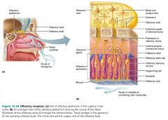
olfactory epithelium |
organ of smell located in the roof of the nasal cavity |

|
|
|
olfactory sensory neurons |
bowling pin-shaped receptor cells of the olfactory epithelium |

|
|
|
supporting cells |
cells surrounding and cushioning the olfactory sensory neurons |

|
|
|
olfactory cilia |
increase the receptive surface area of olfactory sensory neurons |
|
|
|
specificity |
the process of 400 "smell genes" creating combinations for 10,000+ different odors |
|
|
|
- olfactory nerve (CN I) - olfactory bulb - olfactory tract - olfactory cortex |
trace the path of smell as it enters the nose (4) |
|
|
|
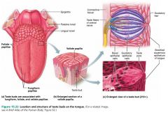
taste buds |
sensory organs for taste located on the tongue |

|
|
|
papillae |
peglike projections of the tongue mucosa that make the tongue surface slightly abrasive; house taste buds |

|
|
|
1. fungiform 2. foliate 3. vallate |
3 types of papillae |

|
|
|
1. gustatory epithelial cells 2. basal epithelial cells |
2 cell types found in taste buds |

|
|
|
gustatory epithelial cells |
receptor cells for taste; taste cells |

|
|
|
gustatory hairs |
long microvilli that project from the tips of gustatory epithelial cells; sensitive portion of the cell |
|
|
|
basal epithelial cells |
stem cells that become new gustatory epithelial cells |

|
|
|
1. sweet 2. sour 3. salty 4. bitter 5. umami |
5 basic taste sensations |
|
|
|
sweet |
taste sensation elicited by sugars and some amino acids |
|
|
|
sour |
taste sensation elicited by acids (hydrogen ions) |
|
|
|
salty |
taste sensation elicited by sodium |
|
|
|
bitter |
taste sensation elicited by alkaloids or rotten foods |
|
|
|
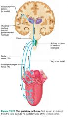
saliva |
must dissolve chemicals before they can be tasted |
|
|
|
1. facial nerve (CN VII) 2. glossopharyngeal (CN IX) |
2 main cranial nerves that carry taste information |
|
|
|
1. external ear 2. middle ear 3. internal ear |
3 main areas of the ear |
|
|
|
1. auricle (pinna) 2. external acoustic meatus (auditory canal) 3. tympanic membrane (eardrum) |
3 main parts of the external ear (outer ear) |
|
|
|
auricle (pinna) |
elastic cartilage surrounding the opening of the external acoustic meatus (auditory canal) |
|
|
|
external acoustic meatus (auditory canal) |
a short, curved tube that extends from the auricle to the eardrum; contains hairs, sebaceous glands, and ceruminous glands |
|
|
|
tympanic membrane (eardrum) |
thin, translucent connective tissue membrane between the outer and middle ears |
|
|
|
1. ossicles 2. oval window 3. pharyngotympanic tube (auditory tube) |
3 main parts of the middle ear (tympanic cavity) |
|
|
|
ossicles |
send vibration from the tympanic membrane to the oval window; three smallest bones in the body |
|
|
|
malleus |
the ossicle secured to the tympanic membrane (eardrum) |
|
|
|
incus |
middle ossicle |
|
|
|
stapes |
ossicle that fits into the oval window |
|
|
|
oval window |
opening in the lateral wall of the inner ear that eventually leads to the cochlea |
|
|
|
pharyngotympanic tube (auditory/eustachian tube) |
equalizes pressure in the middle ear with external air pressure; runs from the middle ear to the nasopharynx |
|
|
|
1. cochlea 2. vestibule 3. semicircular canals |
3 main parts of the inner ear (labyrinth) |
|
|
|
cochlea |
spiral bone cavity filled with endolymph |
|
|
|
endolymph |
fluid that conducts sound vibrations |
|
|
|
organ of Corti (spiral organ) |
receptor organ for hearing, located in the cochlea duct |
|
|
|
vestibule |
egg-shaped cavity between the cochlea and the semicircular ducts |
|
|
|
semicircular canals |
set of three canals (anterior, posterior, lateral) that attach to the vestibule of the inner ear |
|
|
|
crista ampullaris |
equilibrium receptors at the base of the semicircular canals that respond to rotational movement of the head |
|
|
|
sound |
pressure disturbance produced by a vibrating object and propagated by the molecules of the medium |
|
|
|
frequency |
the number of sound waves that pass a given point in a given time; measured in Hz (hertz) |
|
|
|
amplitude |
height of the sound wave, representing the intensity of the sound; measured in dB (decibels) |
|
|
|
differential stimulation |
the process of different pitches stimulating different areas of hair cells |
|
|
|
amplification |
pressure hitting the oval window is 20x greater than on the eardrum, due to its smaller size |
|
|
|
cochlear hair cells |
cilia that protrude into the endolymph and are vibrated; open K+ channels |
|
|
|
cochlear nerve |
nerve serving the auditory pathway; leads to the inferior colliculi (of the midbrain) and the thalamus, before being routed to the temporal lobe |
|
|
|
conduction deafness |
deafness caused by interference with conduction of vibrations to the inner ear, due to blockage of the external ear or ossicles fusing together |
|
|
|
sensorineural deafness |
deafness caused by damage to the hair cells (loud noises) or neural component problem (hair cells to cortex) |
|
|
|
static equilibrium |
linear changes in head position |
|
|
|
dynamic equilibrium |
rotational acceleration changes in head position |
|
|
|
maculae |
equilibrium receptors of the inner ear that respond to change in position of the head |
|
|
|
1. vestibular nuclei 2. cerebellum |
2 destinations of the equilibrium pathway |
|
|
|
motion sickness |
sickness caused by a mismatch of vision and equilibrium signals; nausea and vomiting |
|