![]()
![]()
![]()
Use LEFT and RIGHT arrow keys to navigate between flashcards;
Use UP and DOWN arrow keys to flip the card;
H to show hint;
A reads text to speech;
108 Cards in this Set
- Front
- Back
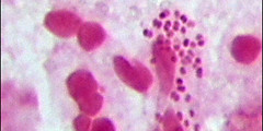
- Pathogenic Gram negative diplococci in kidney shape
- Intracelullar |
Neisseria gonorrhoeae
-Causes: Gonorrhoea, a purulent infection of urethra and endocervical mucous membrane - Transmitted sexually or at childbirth -Agar Chocolate, Thayer-Martin, Martin Lewis, Tray GC and - Coagulase +, Oxidase + |
|
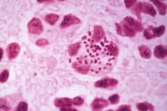
- Can be normal flora in nasopharynx and mouth, but its usually pathogenic
- Gram negative intracellular diplococci
|
Neisseria meningitidis
-Causes Meningitis
- appear grey or creamy in BA
- some can be encapsulated - colonies are mucoid
|
|
|
Who am I?
What disease to I cause?
PS: I am a Gram positive cocci found as plain chains sometimes normal flora of skin and throat or mouth. I belong to Lancefield group A.
|
Streptococcus pyogenes -Causes strep throat (a.k.a. pharyngitis), puerperal fever (sepsis after childbirth), Scarlet fever (rash because of antibiotic therapy), erysipelas (a form of cellulitis accompanied by fever and systemic toxicity) wound infections and eating- flash disease, necrotizing fasciitis, myositis and streptococcal toxic shock syndrome, rheumatic fever and acute glomerulonephritis. -Beta hemolytic (important Group A pathogen) - Pus producing infections - Bacitracin and SXT resistent |
|
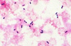
Who am I? What disease to I cause? PS: - Gram positive, Large lancet-shaped diplococci, have a polysaccharide capsule |
Streptococcus pneumoniae -Alpha hemolytic = greenish colour on BA = partial hemolysis -Not normal human flora, but resides asymptomatically in the nasopharynx of healthy carriers. -Common cause of pneumonia, middle ear infections, endocarditis, and meningitis. - Optochin sensitive - distinguishes it from other alpha-hemolitics.
|
|
Who am I? What disease to I cause? PS: Gram positive cocci in tetrads clusters, colonies are mucoid, glistening, smooth and circular, greyish on NA. - Catalase + |
Staphylococcus epidermidis - Alpha-hemolitic = green hemolysis on BA = partial hemolysis - Normal flora of skin of forehead, scalp, eyebrows, face and ears. -Opportunistic infections, infections from catheters and stitches, acne pimples.
|
|
Who am I? Gram positive cocci found in pairs or clusters. Colonies can be yellow orange or white, smooth, raised and circular. - Normal human flora of nasal membranes and skin. |
Staphylococcus aureus -Beta hemolytic - total hemolysis = clear on BA -Causes wound and skin infections (pimples), endocarditis, abscesses in bones, food poisoning, sinusitis, shock toxic syndrome, cellullitis, MRSA. - Differentiate form other Staphylococcus because it is Coagulase + |
|
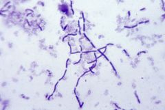
Who am I? |
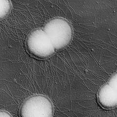
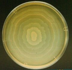
Streptomyces venezuelae
- gram positive rod
-Note long chains of circular conidia (reproductive spores - not endospores) |
|
What are the white coloured capsules? |
Endospores in Bacillus subitilis |
|
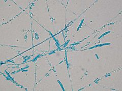
Who am I? |
Penicillin, a.k.a. "bread mold"
-Note tree-like structure -From top to bottom the structure goes: Conidia, Conidiophore, hyphae.
Scotch tape method = lactophenol cotton blue dye |
|
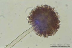
Who am I?
What disease can I cause?
Where am I found outside the human body? |
Aspergillus niger
Causes Aspergillosis in lungs (a.k.a. Farmer's lung), as well as skin, ear, sinus infections -Note chains of conidia that create the "dandelion" look-like and the stem-like hyphae. -The entire structure is called a conidiophore (black) -Found in soil, external auditory canal |
|
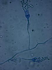
Who am I? What disease can I cause? Where am I found outside the human body? |
Microsporum canis
-Causes "ringworm", a.k.a. tinea capitis/corporis -Found in dogs and cats -Note spindle-shaped macroconidia and small circular microconidia |
|
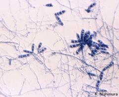
Who am I? What diseases do I cause? Where do I live outside the body? |
Epidermophyton floccosum
- dermatophyte - skin, nails, and hair - Causes tinea pedis (a.k.a. Athlete's foot), tinea cruris (skin upper tight in males), and onychomycosis. -Common on wet gym floors -Note thread-like hyphae and terminal macroconidia triads. There are no microconidia. |
|
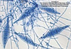
Who am I? What disease do I cause? Where do I live outside the body? |
Trichophyton mentagrophytes
- skin, nails and hair - Causes tinea pedis (a.k.a. Athlete's foot) -Common on wet gym floors -Note threadlike hyphae, small circular microconidia in clusters like grapes, and bulbous macroconidia that not end in triads. |
|
![Who am I? (This is eosin methylene blue [EMB] agar)](https://images.cram.com/images/upload-flashcards/66/69/31/7666931_m.jpg)
Who am I?
(This is eosin methylene blue [EMB] agar) |
Escherichia Coli
-Only microorganism that turns metallic green on EMB agar.
-Lactose fermentor |
|
|
Eosin Methylene Blue (EMB) Agar
What am I differential and selective for? What colors do I turn, and what do they mean? |
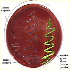
-Selective for gram negative rods (dye inhibit gram positive to grow) - Differentiate from lactose fermentors, specially E.coli - metallic -Fmgreen sheen -Fermentors turn purple, - non-fermentors turn brown. |
|
|
What am I differential for? What colors do I turn, and what do they mean?
Blood Agar |
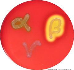
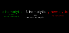
-Tests for hemolysis (breakdown of blood cells by m/o's)
-3 types of hemolysis shown on the agar: Alpha hemolysis (green color - indicates partial hemolysis, e.g. E. coli) Beta (clear - indicates complete hemolysis, e.g. Strep. pyogenes), Gamma (colonies are white colored, no hemolysis at all, e.g. Staph. epidermis) |
|
|
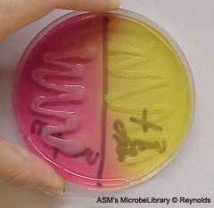
MacConkey (MK) Agar
What am I differential an selective for?
What colors do I turn, and what do they mean? |
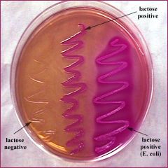
- selective for gram negative rods - differential for lactose fermentators (pink) and non lactose fermentors (yellow-brown)
-E. coli and Yersinia are fermentors |
|
|
Mannitol Salt Agar
Selective? Differentiator? |
- Select m/o that can grown in high concentration of salt like Staphylococcus - Differentiate between Mannitol fermentors -Fermentors = yellow = S. aureus. -Non-fermentors = pink = S. epidermis
|
|
|
DNAse Agar
confirmatory for? |
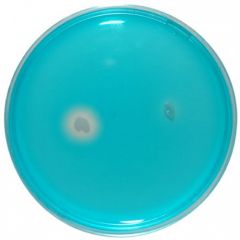
- Tests for breakdown of DNA by m/o -Clear zone = positive for DNA breakdown -Confirmatory for Staph. aureus |
|
|
Hektoen Enteric (HE) Agar
Selective and differential for?
What colors do I turn, and what do they mean? |
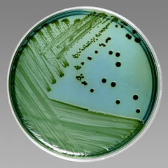
-Selective for gram negative Differentiates Salmonella and Shigella = non fermentors -Shigella = green colonies -Salmonella = colonies with black due to production of ferrous sulfide |
|
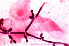
Who am I?
What disease do I cause?
Am I normal flora? |
Candida Albicans
-Causes candidiasis (a.k.a. yeast infection) -Normal flora of the human GI tract, skin, mouth, and vagina. -Gram positive, Opportunistic pathogen, kept in check by lactobacilli - bulb-like chlamydospores, stem-like pseudohyphae, and budding yeast cells (the dark pink, branch-like structure is the yeast infection) |
|
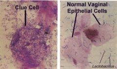
What is a "clue cell"? |
Clue cells are epithelial cells of the vagina that get their distinctive stippled appearance by being covered with bacteria. - They are a medical sign of bacterial vaginosis, particularly that caused by Gardnerella vaginalis |
|
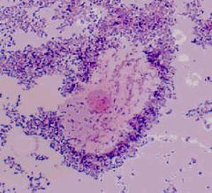
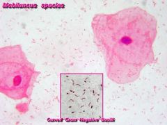
Who am I?
What disease is this called? |
Gardnerella vaginalis -Gram negative to variable, pleomorphic, non-motile rod, beta-hemolitic on Vaginalis agar. -Major causes of vaginosis - infection is characterized by a foul, fishy smelling, thin gray vaginal discharge, and an increase in vaginal pH from around 4.5 to over 5.5 -Note large epithelial "clue cell" covered in bacteria, and Mobiluncus (slender curved bacilli not attached to cell surface) |
|
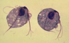
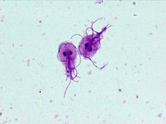
Who is this pear shaped flagellated protozoan? |
Trichomonas vaginalis (trophozoite) -Inhabits urogenital system of females and males. Sexually transmitted. -Note wavy undulating membrane, dark staining nucleus, 4 anterior flagella, and the axostyle that extends from nucleus to outside of the membrane, forming a "tail". A parabasal body lies between nucleus and undulating membrane. - accounts for 5-10% of vaginal infections - is STD - only trophozoite, does not survive outside body |
|
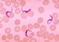
Who am I? What disease do I cause? Who are my hosts? |
Trypanosoma brucei gambiense
-Causes Sleeping Sickness (a.k.a African Trypanosomiasis) -Intermediate Host = Tsetse fly (Glossinia) injects trypomastigostes in humans -Definitive Host = Humans -Note flagella, undulating membrane, and central nucleus , presence of RBCs around. |
|
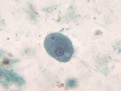
Who am I? What stage am I in? What disease do I cause? Where do I live outside of the human body? Where do I live in the body? |
Entamoeba histolytica (cyst)
-Protozoan: amoeba -Causes amoebiasis (a.k.a. amoebic dysentery) -Lives in contaminated food and water -Becomes trophozoite in human intestine -Turns into cyst when leaving the host -Note the 3 eye-like nuclei, darker coloured chromatoid bodies (RNA accumulations) |
|
Who am I? What stage am I in? What disease do I cause? Where do I live outside of the human body? Where do I live in the body? |
Entamoeba histolytica (trophozoite)
-Protozoan: amoeba -Causes amoebiasis (a.k.a. amoebic dysentery) severe abdo cramps and diarrhea, pseudopodes to move -Lives in contaminated food and water -Becomes trophozoite in human intestine -Turns into cyst while outside a host -Note eye-like nucleus, with a dark staining karyosome in its center, and peripheral chromatin beads |
|
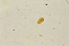
Who am I? What stage am I in? What disease do I cause? Where do I live outside of the human body? |
Giardia lamblia (trophozoite)
-protozoa: flagellate pear-shaped protozoa with two nuclei, giving appearance of eyes. - note: central axis is a structure called axostyle that crosses to ends in the flagellas (provides support and anchors for the organism) A parabasal body in the horizontal looks like a smily-lips above the nuclei. - Causes: Beaver fever or travellers diarrhea - ingestion of contaminated water - bearver is reservoir - 10% infection in USA |
|
Who am I? What stage am I in? What disease do I cause? Where do I live outside of the human body? Where do I live in the human body? |
Giardia lamblia (cyst) -Protozoan: flagellate -Causes Giardia (a.k.a. beaver fever/traveller's diarrhea), moves via whiplike appendages - flagellas -Beaver and wild animals are reservoir, lives in contaminated water -Lives in human GI tract -Note two sets of eye-like nuclei, and darker-staining curved parabasal body - boil water |
|
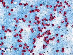
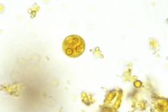
Who am I? What disease do I cause? Where do I live outside of the human body? Where do I live in the body? |
Cryptosporidium parvum
-Protozoan: sporozoan -Causes cryptosporidiosis in AIDS pt (profuse, watery diarrhea and epigastric cramping 10-15 days) -Inhabits contaminated water sources -Infests small intestine -Note large, pink oocysts and smaller, circular, dark sporozoites. - cysts are very resistant to water treatment with chlorine. |
|
|
Toxoplasma gondii ( tachyzoite) |
|
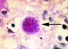
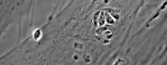
Who am I? What stage am I in? What disease do I cause? Where do I live in the body? Who are my hosts? |
Toxoplasma gondii (bradyzoites) -Protozoan: bradyzites -Causes toxoplasmosis -Intermediate hosts = rodents or humans (harbour 2 of the parasite's 3 stages - bradyzoites or tachyzoites) -Definitive host = cats (harbours merozoites) - dangerous for pregnant worn - fetus brain and eye damage -Note many small circular bradyzoites within the large circular tissue cyst |
|
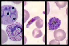
Who am I? What stages am I in? What disease do I cause? Where do I live in the human body? What am I transmitted by? What is my disease progression? |
Plasmodium
-Protozoan: plasmodium -Causes malaria - 1 million of death worldwide -Transmitted by Anopheles mosquito = definitive host -Stages from left to right: signet ring/trophozoite, macrogametocyte, schizont -Disease progression: Sporozoites injected into host by mosquito; then enter hepatocytes of the liver, where schizonts are formed; these burst to release merozoites, which feed on hemoglobin and become trophozoites; can then form more merozoites or microgametocytes and macrogametocytes to be taken up again by mosquito |
|
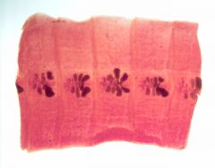
Who am I?
Who are my hosts? |
Cross section of Taenia saginatus, a.k.a. "beef tapeworm"
-Intermediate host = cows -Determinate host = humans -Note branched uterus within the mature proglottid |
|
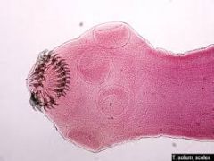
Who am I? Who are my hosts? |
Taenia solium, a.k.a. "pork tapeworm"
-Intermediate host = pigs -Determinate host = humans -Note the hooks in the centre = rostellum and four circular suckers |
|
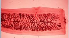
What is this a cross section of? What structures can you see? |
Cross section of Diphyllobothrium latum, a.k.a. "fish tapeworm"
-Note rectangular sections called proglottids, bilobed ovary on the posterior section of the proglottid with the winding uterus and tube-like vagina - vit b12 deficency in humans - fish is 2nd intermediary host - human is definitive host - eats the fish with plerocercoid larvae |
|
Who am I? Who are my hosts? What stage am I in? |
Echinococcus granulosus, a.k.a. "dog tapeworm" (adult stage) -Intermediate host = humans ingest eggs hanging on fur of dogs -Determinate host = dogs, wolf or coyote. - larvae in mucosa goes to blood and form hydatid cysts in brain, liver. Its explodes and larva's in blood produce anaphylactic shock (massive immune response) -Note rostellum protruding from scolex with suckers, and dark staining ovary on the posterior mature proglottid - most dangerous and smallest - 5mm |
|
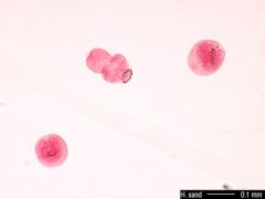
What is this? |
Hydatid sand of Echinococcus granulosus
-Note circular scolices (black rings) |
|
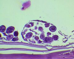
What is this? |
Hydatid cyst of Echinococcus granulosus in brain tissue
-Note hydatid cyst (fluid filled bag) and purple colored circular brood capsules inside it |
|
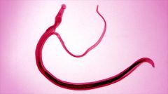
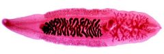
Who am I? What is going on in the picture? What disease do I cause? Who are my hosts? |
Schistosoma species (picture shows female and male in copula)
-protozoa - fluke - Causes cercarial dermatitis (swimmer's itch) -Intermediate host: snails -Definitive host: humans -Note thicker male fluke and thin female in copula |
|
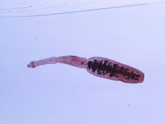
Who am I?
What stage am I in? |
Schistosoma mansoni (egg)
-Note the spines present on egg |
|
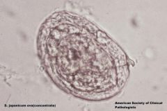
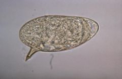
Who am I? What stage am I in? |
Schistosoma japonicum (egg)
-Note very small subterminal spines present on egg compared to S. mansoni eggs. |
|
Who am I? Where do I live in the human body after infection? Who are my hosts? |
Clonorchis sinensis, a.k.a. "Chinese river fluke"
-Infects human pancreas and liver -Intermediate hosts: snails (cercaria) and fish -Definitive host: humans -Note oral sucker on anterior end, followed by two caeca branches, with a long, dark staining uterus followed by dark pink testes at the posterior end. |
|
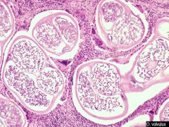
Who am I? What disease do I cause? Who are my hosts? |
Onchocerca volvulus
-Causes river blindness (a.k.a. onchocerciasis) -Intermediate host: black fly (Simulium) -Definitive host: humans -Note adult worms present inside the parasite-induced circular nodules surrounded by subcutaneous tissue |
|
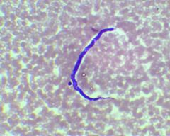
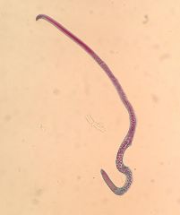
Who am I? What disease do I cause? Who are my hosts? |
Loa loa (a.k.a. "eye worm")
-Causes Calabar swellings (inflammatory reactions) in the eye -Intermediate host: deer fly (Chrysops species) -Definitive host: humans -Note dark purple color and dark staining nucleus that extends to the tail of the sheathed microfilariae |
|
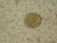
Who am I? What stage am I in? |
Ascaris lumbricoides (egg)
-Most common helminth infection -Causes Ascaris pneumonitis (a.k.a. Loeffler's syndrome) -Note ovoid shape, yellow-brown color, with thick, transparent shell and wavy membrane. |
|
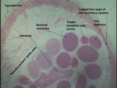
Who am I? What stage am I in? Am I male or female? What disease do I cause? Can you identify the different structures? |
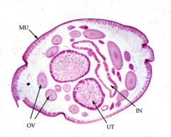
Ascaris lumbricoides (female adult)
-Causes Ascaris pneumonitis (a.k.a. Loeffler's syndrome) -Note uterus (UT), ovaries (OV), intestine (IN), longitudinal muscle (MU), and cuticle (outer membrane) |
|
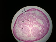
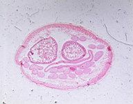
Who am I? What stage am I in? Am I male or female? What disease do I cause? Can you identify the different structures? |
Ascaris lumbricoides (male adult)
-Causes Ascaris pneumonitis (a.k.a. Loeffler's syndrome) -Note testis, body cavity (white space), intestine, cuticle (outer membrane), and muscle. |
|
Who am I?
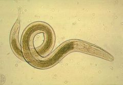
What stage am I in? |
Enterobius vermicularis, a.k.a. "pinworm" (adult stage)
-Note thin spicule at the end of the pin shaped tail with dark-staining eggs inside the worm.
|
|
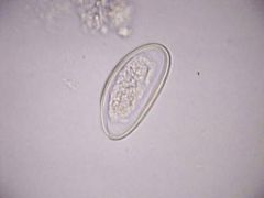
Who am I? What stage am I in? |
Enterobius vermicularis, a.k.a. "pinworm" (egg stage)
-Note ovoid shape, and flattened area on one side |
|
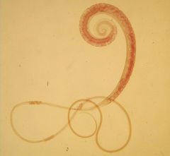
Who am I? What stage am I in? |
Trichuris trichiura, a.k.a. "whipworm" (adult stage)
-Note thickened posterior with dark-staining eggs and whip-like anterior. |
|
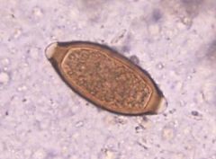
Who am I? What stage am I in? |
Trichuris trichiura, a.k.a. "whipworm" (egg stage)
-Note oval shape and distinctive brown shell with translucent knobs at each end. |
|
Who am I? What disease do I cause? Who are my hosts? What stage am I in? |
Trichinella spirallis, a.k.a. "porkworm" (adult)
-Causes trichinosis -Intermediate host: bears/pigs -Definitive host: pigs/humans -Note long, thin shape |
|
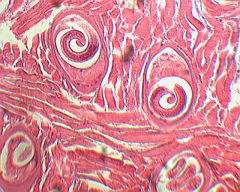
Who am I? What disease do I cause? Who are my hosts? What stage am I in? |
Trichinella spirallis, a.k.a. "porkworm" (cyst)
-Causes trichinosis - nauseas, muscle pain, fever, encephalitis -Intermediate host: bears/pigs -Definitive host: pigs/humans -Note oval shaped cyst that is embedded in muscle tissue, containing a coiled up larva. |
|
Who am I? What disease do I carry? |
Glossina, a.k.a. "Tsetse fly"
-Transmits African sleeping sickness (Trypanosoma gambiense) -Note slender, horizontally projecting proboscis with hooked legs |
|
Who am I? What disease do I carry? |
Simulium fly, a.k.a. "Blackfly"
-Transmits onchocerciasis (O. Volvulus) -Note dark color, and ten-segmented abdomen with a humpbacked thorax. |
|
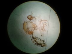
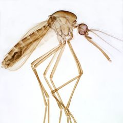
Who am I? What disease do I carry? |
Anopheles mosquito
-Carries malaria (a.k.a. plasmodium) -Lives in Africa and South America -Note dark colouring head and long palpi (2) and proboscis |
|
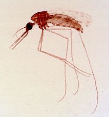
Who am I? What disease do I carry? |
Culex mosquito
-Carries West Nile Virus (a.k.a. Flavivirus) -Lives in North America -Note light coloring and very short palpi |
|
Who am I? What disease do I carry? |
Cimex lectularius, a.k.a. "Bedbug"
-Can sometimes transmit MRSA -Note flat, oval body with a short, wide head and a hairy body |
|
Who am I? What disease do I carry? |
Chrysops niger, a.k.a. "deer fly"
-Transmits Loa loa (the dark purple worm) -Note the large conical labellum (mouthpart for cutting skin and sucking blood) and wings with evenly distributed veins. |
|
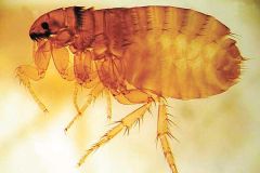
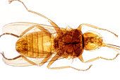
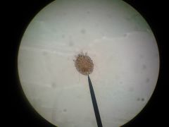
Who am I? What disease can I carry? |
Ctenocephalides canis, a.k.a. "dog flea"
-Causes dermatitis, mostly affects dogs and cats -Note segmented abdomen, strong hind legs, and tuft of hair behind head. |
|
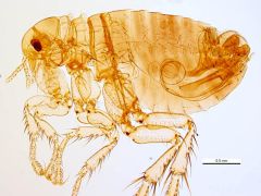
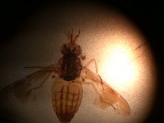
Who am I? What disease do I carry? |
Pulex irritans, a.k.a. "human flea" (Pulga)
-Intermediate host for D. caninum (dog tapeworm) cause itching -Note segmented abdomen, strong hind legs, and the absence of hair behind the head. |
|
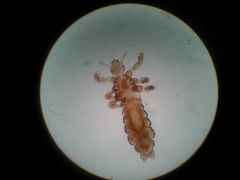
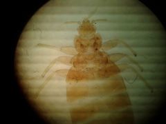
Who am I? |
Pediculus humanis, a.k.a. "head lice"
-Note segmented abdomen, and longer body shape |
|
Who am I? |
Phthirus pubis, a.k.a. "crab lice"
-Note crab-like body, with short head and powerful claws - found in pubic hair, transmitted sexually |
|
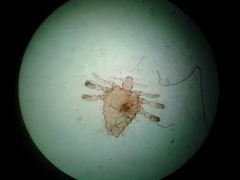
Who am I? |
Sarcoptes scabiei, a.k.a. "itch mite"
-Note small size, simple eyes, no antennae, and four legs, with streaming "tails" posteriorly - very membranous body with numerous long hairs
|
|
Who am I? What disease do I carry? |
Amblyoma americanum, a.k.a. "hard tick"
-Transmits spotted fever (Rickettsia rickettsi) -Note crescent-moon shape marking on back and spider-like appearance |
|
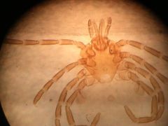
Who am I? What disease do I carry? |
Argas persicus, a.k.a. "soft tick"
-Transmits rabies and relapsing fever (Borrelia) -Note sand-dollar (bolacha do mar) appearance and egg-like shape |
|
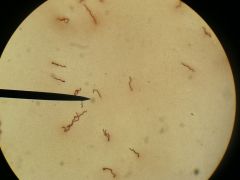
Who am I? |
Borrelia burgdorferi, a.k.a. "Lyme's disease"
-Note long Gram-negative spirochetes |
|
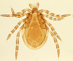
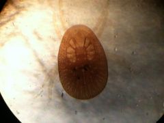
Who am I? What disease do I carry? |
Ixodes dammini, a.k.a. "Deer tick"
-Transmits Lyme's disease (Borrelia burgdorferi) -Note reddish brown body, spider-like appearance, and bulb-shaped thorax
corpo parece ter armadura |
|
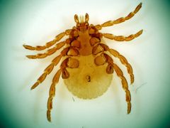
Who am I? What disease do I carry? |
Dermacentor andersoni, a.k.a. "Wood tick"
-Transmits Rocky Mountain spotted fever (R. rickettsii) -Note oval shape, eight segmented legs, and many lil rectangles shaped on edge of thorax |
|
|
Describe Gram stain.
Which is the mordant and what's it used for?
What's the critical step on this technique? |
- Gram staining is based on the ability of bacteria cell wall to retaining the crystal violet dye during solvent treatment. - The cell walls for Gram-positive microorganisms have a higher peptidoglycan and lower lipid content than gram-negative bacteria. - All bacteria cell walls are stained by the crystal violet. - Iodine is subsequently added as a mordant to form the crystal violet-iodine complex so that the dye cannot be removed easily. - Alcohol - acetone decolorizer dissolves the lipid layer from the gram-negative cells, removing the lipid layer along with Crystal violet from Gram Negative, but dehydrates the thicker Gram-positive cell walls, closing the pores as the cell wall shrinks holding violet-iodine inside the bacteria that remain stained purple. - The he decolorization is critical step because a prolonged exposure to the decolorizing agent will remove all the stain from both types of bacteria. - Finally, a counterstain of basic safranin is applied to the smear to give gram-negative bacteria a pink color. |
|
|
What is negative staining?
When is it useful? |
- Negative stain creates a dark background of suspended carbon particles on which transparent organisms are easily visible.
- India ink (nigrosine) is mixed with the sample and spread in the slide.
- Is useful for viewing bacterias difficult to satin such as spirochetes
|
|
|
Explain the Acid-fast Stain?
What's used for? Give examples. |
- Is used to stain bacterias that stain poorly on Gram technique because of the large amount of wax layer of mycolic acid. - examples of bacterias are Mycobacterium tuberculosis, Mycobacterium leprae, and M. phlei. - Carbol-fuchsin is used to stain the smear placed on top of a boiling water bath, so heat help the solvent to stain the cells (4 min) - Acid-Alcohol is used to decolourize - Methilene-blue is counterstain used to differentiate non-acid fast bacteria. - The bacteria staining pink is the Acid-fast, because it was not readily decolourized by acid-alcohol and kept carbol-fuchsin. |
|
|
What's Brownian motion?
Give examples |
Brownian motion is a vibratory movement of bacterias and particles suspended in liquid caused by random collisions with molecules of water.
It is not true motility. |
|
|
What are the types of Flagellar motion? |
The Flagellar motion is a mechanism of bacterial motility.
2 types: Peritrichous and Polar
Peritrichous flagella: attached across the whole surface of the bacterium and radiating in all directions
Polar flagella: attached at the ends of the bacterium. Polar Monotrichous: single flagella at one end; Lophotrichous: more than one flagella at one end; Amphitrichous: more than one flagella at both ends. |
|
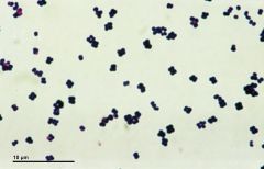
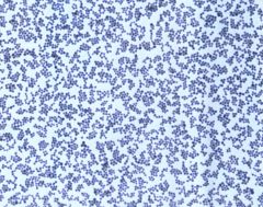
Who am I?
I am a purple, large cocci, usually found in tetrads? |
I am Micrococcus sp
- I a Gram positive bacteria, nonmotile, non-pathogenic aerobe found in skin Micrococcus luteus: tetrads and clusters, colonies smooth, golden yellow and convex on NA Micrococcus lylae: tetrads, colonies non-pigmented, circular, smooth and convex on NA Micrococcus roses: pairs and tetrads, colonies are pink or red on NA
|
|
|
Who am I? Am I normal flora? PS: I am the predominant cause of urinary tract infection in young women |
Staphylococcus saprophyticus - Gram positive cocci found in pairs or isolated - Alpha-hemolitic - no hemolysis - rarely present on skin - transient flora - predominant staphylococcal infection of GU - cystitis, urethritis and pyelonephritis in young women - great ability to hold on to uroepithelial cells - Novobiocin (NB 5) Resistent = identification |
|
|
Who am I? PS: I am the 2nd most frequent causes of nosocomial infections and septicemia. |
Staphylococcus haemolyticus - gram positive cocci found in skin. - in pairs or tetrads, colonies similar to S. saprophyticus. - Beta-hemolitic = greenish = parcial hemolysis on BA - 2nd most frequent cause of septicemia, native valve endocarditis, peritonitis, UTI, wound, bone, and joint infections - Can colonize central venous catheters due to health care manipulation. |
|
|
- Facultative anaerobes or microaerophilic - Gram positive cocci in chains - Lancefield classification from A to O - Can cause hemolysis - Major causes of bacterial pneumonia and sore throats - White colonies on BA - Catalase negative |
Streptococcus sp |
|
|
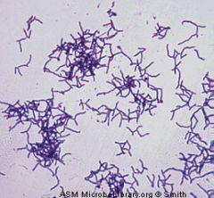
- I am a gram + cocci in short chains
- I vary form and this contributes to my name - in acidic environment or from oral samples I appear as short rod.
- I contribute to tooth decay
- viridans group |
Streptococcus mutans |
|
|
- I am a gram positive cocci found on cheek mucosa
- occasionally a pathogen in ulcerated teeth, sinuses, and blood/heart lesions in subacute endocarditis.
- alpha-hemolitic = viridans group |
Streptococcus mitis |
|
|
- Gram positive isolated from human faeces
- causes UTIs and endocarditis
- football shape in short chains
- Lancefield group D |
Enterococcus faecalis |
|
|
- Gram negative, kidney bean shaped diplococci,
- normal flora of mucus membranes
- non-pathogenic
- gamma - hemolitic |
Neisseria mucosa Neisseria subclava Neisseria sicca |
|
|
Gram negative cocci
Formally called Neisseria
Normal flora of upper respiratory tract of healthy individuals
Catalase + |
Moraxella catarrhalis
Implicated in infections of middle ear, maxillary sinus, meningitis and endocarditis |
|
|
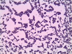
Gram positive rods
common in moist areas of skin
catalase +
mucoid grey colonies |
Corynebacterium
sometimes produces metachromatic granules and curved rods |
|
|
Small Gram positive rods
Found in forehead, scalp, and near sweat glands
Yellow orange or red colonies |
Propionibacteria
causes acne |
|
|
Large Gram positive rods in vagina
non-sporing
fermentative (glycogen in vagina = lactic acid = ↓ pH = protection)
|
Lactobacillus - Döderlein |
|
|
Gram positive rod, but tend to stain pink!
Predominant anaerobic
Causes vaginoses
Curved arranged in pairs = gull wings appearance |
Mobiluncus
- Usually in association with Gardnerella - clue cells |
|
|
Do not have cell wall
Found in vagina and cervix
less common cause of vaginoses |
Mycoplasma hominis |
|
|
This vaginose accounts for 30 - 35% of the vaginal infections.
Common in diabetics and pregnant women, or due to use of oral contraceptive and antibiotic therapy that inhibits normal vaginal flora
|
Candidiasis |
|
|
Catalase test |
Catalase enzyme is found in aerobic organisms, breaks down H2O2 to water and O2
Positive = bubbling O2 --- Staphylococcus and Micrococcus, Neisseria and Moraxella
Negative = no gas release --- Streptococcus |
|
|
Oxidase test |
Oxidase is a cytochrome oxidase enzyme that oxidize purple/blue reagent (redox dye).
- Distinguish Staphylococcus from Micrococcus
- Positive - deep blue/black = Micrococcus, Neisseria and Moraxella
- Negative - no colour = Staphylococcus |
|
|
API Staph-trac |
API Staph-trac = identification card for species isolated from skin, nose and throat, suspected of Staphylococcus. - series of substrate test such as fermentation of GLU, FRU, MNE, MAL. LAC, MAN (positive is yellow) |
|
|
Flow chart for Identification of Skin, Nose, Throat |
1- Cellular morphology = if Gram + in chain go to step 2 and 4 because is Strep. 2 - catalase test = positive - Micrococcus final test (differentiate species by colony colour), Staphy. Moraxella or Neisseria. - Negative is Strep go to step 4. 3- Oxidase test = positive is Moraxella, and Neisseria negative is staph - step 5 4- Blood agar hemolysis = Alpha - Optochin R is S. mitis or S. mutans. Alpha Optochin S - is S. pneumoniae. Beta Bacitracin R is group B (S. agalatiae) or C. Beta Bacitracin -S is S. pyogenes. 5- Blood agar + Mannitol + Coagulase = beta hemolysis + fermentor + coagulase + = S. aureus. Coagulase negative is S. haemolyticus
|
|
|
PCR for S. aureus |
1- Denaturation = 95C 2- Annealing = 50-60C - primers binding 3- Elongation = 72C 4- New denaturation for new synthesis
DNA is fragmented by Taq enzyme and fragments go to gel electrophoresis.
Fragments 241pb and 108pb identify S. aureus |
|
|
Gram negative Rod
Peritrichous flagellated
Normal Flora of intestines
Mannitol fermentor, aerobe or facultative anaerobe non-sporing
Can cause mild diarrhea to haemorrhagic colitis |
E. coli
E. coli O157:H7 is the most dangerous and identificated in diarrhea cases. |
|
|
Gram Negative Rod
Non- motile pathogen invading mucosa
Bacterial dysentery with cramping and severe abdo pain and bloody mucoid diarrhea
Very common cause of diarrhea in USA |
Shigella sp (sonnei or flexneri) |
|
|
Gram negative rod
Motile peritrichous fagella
Pathogen causing severe food poisoning worldwide
|
Salmonella sp
Slamonella typhi causes typhoid fever |
|
Swarming growth
motile gram negative rod
found in faeces and soil
secondary wound infections
|
Proteus sp
Proteus mirabilis and vulgaris cause UTIs |
|
|
gram negative rod causing important nosocomial infections
aerobe or facultative anaerobe non-sporing
found in soil, dairy, water and sewage |
Enterobacter sp |
|
|
Large mucoid colonies in MacConkey agar
fermentor MAC = pink colonies
aerobe or facultative anaerobe non-sporing
Gran negative, non- motile, capsulated
Causes pneumonia characterized by consolidation and cavitation of lungs |
Klebsiella pneumoniae |
|
|
Obligate aerobes, gram negative slightly curved rods
non fermentor
one or several polar flagella
Infects burns lesions and can causes UTIs, very resistant to antibiotics |
Pseudomonas aeruginosa |
|
|
Spirochete curved gram negative rod, with single polar flagella, moves like corkscrew
needs 3-15% O2 to grow (microaerophilic)
causes enteritis, found in unpasteurized milk and poorly cooked poetry and beef. |
Campylobacter jejuni |
|
|
Gram negative curved rods
Moves by lophptrichous or monotrichous polar flagella.
Facultative anaerobe, lives in freshwater contaminated by human faeces
causes a acute diarrhea that results in rapid dehydration |
Vibrio choleare
causes cholera |
|
|
gram negative rod that causes whooping cough - highly communicable disease by droplets |
Bordotella pertussis
causes Pertussis |
|
|
API 20E card |
Identification system that provides biochemical identification of the Enterobacterias |
|
|
m-FC Agar |
Selective and differential media
Inhibit growth of other bacteria and while identifying coliforms by the colour
For Fecal Coliform = blue = E.coli
Yellow, Gray, or Pink = total coliforms (Enterobacter, Salmonella, Klebsiella) |
|
|
m-ENDO agar |
Selective and differential for total coliforms
Green metallic sheen = coliforms (total and fecal)
Red, pink or colourless = non- coliforms |