Reading...
![]()
Play button
![]()
Play button
![]()
Use LEFT and RIGHT arrow keys to navigate between flashcards;
Use UP and DOWN arrow keys to flip the card;
H to show hint;
A reads text to speech;
89 Cards in this Set
- Front
- Back
|
absolute reticulocyte count =
|
% reticulocytes x RBC count
|
|
|
sample tubes color codeing
lavender = green = gray = blue = |
lavender = EDTA
green = heparin gray = fluoride (inhibits glucose metabolism blue =citrate (coagulation tests) |
|
|
inadequate filling of lavender top tube results in excess ________, which causes ____________ to shrink, resulting in an erroneous decrease in _______
|
EDTA
erythrocytes PCV and MCV |
|
|
for collection of serum allow blood to clot for ____
|
15- 30min
|
|
|
what is in the buffy coat
|
leukocytes
nucleated erythrocytes platelets |
|
|
yellow plasma means
|
suggestive of icterus. In large animals may be due to carotene pigments associated with diet
|
|
|
white plasma means
|
lipemia (chylomicrons) may be due to postprandial collection or diseases associated with abnormalities in lipid metabolism
|
|
|
what can artificially increase plasma protein concnetration when read by refractometry
|
lipemia
urea glucose cholesterol |
|
|
increased albumin always due to ___________
|
dehydration
|
|
|
both total protein and PCV increased, suggests ________
|
dehydration
|
|
|
If total protein and PCV decreased, suggests ________
|
blood loss
|
|
|
Inceased globulin means
|
plasma cell tumor or infection or vaccination
|
|
|
MCHC increases =
|
always artifactual.
hemoylsis lipemia heinz bodies |
|
|
MCHC decreases =
|
may be due to iron def. but not unless very severe
most likely due to presence of many reticulocytes |
|
|
leukemia terminology
red cell leukemia = neutrophil = monocytes= combo of red cells and neuts or monos= combo of neuts and monos= platelets= |
red cell leukemia =erythremic myelosis
neutrophil =granulocytic leukemia monocytes=monocytic leukemia combo of red cells and neuts or monos=eryhroleukemia combo of neuts and monos=myelomonocytic leukemia platelets=megakaryocytic leukemia |
|
|
Neutrophil "toxic" changes
|
due to accelerated rate of production seen with inflammation, which results in persistence of ribosomes
1.increased basophilia of cytoplasm 2.presence of doyle bodies 3.cytoplasmic vacuolation |
|
|
neutrophil hypersegmentation
|
relatively unimportant finding. is a result of normal aging which may occur in vivo (circulating longer usually as a result of corticosteriods) or in vitro, as a result of aging prior to making blood flim
|
|
|
neutrophil degeneration
|
term usually used to describe neutrophils that are not in circulation
marked cytoplasmic vacuolation and nuclear swelling. leading ot lysis. can be seen in very old blood sample (day or so prior to making blood flim) |
|
|
pelger-huet anomaly
|
inherited neutrophil abnormality. Mature hyposegmented neutrophils are seen in heterozygotes. Neutrophils function normally, and affected animals are healthy. Typically, no segmented neuts are seen. esinophils are also affected. Importance of recongizing is to prevent misidentifaction of a left shift
|
|
|
birman cat neutorphil granulation anomaly
|
inherited neutrophil abnormality. Neutrophils from affected cats contain fine eosinophilic to magenta-colored granules. autosomal recessive. Neutrophil function is normal. This granulation must be distingished from toxic granulation, which is rare, and from that seen in neutrophils from cats with mucopolysaccharidosis
|
|
|
mucoplysaccharidoses
|
neutrophils contain numerous distinct, dark purple or magenta colored granules and vacuoles. MPS is a group of heritable, lysosomal storage disorders caused by a deiciency of lysosomal enzymes needed for the stepwise degradation of glycosaminoglycans. Common features include dwarfism (except feline MPS I), severe bone disease, degenerative joint disease including hip subluxation, facial dysmorphia, hepatomegaly (except feline MPSVI),corneal clouding, enlarged tongue, heart valve thickening, excess urinary excretion of glycosaminoglycans, and metachromatic granules in blood leukocytes. The disease is progressive, with clinical signs becoming apparent at 2 to 4 months of age. Affected animals live several years, but locomotor difficulty is progressive.
|
|
|
Chediak-Higashi syndrome
|
Neutrophils in cats affected by this syndrome have large, fused, 2.0 um lysosomes that stain lightly pink or eosinophilic within the cytoplasm. Cats tend to bleed because platlet function is abnormal.Neutophils are also abnormal, but cats are generaly healthy. autosomal recesive and affects cats of persian ancestry
|
|
|
Inherited lymphocyte abnormalities
|
MPS and GM2 gangliosidosis may result in cytoplasmic granulation or vacuolation. Others result in lymphocyte cytoplasmic vacuolation only (alpha mannosisdosis; niemann pick disease types A,B,C; acid lipase deficiency and fucosidosis) All of the above except MPS and acid lipase deficiency result in severe progressive neurologic disease
|
|
|
acquired lymphocyte vacuolation
|
ingestion of plants containing swainsonine (such as locoweed), which reults in inhibition of lysosmal enzymes, resulting in disease similar to alpha mannosidosis
|
|
|
neutrophil excitement respones
|
fight or flight- increased blood flow through microcirculation results in shift of leukocytes from marginated pool to the circulating pool. Can result in 2 fold leukocyte concentrations. lymphocytosis is most prominent feature of feline excitement response. seldom occurs in dogs
|
|
|
leukocyte stress response
|
occurs with illness, pain, metabolic disturbances or endogenous or exogenous corticosteroids
Lymphopenia, neutrophilia(can be 2 fold upper limit of reference range), eosinopenia |
|
|
Lack of steriod response in a sick animal should trigger consideration of _________
|
hypoadrenocorticism
|
|
|
Causes of Neutrophilia
|
1. inflammation- should see left shift or concentration greater than 2 x upper limit
2.excitement- should also see lymphocytosis and no left shift 3.stress- should also see lymphopenia, and no left shift 4. can be combo |
|
|
causes of lymphocytosis
|
1.excitement response
2.neoplastic lymphoproliferative disease 3.antigenic stimulation(rare cause of lymphocytosis in domestic animals except canine ehrlichiosis) with ehrilchiosis will also probably see large granular lymphocytes, gammopathy |
|
|
causes of lymphopenia
|
1.steroid response
2.acute viral infections 3.Immunodeficiency (rare) example combinded immunodeficiency syndrom of Arabian foals |
|
|
Causes of monocytosis
|
1.Inflammation
2.stress response |
|
|
causes of eosinophilia
|
1.parasitism
2.hypersensitivity 3.lesions producing eosinopil chemoattraactants, such as mast cell tumor 4.specific examples heart worms, hook worms, dermatitis, asthma |
|
|
causes of basophilia
|
usually accompanies eosinophilia
|
|

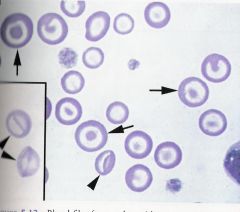
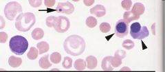
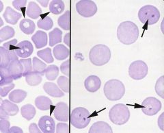
Blood flim from an anemic dog. describe the abnormalities
|
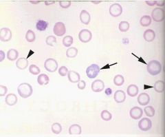
1.lack of density is suggestive of a marked anemia
2.Most of the erythrocytes are small and hypochromic (arrowheads) which is evidence for Iron def. anemia 3.Numerous polychromatophilic erythrocytes (arrows) which is evidence for regeneration |
|

Dog blood flim describe the abnormalities
|
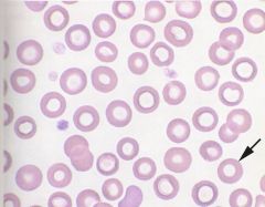
numerous torocytes or punched out cells. Not true hypochromasia. Note wide rim of hemoglobin. This is an insignificant finding
|
|
|
Erythocytes fragments are termed ________
|
schistocytes
|
|
|
cause of schistocytes
|
usually result from shearing of the red cell by intravascular trauma. Seen in animals with Disseminated Intravascular Coagulopathy (DIC). Also seen in dogs with hemangiosarcoma (common to also see numerous acnthocytes if hemangiosarcomea).
Also seen in animals with Iron def. anemia- thought to result from keratocytes which fragment |
|
|
iron def. erythrocytes initially develop an apparent _______ or _______, which is thought to represent an oxidative injury and which inner membrane surfaces crosslink across the cell. Lesions enlarge and break open to form cells with one or more spicules. When one spicule is present it's called _______ and when two or more are present it is called _____
|
blister
vacuole apple-stem cell keratocyte |
|

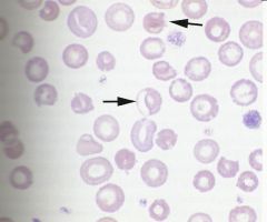
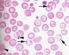
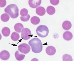
Blood flim from a cat. describe the abnormalities
|
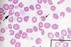
erythrocyte membrane abnormalities
1. blister cells (small arrow) 2.keratocytes (large arrows) |
|
|
granulopoiesis
|
neutrophil production
|
|
|
causes of aplasic anemia
|
drugs
chemicals toxins estrogen viral neoplasic |
|
|
endonenous estorgen may result in bone marrow suppression from _______ tumors in males and _____tumors or _______ in females
|
sertoli cell
granulosa cell cystic ovaries |
|
|
aplastic anemia in cattle has been associated with grazing of __________ and ingestion of soy bean meal contaminated with ______ solvent. ________, a commonly used solvent has been assoicated with aplastic anemia and leukemia
|
Bracken fern
trichloroethylene benzene |
|
|
________ (toxin) has been associated with bone marrow suppression in horses and cattle, and ________ has been reported to cause aplastic anemia in pigs
|
Mycotoxins
aflatoxin B |
|
|
infectious causes of aplastic anemia
|
FeLV
ehrlichia canis EIAV parvo virus in dogs |
|
|
causes of pure red cell aplasia
|
dogs- IMHA
dogs and hores treated with human recombinant erythropoitin FeLV SUBGROUP C |
|
|
causes of red cell hypoplasia
|
anemia of inflammatory disease
anemia of chronic renal failure anemia associated with endocrine disease anemia associated with nurritional deficiencies |
|
|
anemia caused by ____________ is the most common and is usually mild, nonregeneritive and normocytic
|
inflammatory disease
|
|
|
clinical finds of anemia of chronic renal failure
|
anemia is usually moderate to severe, nonregeneritve, and normocytic. Azotemia and decreased urine spefic gravity also present, and severity of anemia usually croulates to severtiy of renal failure
|
|
|
Hypothyroid dogs almost always may a ____, ________, _________ anemia.
|
mild, nonregenertive, normocytic
|
|
|
Some dogs with hypoadrenocorticism have a _____, _________, _________, anemia that is often masked by ___________
|
mild, nonregenerative, normocytic
dehydration |
|
|
most common causes of anemia associated with nutritional deficiencies
|
low Iron
low Cobalt |
|
|
Cobalt def. anemia is usually ________ and _________
|
normocytic
nonregenerative |
|
|
In anemia caused by acute blood loss, by __ hours post bleeding polychromatophilic erythrocytes should be present in the blood.
|
72
|
|
|
Thromboctopenia may result in bleeding when concentration is less than ___________
|
25,000/ul
|
|
|
Blood loss does not cause platelet concentration to fall below ___________
|
100,000/ul
|
|
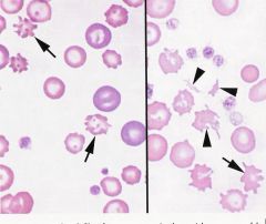
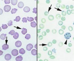
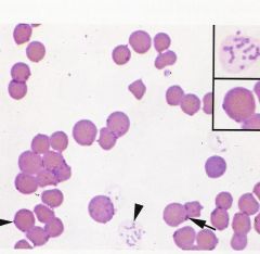
blood flim from an anemic dog with a ruptured hemangiosarcoma of the spleen. describe the abnormalities
|

LEFT: numerous acanthocytes(arrows) note also the polychromatophilic cells which indicate regeneration
RIGHT:Acanthocytes (arrow) schistocytes (arrowheads) |
|

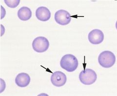
blood flim from an anemic dog. describe the abnormalities
|

Echinocytes (arrows) which can be artifactual because of slow drying blood flim. Also associated with renal disease, lymphoma, rattlesnake envenomation, and cemotherapy in dogs and after exercise in horses. when seen with rattlesanke envenomation is termed type 3 echinocytes- with numerous very fine spicules on all red cells, sometimes spheroechinocytes are formed
|
|
blood flim from a dog with anemia describe the abnormalities
|

echinosperocytes, dog was bitten by a rattle snake
|
|

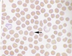
dog blood flim. descibe the abnormalities
|
leptocytes, also called codocytes or target cells. little diagnostic significance. May form if lavander blood tube is not filled enough because of EDTA. also seen in dogs with high cholestrol concentrations
|
|
blood flim from a dog. describe the abnormalities
|

Eccentrocytes (arrows) associated with oxidative damage. often found in conjunction with heinz-bodies
|
|

blood flim from a dog. describe the abmormalities
|

stomatocytes. when only a few are present insigfinicant. Hereditary stomatocytosis has been reported in alaskan malamutes, miniature schnauzers, and the Drentse partrijshond. a;; autosomal recessive. Alaskan Malamutes have condrodysplasia, and only small % of red cells are stomatocytes. Drentse partrijshonds have hypertrophic gastritis, retarded growth, diarrhea, renal cytes, and polyneuropathy. Miniture schnauzers are asymptomatic
|
|

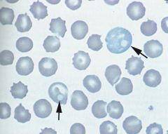
blood flim from a cat LEFT wrights stain, RIGHT brilliant cresyl blue stain.
descibe the abormalities |
Heinz bodies. arrowhead points to a reticulocyte
|
|
|
causes of Heinz body anemia
|
oxidative denaturation.
cats normally have 1 to 2 % Heinz bodies. onions(sheep can adjust) garlic brassica species plant wilted dryed leaves from red maples (horses only) benzocaine zinc copper acetaminophen propofol phenazopyridine phenothiazine phenylhydrazine naphthalene vitamin K methylene blue propylene glycol |
|
|
causes of Howell-Jolly bodies
|
regenerative anemia
splenectomy suppressed splenic function |
|

dog blood flim. descibe the abnormalities
|
Howell-jolly body(arrow)
nucleated red blood cells (arrow head) spherocytes eccentrocytes stomatocytes |
|

blood flim from a dog. descirbe the marked abnormalities
|
siderotic granules or pappenheimer bodies (arrows)rare associated with chloramphenicol, myelodysplasia, and ineffective erythropoiesis
Howell-jolly bodies (arrowheads) |
|

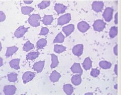
blood flim from an anemic cat. describe the abnormalities
|
spherocytes with mycoplasma haemofelis
|
|
blood flim from an anemic cat. describe the abnormailities
|

cytauxzoon organisms
|
|
blood flims from a dog. describe the abnormitlies
|

LEFT: babesia canis
RIGHT: Babesia gibsoni |
|
blood flim from a dog. describe the abnormalities
|

Spherocytes
mycoplama haemocanis |
|

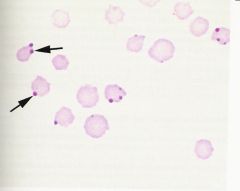
blood flim from a bovine. describe the abnormalities
|
eperythrozoon wenyoni
|
|

blood flim from a bovine. describe the abnormalities
|
anaplasma marginale
|
|
blood flim from a dog. describe the marked abnormalities
|

viral inclusions- distemper
|
|
|
the combination of reticulocytosis and hypoproteinemia is indicative of _____________
|
blood loss
|
|
|
T or F. Platlets are usually increased during Iron def. anemia.
|
T
|
|
|
oral iron is toxic to which speices
|
kittens
|
|
|
Isoerythrolysis
|
occurs in neonates after ingestion of colastrum which contains antibodies against their red blood cells.
|
|
|
What does hypophosphatemia (less than 1mg/dl)cause in animals
|
induced hemolysis, due to inhibiton of erythrocyte glycolysis
|
|
|
hypophosphatemia induced hemolysis associated diseases
|
post parturient hemoglobinuria in cattle
Diabetic cats enteral alimentation in cats |
|
|
Giving unlimited access to water following unavailability, causes what in calves
|
water intoxication induced hemolysis
|
|
|
hereditary erythrocyte membrane defects causeing regeneritve anemia
|
1.Hereditary spherocytosis in cattle and mice
2.Hereditary stomatocytosis in dogs 3.Coomb'd negative chronic intermittent hemolytic anemia in abyssinian and somali cats 4. animals with membrane transport defects |
|
|
metabolic diorders wich cause anemia
|
1.pyruvate kinase deficiency-dogs, cats
2.phophofructokinase deficiency-dogs 3.glucose phosphate degyhrogenase deficiency- american saddle bred colt and dogs 4.hereditary methemoglobinemia-dogs cats horses 5.porphyrias-cattle, cats swine 6.bovine congenital erythropoietic porphyria |
|
|
diagnostic approach to polyctyemia (erythrocytosis)
|
first rule out relative polycythemia by deydration or fuild shifts, and splenic contraction. Consider if patient is excited or dehydrated and then redue CBC. If total protein is also increased animal is likly to be dehydrated.
If relative polycythemia is ruled out then consider secondary absolute polycythemia dur to hypoxemia- should have decreased PaO2 and increased erythropoitin. then consider inappropriate erythropoitin production- normal PaO2 and increased erythropoitin If all other causes are ruled out then consider primary absolute polycythemia (polycythemia vera)- normal PaO2 and normal or decreased erythropoietin |