![]()
![]()
![]()
Use LEFT and RIGHT arrow keys to navigate between flashcards;
Use UP and DOWN arrow keys to flip the card;
H to show hint;
A reads text to speech;
74 Cards in this Set
- Front
- Back
|
1. Nasal Cavity 2. Larynx 3. Trachea A. Hyaline Cartilage 4. Bronchus 5. Bronhiole 6. Lung Tissue: Alveoli 7. Diaphragm |

|
|
|
1. Pulmonary Artery 2. Bronchiole 3. Pulmonary Vein 4. Capillaries 5. Alveoli 6. Respiratory membrane |

|
|
|
Know the diagram |

|
|
|
Know the diagram |
|
|
|
Know the diagram |

|
|
|
Know the diagram |

|
|
|
- Tidal Volume -- Inspiratory Reserve Volume --- Expiratory Reserve Volume ---- Residual Volume ----- Vital Capacity ------ Total Capacity |
- air exchange during quietbreathing -- air forcefully inhaled aftertidal inspiration --- air forcefully exhaled aftertidal expiration ---- air in lungs after forceful expiration ----- total exchangeable air volume ------- Vital capacity + Residual Volume |
|
|
Know this |

|
|
|
- Nervous Control: Respiratory control Center a. Voluntary b. Chemical factors |
- In brain stem: sets baseline rateof breathing - Controlsmuscles for breathing, e.g. diaphragm - Regulated By: a. contraction of skeletal muscle; e.g.hold breath, speech b. H+ , CO2 and O2 |
|
|
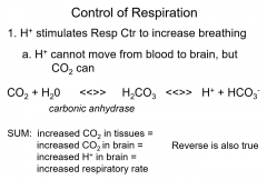
Control of Respiration |
Exercise: Increased muscle activity = increased respiration |
|
|
- Carotid Bodies: chemoreceptors for O2 and H+ A. Decreased O2 B. Increased H+ -- Gut Reflex --- Joint Receptors |
a. Stimulates respiration b. stimulates respiration -- Breathing stops during swallowing --- detect movement and increase respiration |
|
|
know this |

|
|
|
know this |

|
|
|
know this |
|
|
|
- Sinusitis -- Laryngitis --- Bronchitis ---- Pneumonia ----- Emphysema ------ Lung cancer |
- infectionleads to congestion > poordrainage of sinuses -- infection of the larynx --- infection of bronchi ---- infection of lungs, fill with fluid ----- : damage to alveoli walls = increaseddead space = decreased gas exchange;smoking most common cause ------ Awful, smoking |
|
|
Know this |

|
|
|
know this |

|
|
|
know this |
Distal Convoluted Tubule |
|
|
know this |
|
|
|
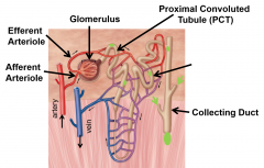
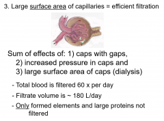
Glomerular Filtration -- Tubular Reabsorption --- Tubular Secretions |
- most plasma pushed from blood into PCT = urine filtrate -- essential molecules, ionsand water move from filtrate back into blood --- specific ions and moleculesmove from blood back into urine filtrate |
|
|
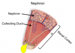
Glomerulus |
- a knot of capillaries with large gaps between endothelial cells = permeable |
|
|
Afferent Arteriole |
- leading into glomerulus has larger diameter thanthe EfferentArteriole = increasedpressure in capillaries |
|
|
know this |
|
|
|
knowy this |

|
|
|
tubular Reabsorption Efficient A. Filtrate made at - B. Urine made at - |
- Mostwater, nutrients andsome ions move fromurine filtrate back into capillaries nearthe proximal convoluted tubules (PCT) a. 125 ml b. 1 ml |
|
|
know this |

|
|
|
Tubular secretions a. creates -- Sum |
- Somechemicals are reabsorbed, but then secretedback into urine filtrate by active transport A. H+, creatinine, penicillin -- urine is rich in waste products and devoid of important nutrients |
|
|
water reabsorption -- Sum |
- 1.Water reabsorbed along with solutes, mostly atPCT = osmosis - More water removed from urinein tubules and collectingducts -- : Dehydrates urine = prevents dehydration of blood and body |
|
|
Diuretics 1. Alcohol 2. Caffeine 3. Medical diuretics a.Decreases what? |
1. inhibitsthe release of antidiuretichormone from pituitary 2. increases glomerular pressure, whichincreases filtrate formation 3. block Na+ reabsorption = morewater in filtrate a. Decreasesblood volume > decreases blood pressure |
|
|
what is this? |

|
|
|
micturition -- External Urethral Sphincter -- relaxed -- Contracted |
- a.Urinefills bladder = increasedpressure - Increased pressure initiatesa spinal reflex - Signals sent to inform brain= urge - Peristalsisbegins in bladdersmooth muscle -- Urine flow -- No flow |
|
|
Renal Failures -- Effects |
- Marked decrease in glomerularfiltration - Many causes: e.g. decreased cardiacoutput -- Water retention = edema -- Acidosis H+ accumulation -- Toxic waste accumulation -- Uremia: Terminal kidney failure |
|
|
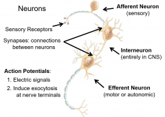
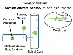
Nervous System - Receive Sensory information --Integrates sensory information --- Generates motor and autonomic ouput |
- olfactory receptors > Smell -- Identifies smell as pumpkin pie --- Eat the pie |
|
|
- Central Nervous Sysem (CNS) -- Peripheral nervous system --- Afferent ---- Efferent |
- Brain and Spinal cord - Encased in bone: cranium vertebral column -- Nerves outside the CNS --- nerves bring sensory to CNS ---- nerves send from CNS to periphery |
|
|
- Cell Body -- Dendrites --- Axon ---- Myelin ----- Nerve terminals |
- nucleus -- Carry action potentials --- Carry AP away from cell body ---- insulates axons to speedconductance of AP ----- site of exocytosisof neurotransmitters |
|
|
read this |
|
|
|
Action potentials and neurotransmission - Nerve (and muscles) cells have what? and do what? -- Stimulus does what and does what? |
-Na+ that can open/close -- opens Na+ channels > the ion moves quicklyinto cell down a steep concentration gradient= depolarization =electric signal |
|
|
Ap Spreads where and what is released? |
Nerve terminal and NT |
|
|
Threshold Stimulus |
- issufficient to open Na+ channelsand start AP |
|
|
- Excitatory NT -- Inhibitory NT --- Summation |
- brings target cell closer to threshold --takes target cell away fromthreshold ---when Excitatory exceeds Inhibitory andthreshold is reached > AP in target cell |
|
|
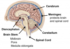
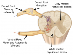
-Grey matter
-- White Matter Regions --- Cerebrum ---- Diencephalon ----- Brain stem ------ Cerebellum |
- Areas of the body -- Areas of myelinated axons --- Sites of thoughts, motor control ---- thalamus and hypothalamus; connections and hormones ----- midbrain, pons and medulla oblongata;primary functions, e.g. respiration, heart rate ------ posterior in brain; motor coordination |
|
|
know this diagram |
|
|
|
4 lobes - Frontal -- Temporal --- Parietal ---- Occipital |
- Motor -- Temporal --- Somatic Senses ---- Vision |
|
|
Gyrus -- Cerebal cortex |
- =ridge; Sulcus =valley >> increase surface area --¼ inch outer edge of gray matter |
|
|
Spinal nerves how many pairs? - Dorsal Roots -- Ventral Roots |
31 - a.afferent,sensory info entersthe CNS -- efferent, autonomic and motorfibers exit CNS |
|
|
know this |
|
|
|
Cranial nerves how many pairs? a. Some are what? and others are what? -- Spinal nerves a how many pairs b. they are all ____ nerves both ___ and ____ |
12 a. sensory/ some are motor some are mixed -- a 31 pairs mixed / sensory and motor |
|
|
know this |
|
|
|
Autonomic system - Efferent -- Effectors --- two opposing divisions are |
- withtwo neurons between CNS and effectors --glands, smooth and caradic muscle --- sympathetic and parasympathetic |
|
|
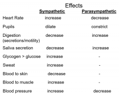
- Sympathetic Division 1. 2. Parasympathetic divison 1. 2. |
1. Epinephrine (adrenaline) orNorepinephrine releasedto effectors 2. Function: prepare body for intensemuscle activity> Fight or Flight 1. Releases acetylcholine toeffectors 2. Function: maintain homeostasis> Restand Repose |
|
|
Sympathetic Parasympathetic Increase or decrease Heart rate Pupils Digestion Saliva Glocogen > Glucose Sweat Blood to skin Blood to muscle Blood pressure |
|
|
|
- Thermoreceptors -- Mechanoreceptors --- Chemorecptors ---- photorecpetors ----- nociceptors |
- Temp -- Force --- Chemicals ---- Light ----- Tissue damage = pain |
|
|
Stimulus Detection - Quality -- Intensity --- Adaptation |
,- of sense determined by receptormodality -- of stimulus detected as frequencyof AP --- decrease in AP despite continuedstimulus application, due to receptor exhaustion |
|
|
Proprioceptors - Muscle Spindle: -- Golgi Tendon OPrgan |
- Stretch causes AP to spinal cord[` - AP stimulate a motor neuron to thatmuscle = contraction= StretchReflex to protect muscle -- a.Pressure> AP to spinal cord > inhibits motor neuron > inhibits muscle contraction -- Protect muscle from tearing |
|
|
Chapter 10 Muscles slide 7-8-9-11-12-15-20 |
go back |
|
|
Nociception |
- Pain receptors are free nerveendings - Detect tissue damage due to releaseof cellcontents, e.g. K+ - Found in skin and most organs - May stimulate sympathetic nervoussystem - |
|
|
- Optic Disk
-- Sclera --- Iris ---- Lens ----- Cornea ------ Cornea ------- Retina ------- Fovea Centralis |
- No rods or cones, blind spot -- Boundary white --- Muscle, controls aperture ---- focus light on retina ----- clear, curved to focus ------ curves len for focus ------- nerve tissue, rods and cones -------- acute vision |
|
|
Photoreceptor cells - - Rods -- Cones |
- Detect white light - 1 billion/retina - Numerous rods synapse with bipolar cell/ enriched in periphery of retina -- detects red, green, or blue -- 3 million/retina -- Synapse on few bipolar neurons -- Centered in retina; fovea centralis |
|
|
Hearing - Sound -- Pitch --- Loudness |
- waves of compressed air -- frequency of waves; cycle/sec; Herz (Hz) --- amplitude of waves; decibel; dB |
|
|
Mechanism of hearing |
- Sound waves move tympanicmembrane which pushes stapes against cochlea -- . Liquid waves in cochlea deflectthe basilar membrane --- Movements activate mechanoreceptors= Hair Cells > AP along cochlear nerve to CNS ---- Decibels detected by amplitude of deflection ----- Herzdetected by location of hair cell stimulated |
|
|
Rotational Equilibrium -- Intertia --- Momentum |
- 3 fluid filled semicircularcanals -- Inertiakeeps fluid from moving when head moves > activates mechanoreceptors = APto CNS --- fluid catches up with moving head > dizzy when stop ---- AP to Cerebellum to coordinate movements while moving; including eyes |
|
|
Gravitational equilibrium |
Utricle and Saccule:fluid filled Tilt head > slides fluid > otoliths deflectHair Cells > AP to CNS Direction of gravity detected by which cells activated info to cerebellum > balance |
|
|
- posterior pituitary -- ADH --- OT |
- Antidiuretic Hormone (ADH) and Oxytocin (OT) are produced in hypothalamus, but released from posterior pituitary -- decrease urine volume -- lack > diabetes insipidis --- Stimulates uterine contractions and lacatition --- synthetic OT used to induce labor |
|
|
Growth Hormone Lack of GH -- Excess GH |
dwarfism ifduring development --giantismduring development; acromegaly in adults |
|
|
- Follicular cells -- Hyposecretion --- Hypersecretion |
- Secrete Triiodothryonine (T3; 3I)and Thyroxine (T4; 4 I) - Effects: increased metabolism ofall cells; increased heartrate; tissue growth in children - Secretion increased by TSH from anterior pituitary -- cretinism in children; myxedema in adults > lethargy --- increased BMR, Graves disease |
|
|
Chapter 11 - 8-12-13-15-16 |
no |
|
|
- ParafollicularCells: |
- Secrete Calcitonin(CT): - Effects: puts Ca2+ into bone to reduceblood Ca2+ - Secretion increased by high blood Ca2+ - hyposecretion:osteoporosis |
|
|
- Androgens |
- Effects: sexual development;primarily in males |
|
|
Diabetes - type 1 -- type 2 |
- juvenile onset; lack of insulin --adult onset, over weight, insulinresistance |
|
|
Glucagon --Effects |
- Secretion from a-cells dueto low blood sugar -- Release glucose from glycogen inliver -- Glucose synthesis in liver -- Increased lipolysis: TG > fattyacids > energy -spares glucose use for energy |
|
|
Ovaries - Secrete _____ and _________ - Effects Testes -- Secretes __________ -- Effects |
- estrogens and progesterone - Female sexual development, Secondary sex characteristics -- testosterone -- male sexual development -- Secondary sex characteristics |
|
|
Pineal gland - Secretes ____ -- effects |
- melatonin -- Controls sleep cycle Aing: Decreased melatonin disrupts sleep |
|
|
Origin of life 4 stages - Stage 1small organic molecules -- stage 2 Macromolecules ---Stage 3Protocells ---- Stage 4 Living cells |
-Simplemonomers evolved from inorganic compounds -- Monomers joined to form polymers ---Membranes enclosed macromolecules >form cell precursor ---- Protocells develop ability to replicate |
|
|
a |
b |