![]()
![]()
![]()
Use LEFT and RIGHT arrow keys to navigate between flashcards;
Use UP and DOWN arrow keys to flip the card;
H to show hint;
A reads text to speech;
46 Cards in this Set
- Front
- Back
|
hydrophobic effect |
hydrophobic molecules tend to cluster together, causing the water to not be constrained among the molecules, leading to an overall incr entropy (favored) |
|
|
phospholipids, phospholipid bilayer |
- main component of cell membrane phosphilipid bilayer: - hydrophilic head, hydrophobic tail - solves the containment problem |
|
|
macromolecules |
formed by COVALENTLY joining together single-unit monomers to form polymers |
|
|
monomer ---> polymer |
monosaccharides ---> polysaccharides amino acids ---> proteins nucleotides ---> nucleic acids |
|
|
synthesizing a polymer (1 + P) |
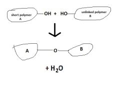
by dehydration rxns |
|
|
sugars (structure, example) (4) |
- multiples of CH2O - have carbonyl and hydroxyl groups - illustrated as rings - glucose is the most common sugar |
|
|
polymers of glucose |
- glycogen - starch - cellulose - chitin |
|
|
nucleotides, structure (4 + P) |
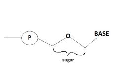
- monomers of nucleic acids - 3 main parts: - phosphate group (negatively charged) - sugar group - base (e.g. A, T, G, C) |
|
|
DNA vs RNA (as nucleotides) |
- both store, transmit, express genetic info - the sugars are diff b.t them (RNA ribonucleic acid, DNA deoxyribo) |
|
|
bases (base pairs) |
- adenine - cytosine - guanine - thymine - uranine base pairs: A-T/U, C-G |
|
|
base pair directionality (2 + P) |
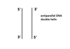
5' to 3' DNA double helix is antiparallel (P) |
|
|
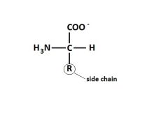
amino acids (basic structure) (P) |
|
|
|
amino acid side chains (3) |
- dictate structure of protein (and therefore function) - polar (hydrophilic) vs nonpolar (hydrophobic) - acidic (- charged) vs basic (+ charged) |
|
|
amino acid structures (primary through quarternary) |
1* : long chain of amino acids, basic structure linked by covalent bonds 2* : string of amino acids that take on alpha helices or beta sheets 3* : everything folded up into a 3D structure 4* : association of two or more 3* structures |
|
|
sickle cell anemia (3) |
- affects hemoglobin, which transfers O2 through bloodstream - amino acid change from Glu to Val, changing structure of beta subunit and exposing a hydrophobic region => causes molecules to crystallize, reduces capacity for cell to carry O2 |
|
|
functions of proteins (7) |
- speed up chem rxns (enzymatic) - provide structural support (structural) - e.g. actin & myosin (contractile & motor) - cellular communication (receptor, hormonal) - transport across cell membrane (transport) - activate antibodies, destroy viruses (defensive) - store amino acids (storage) |
|
|
endomembrane system, main functions (5) |
- organelles located outside of the nucleus main functions: - synthesize some proteins - transport proteins into membranes & organelles - lipid metabolism & movemen - poison detox |
|
|
nucleus (2) |
- DNA located here in the form of chromosomes - makes mRNA that codes for proteins and leaves nucleus to form polypeptides |
|
|
ribosome (3) |
- protein factories - free ribosomes (not attached to ER) are located in cytoplasm and make cytoplasmic proteins - bound ribosomes make proteins that ultimately end up outside the cell |
|
|
3 main ribosome pathways |
1) cytoplasmic: ribosome stays in cytoplasm, makes cytoplasmic proteins
2) signal sends ribosome from cytoplasm to some other organelle via a transport vesicle (e.g. mitochondria)
3) ribosome sent to ER where it finishes making protein (see next flashcard) |
|
|
rough ER ribosomes |
1) resident ER proteins
2) travel to golgi body - vesicles transfer proteins from golgi body either to the plasma membrane (infuses with it) or outside the membrane to be secreted (via exocytosis) - some vesicles become lysosomes if the protein inside wills it |
|
|
golgi body |
- like a warehouse - reads signals and ships proteins out as needed |
|
|
smooth ER (4) |
- hormones are made here - makes lipids that are transported to the cell membrane - detoxifies drugs in the liver - peroxisomes: involved with redox rxns to detoxify |
|
|
lysosomes (4) |
- lysosomes are formed from vesicles - sacs of enzymes that break down molecules - fuse with macrophages & dumps enzymes to digest the things inside - involved with autophagy |
|
|
mitochondria & chloroplasts (3) |
- powerhouse of the cell - convert energy into forms that cells can use (e.g. ATP) *plants have both chloroplasts AND mitochondria |
|
|
endosymbiotic theory & evidence for it (4) |
- belief that mitochondria & chloroplasts were originally photosynthetic organelles that were engulfed by bacteria
evidence for theory: - mitochondria & chloroplasts have their own DNA - they have two membranes - they are of similar size to bacteria |
|
|
cytoskeleton, its functions, its fibers (5) |
- network of fibers in the cell - gives cells structure, support, shape - allows cells to move (*when cells are moving, the cytoskeleton is actively changing and rearranging) - fibers: microtubules, actin filaments - cilia and flagella (are made up of these microtubular filaments) |
|
|
fluid mosaic model (3) |
- fluidity of the membrane - saturated vs unsaturated tails *proteins can move in the membrane |
|
|
saturation vs unsaturation (fluidity), cholesterol |
- saturated: no double bonds, pack together - unsaturated: kinks in chains due to double bonds, prevent packing *therefore, unsaturated tails make membrane more fluid - cholesterol: as T incr, fluidity inhibited as T decr, fluidity incr => cholesterol helps regulate fluidity of membrane against large changes in fluidity due to temp changes |
|
|
microtubules |
- cellular highways: form "tracks" utilized for movement in the cell - cilia & flagella: help organelles to move around |
|
|
integrin & microfilament protein pathways |
- integrins: integral membrane protein; goes through ER - microfilaments: inside the cytoplasm; not part of endomembrane system |
|
|
tight junctions, examples (3) |
- assemblies of proteins - e.g. sheets of cells in intestines or skin - form seals & prevent fluid from moving across a layer of cells (i.e. so you don't "leak") |
|
|
gap junctions (2) |
- tiny holes that allow some things to go from one cell to another - one form of cellular communication |
|
|
how do things get across membranes? |
1) diffuse directly through membrane 2) travel through membrane via proteins |
|
|
diffusion, osmosis |
- from higher conc to lower conc, eventually reach equilibrium - osmosis: diffusion of water across a semi-permeable membrane - larger molecules can't pass through - water goes to side w/ lower conc of water |
|
|
red blood cells and water |
hypotonic: put cell into water, cell pops b/c water goes inside; [H2O]cell < [H2O]sol'n isotonic: put cell into water, cell intact; [H2O]cell = [H2O]sol'n hypertonic: put cell into water, cell shrivels up b/c water goes outside; [H2O]cell > [H2O]sol'n |
|
|
diffusion directly through membrane |
- middle of membrane very hydrophobic (bilayer's tails) - nonpolar molecules (e.g. steroids) will pass |
|
|
channel proteins, examples (5) |
- provide hydrophilic environment for other molecules to pass through membrane - many proteins have alpha helix structures to help facilitate movement - e.g. potassium channels - e.g. aquaporins: channels which allow passage of lots of water through open pores *channel proteins are porous and function to allow many things to go across quickly |
|
|
carrier proteins (3) |
- undergo a physical conformational change to get things from one side of the membrane to the other - once protein picks up and binds to a thing, triggers conformational change and releases thing onto other side of membrane - almost all of these proteins are very selective |
|
|
passive transport |
- things diffusing down a concentration gradient by themselves - facilitated diffusion via protein |
|
|
active transport (3) |
- requires energy (ATP) - secondary active transport driven by coupled carriers (vs primary active transport) - things not only going down their conc gradient but also up their gradient |
|
|
sodium-potassium pump (steps + 1) |
1) sodium binds to protein on side A with help of ATP 2) binding causes protein to change shape, one P group left on protein from ATP 3) sodium gets released on side B 4) potassium then binds to protein on side B 5) binding causes change in shape of protein 6) potassium gets released on side A, P group goes away
*both ions are moving from an area where they are low conc to where they are high conc (i.e. against their conc gradient) |
|
|
secondary active transport |
- another form of active transport; main focus to drive an ion against its gradient - relies on coupled carriers - energy needed comes from a gradient that was already set up by a separate protein |
|
|
sucrose & proton coupled carrier |
1) proton pump as normal primary active transporter (fueled by ATP): pumps protons against their gradient from side A. side B now has more [protons] 2) sucrose-proton co-transporter takes advantage of the fact that the protons want to go back to side A & drags sucrose (against its gradient) along with the returning protons to side A 3) when protons reach equil, sucrose stops being transported to side A |
|
|
how big things get across cell membranes: phagocytosis & endocytosis |
phagocytosis: "cellular eating" - lysosomes fuse with bacteria inside a vesicle, digest it
endocytosis: "cellular drinking" - cell creates a bubble to take up liquid from outside, bubble pinches off from membrane into cell (becomes vesicle) , now vesicle has extracellular fluid inside |
|
|
receptor-mediated endocytosis (e.g. LDL) |
1) LDL binds to its receptor, gets caught in the sac (bubble) 2) sac buds off into a vesicle, goes into the cell 3) LDL has now been brought into the cell |