Reading...
![]()
Play button
![]()
Play button
![]()
Use LEFT and RIGHT arrow keys to navigate between flashcards;
Use UP and DOWN arrow keys to flip the card;
H to show hint;
A reads text to speech;
85 Cards in this Set
- Front
- Back
|
What would be considered the box and window of the eyes
|
Scalera and cornea
|
|
|
Characteristics of the sclera
|
- opaque
- protects the eye – Ankers extrinsic muscles – Continues with dura where the optic nerve enters |
|
|
Characteristics of the cornea
|
– Transparent
– lets light enter the eye – Many nerve endings [pain receptors] |
|
|
What are the three layers of the ocular apparatus
|
– Outer layer
– Middle layer [Uvea] – Inner layer |
|
|
What are the components of the outer layer
|
– Schlera
– Cornea |
|
|
What are the components of the middle layer
|
– Choroid
– Ciliary body – Suspensory ligament – Lens – Iris – Pupol |
|
|
Function of Choroid
|
– Pigmented layer
– Reduces light scatter within the eye |
|
|
Which structures are part of the focus mechanism
|
– Ciliary body [smooth muscle, maintains tension on the ligaments and changes SHAPE of lens]
– Suspensory ligament [Extends from ciliary body to the lens] – Lens [Acellular, vascular translucent structure, changes FOCAL LENGTH] |
|
|
Which structures are part of the aperture mechanism
|
Shutter = Iris [Pigmented part of the eye between the cornea and the lens]
Aperature = Pupil [Regulates amount of light entering eye] |
|
|
What are the components of the inner layer (retina)
|
– Retinal pigment epithelium
– Retina [photoreceptors and neurons] |
|
|
Describe the outer pigmented layer of the retina
|
Absorbs light and prevented from scattering
|
|
|
Describe that inner neural layer of the retina
|
Contains photoreceptors [Rods and cones] and neuron [bipolar cells, amacrine cells and ganglion cells]
|
|
|
Blood supply to eye and it's divisions
|
Ophthalmic artery
– Posterior ciliary arteries: supplies outer Redknot and optic disc [choroidal circulation] – Central retinal artery: Supplies inner retina |
|
|
Scotopic vision: characteristics
|
– photoreceptors = rods
– High sensitivity in dark – Saturate as light increases – Low spatial resolution |
|
|
Low versus high spatial resolution
|
Low: input from MANY rods are summated by one bipolar, so cells can't easily tell where input is coming from
High: each con synapses with ONE single bipolar self |
|
|
Phototopic vision: characteristics
|
– Photoreceptors: cones
– Best in bright light – Adapt to background to give wide dynamic range – Color vision – High spatial resolution |
|
|
Three types of cones + wavelengths of each
|
Classified based on type of photo pigment [opsin]
– R (long - 564nm peak sensitivity) – G (medium - 534nm) – B (short - 420nm) |
|
|
Components that make up photo pigments
|
– Opsin
– Retinal [vitamin a derived molecule] |
|
|
Describe steps of photo transduction
|
1) Light [a photon] converts 11– cis-retinal to all-trans-retinal causing a change in shape of the photo pigment
2) Activation of the 100 molecules a G-protein transducin 3) Each transducin will activate a cGMP phosphodiesterase molecule 4) Each phosphodiesterase breaks down 1000 molecules of cGMP 5) Closure of sodium channels in the cell membrane 6) Hyperpolarization of cell |
|
|
Distribution of photoreceptors in the fovea
|
– Center of retina
– Highest resolution – Only cones |
|
|
Distribution of photoreceptors in the periphery
|
– Both rods and cones
– Decline with increasing eccentricity |
|
|
What is the physiological blind spot
|
Area on retina with no photoreceptors called the optic disc.
– Located near optic nerve entry |
|
|
What is the three neuron relay
|
photoreceptor --> bipolar cell --> retinal ganglion cell --> lateral geniculate nucleus (LGN) and optic radiation
|
|
|
Characteristics of bipolar cells
|
– Separate bipolar cells for rods and cones
– Receive input from horizontal and amacrine cells in the retina |
|
|
Signal transduction from photoreceptor to bipolar cell
|
1. hyperpolarization of photoreceptor decreases release of glutamate
2. OFF cone bipolar cells are hyperpolarized by this decrease 3. ON cone bipolar cells are depolarized 4. result is a graded excitatory postsynaptic potential (EPSP) 5. release of glutamate and activation of retinal ganglion cell |
|
|
ON versus OFF bipolar cells
|
ON cells are inhibited by glutamate, hence light which reduces glutamate release causes ON cells to become active --> EPSP
OFF cells are excited by glutamate --> no EPSP |
|
|
Described lateral inhibition
|
– Bipolar cells receive inputs from horizontal and amacrine cells in the retina that create lateral inhibition by light in neighboring regions of the retina --> stronger response by central cell
– This gives rise to the "center surround" receptive field organization evident in the retinal ganglion cells to which bipolar cells project |
|
|
Two most important types of retinal ganglion cells
|
– Magnocellular RGC (LARGE)
– Parvocellular RGC (small) |
|
|
Characteristics of magnocellular RGC
|
– Large cell bodies and long dendrites
– Large receptive fields – Low spatial resolution – Respond transiently to onset and offset a flight [high temporel resolution] |
|
|
Characteristics of parvocellular RGC
|
– Small cell bodies
– Short dendrites – Small receptive fields – High spatial resolution – Sustained response to onset of light [low temporal resolution] |
|
|
What is color oponency
|
Some cones prefer to be fired by red colours, but will still fire less strongly for green objects.
What is important is to compare what is going on in green cone vs red cone. Any one cone by itself can’t tell you what colour is out there, it only tells the colour by comparing what is going on with another cone (in that region) with a different wavelength |
|
|
Central scotoma
|
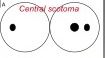
–slicing of arching fibers coming from retina towards optic disc
– As fovea is located in the center of eye, this will create a blind spot in the areas from which the fibers originated |
|
|
Ceca-Central scotoma
|
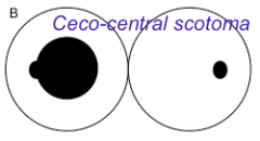
– Cutting of fibers coming from fovea almost at entrance to optic disc
– Would result in merging of physiological blind spot with pathological blind spot |
|
|
(Nasal) Arcuate defect
|
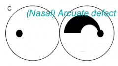
– Severing of fibers that have a rainbow arch shape
– Arch starts from one spot and moves nasally – Would present a problem in your Temporel visual field |
|
|
(Temporal) Wedge Defect
|
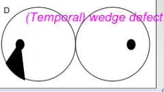
– Fibers leaving blank spot and moving superiorly are cut
– Will result in a wedge defect on your lower visual field |
|
|
Describe the path of fibers leaving the RGC and moving towards the brain
|
– RGC gather together at the optic disc to form the optic nerve
– Optic nerves pass into the cranium through the optic canal – Converge above the pituitary at the optic chiasm – Axons from the nasal half of each retine (temporal hemifeild) decussate in this structure – Optic track continues from Arctic has, but now each tract presents information from the contralateral nasal hemifield of each eye – Track terminates in the LGN |
|
|
Is that optic nerve part of the peripheral or central nervous system?
|
- Optic nerve is actually component of the central nervous system
- it's myelin is produced by oligodendrocytes |
|
|
Possible destinations for axons traveling in the optic nerve
|
– Striate cortex
– Preoptic nucleus – Superior caliculus – Nucleus of the optic tract and accessory optics system – Suprachiasmatic nuclei |
|
|
Role of preoptic nuclei
|
Pupil light reflex
(pretectal nuclei in midbrain) |
|
|
Role of superior caliculus
|
Guide rapid eye and head orientation [mediates reflective eye movements]
|
|
|
The role of nucleus of optic tract and accessory optics system
|
Supplement vestibular information innkeeping gaze stable
|
|
|
Role of suprachiasmatic nuclei
|
Diurnal regulation of homeostasis [regulates sleep/wake cycles]
Located in hypothalamus |
|
|
Bilateral hemianopia
|
– Caused by lesions in the optic chiasm
– Almost always due to compression by a mass, often a pituitary tumor |
|
|
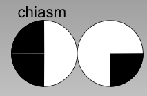
Homonymous hemianopia
|
![– Caused by lesions of the optic tract
– Usually partial
- Usually incongruous [different and extent between the two arts]](https://images.cram.com/images/upload-flashcards/574985/1269059_m.png)
– Caused by lesions of the optic tract
– Usually partial - Usually incongruous [different and extent between the two arts] |
|
|
What would be observed in an endoscopy in loss of supply to inner retinal artery
|
If you lose blood supply to central retinal artery you will lose supply to inner retinal layer resulting in grayness with cherry red spot
|
|
|
Describe the layers of the lateral geniculate nucleus
|
– 4 where Parkthrough cellular RGC axons terminate
– 2 where magnocellular RGC terminate – Organize in` alternation |
|
|
Cortical input to LGN
|
LGN is modulated by cortical input, which may refine perception and mediate effects of visual attention
|
|
|
Retinotopic (spatial) arrangerment
|
– Central field in lateral aspect of mid radiation
– Lower field in upper radiation – Upper field in lower radiation |
|
|
Meyer's loop
|
Forward growth of the anterior horn of the lateral ventricle during development has displaced the lower fibers [from the upper retina] anteriorly, to form the Meyer's loop
|
|
|
Magnocellular information from CONTRALATERAL eye terminates in which layer of the LGN
|
1
|
|
|
Magnocellular information from IPSILATERAL eye terminates in which layer of the LGN
|
2
|
|
|
Parvocellular information from CONTRALATERAL eye terminates in which layer of the LGN
|
4, 6
|
|
|
Parvocellular information from IPSILATERAL eye terminates in which layer of the LGN
|
3, 5
|
|
|
From the LGN where does the information go
|
To the visual cortex via optic radiations
|
|
|
Other names for the visual cortex
|
– Striated cortex
– VI – Calacrine |
|
|
Define: retinotopic
|
Specific for location of stimulus on retina
|
|
|
Describes a highly retinotopic representation of the striated cortex
|
– Fovea at the occipital pole/peripheral field just posterior to parieto-occipital fissure
– lower field in upper calacrine bank/ upper field in lower calacrine bank |
|
|
Homonynous superior quadrartanopia
|
caused by lesions in meyer's loop
– Commonly seen after Temporel lobectomy done to treat epilepsy |
|
|
Describe the processing that occurs in the striated cortex
|
Instead of spots of light, information is starting to be represented as linear segments and boundaries
– Cells have preference for lines of a specific orientation – Lines are organize in a regular array of orientation columns |
|
|
Cortical magnification
|
Refers to the fact that more is devoted to central vision and prayerful vision
|
|
|
Described the defects that would occur as a result of lesions in the following areas of the striated cortex:
Occipital pole Everything but the pool Superior bank Inferior bank |
![Occipital pole : Hemi-central scotoma
Everything but the pool: macula sparing hemianopia
Superior bank: inferior quadrantanopia
Inferior bank: Superior quadrantanopia [can be confusing deletions of meyer's loop]](https://images.cram.com/images/upload-flashcards/574985/1269062_m.png)
Occipital pole : Hemi-central scotoma
Everything but the pool: macula sparing hemianopia Superior bank: inferior quadrantanopia Inferior bank: Superior quadrantanopia [can be confusing deletions of meyer's loop] |
|
|
Describe the extra-striated cortex
|
- After V1, visual information fence out into large array of additional cortical regions arranged in a hierarchy
– Ascending the hierarchy, information becomes less retinotopic and more specific for other special visual property |
|
|
The extrastriate array can be divided into 2 broad streams
|
WHERE stream: Dorsal [ occipito-parietal]
WHAT stream: Ventral [ occipito-temporal] |
|
|
Role of dorsal stream
|
– Motion processing
– Depth perception – Visual spatial data for attention – Eye and hand movement |
|
|
Role of central stream
|
– Color processing
– Object recognition [example words and faces] |
|
|
Define: Akinetopsia
|
Problem seeing motion [rare]
|
|
|
Define: Asteropsis
|
Neglect for the opposite side of space
|
|
|
Balint's syndrome
|
Ocular motor apraxia = Poor targeting of eye movements to targets
|
|
|
Define: Optic Ataxis
|
Misreaching for visual objects
|
|
|
Define: Simultangnosia
|
Inability to attend to more than one object at a time
|
|
|
Legions of various components of the dorsal stream can cause which disorders
|
- Simultangnosia
- Optic Ataxis - Balint's syndrome - Asteropsis - Akinetopsia |
|
|
Lesions of various components of the ventral stream can cause which disorders
|
- acromatopsia
- agnosia - alexia |
|
|
Define: acromatopsia
|
Cerebral loss of color vision
|
|
|
Define: Agnosia
|
Issues with general visual object identification for example man who mistook his wife for a hat
|
|
|
Define: Alexia
|
Impaired recognition of words
|
|
|
Conjunctivitis
|
Inflammation of the superficial covering of the sclera
– Most commonly a viral or infectious event - relatively benign |
|
|
Uvetis
|
– More serious and conjunctivitis
– Associated with systemic diseases like sarcoidosis, RA, MS, tumors, infections, AIDs – Can be segmental |
|
|
Anterior ischemic optic neuropathy
|
- due to ischemia of the posterior ciliary arteries
- result in a swollen infarcted disk |
|
|
Central renal artery occlusion
|
– Results in a pale retina with a cherry red spot a fovea
|
|
|
Opsin mutations
|
- Cause different types of in congenital color blindness
- more prevalent in men – Signs and symptoms depend on which photoreceptors involved |
|
|
Describe the main types of opsin mutations
|

- cone dystrophy, macular degeneration: central scotoma with poor central daytime vision
- retinitis pimentosa: disease of the rods, cause ring schotomata and nyctalopia (night blindness) |
|
|
Where's the density of rods the greatest
|
Around 20°
|
|
|
Where is the density of cones the greatest
|
At 0°
|
|
|
Where is their only cones
|
At 0°
|
|
|
Where is there predominantly rods
|
From ~5° onwards
|