Reading...
![]()
Play button
![]()
Play button
![]()
Use LEFT and RIGHT arrow keys to navigate between flashcards;
Use UP and DOWN arrow keys to flip the card;
H to show hint;
A reads text to speech;
16 Cards in this Set
- Front
- Back
|
What are the basal ganglia?
|
The basal ganglia are a complex of subcortical telencepalic nuclei that integrate cerebellar and cortical influences for motor planning and execution, and provide inhibition of involuntary motor behaviors.
|
|
|
Explain the excitatory pathway.
|
Cortex (stimulates) → Striatum (inhibits) → "SNr-GPi" complex (less inhibition of thalamus) → Thalamus (stimulates) → Cortex (stimulates) → Muscles, etc. → (hyperkinetic state)
|
|
|
Explain the inhibitory pathway.
|
Cortex (stimulates) → Striatum (inhibits) → GPe (less inhibition of STN) → STN (stimulates) → "SNr-GPi" complex (inhibits) → Thalamus (is stimulating less) → Cortex (is stimulating less) → Muscles, etc. → (hypokinetic state)
|
|
|
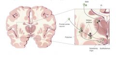
Where does the Striatum project?
|
GPe: main target of axons from striatum.
GPi: important output to STN, SN, and VLo STN: to GPi: involved in local feedback loops. SN: pars compacta |
|
|
What is unique about the basal ganglia's motor functionality? What affects does this feature have on the basal ganglia?
|
They have no direct input or output with the spinal cord. This means the basal ganglia are largely mediated by their sole source of input, the cerebral cortex.
|
|
|
What does the straitum consist of?
|
The striatum consists of three important subdivisions: the caudate nucleus, the putamen, and the ventral striatum (which includes the nucleus accumbens). Except at its most anterior pole, the striatum is divided into the caudate nucleus and putamen by the internal capsule, a major collection of fibers that run between the neocortex and thalamus in both directions. All three subdivisions of the striatum have a common embryological origin.
|
|
|
What other part of the basal ganglia is related to the internal Globus Pallidus?
|
The internal pallidal segment is related functionally to the pars reticulata of the substantia nigra, which lies in the midbrain on the medial side of the internal capsule. The cells of the internal pallidal segment and pars reticulata use γ-aminobutyric acid (GABA) as a neurotransmitter. Just as the caudate nucleus is separated from the putamen by the internal capsule, the internal pallidal segment is separated from the substantia nigra.
|
|
|
What is the part of the substantia nigra, other than the pars reticulata?
|
In addition to its reticular portion, the substantia nigra also has a compact zone (pars compacta). This zone is a distinct nucleus that lies dorsal to the pars reticulata although some of its neurons lie within the pars reticulata. The cells of the pars compacta are dopaminergic.
|
|
|
Where is the subthalamic nuclei and where does it have connections to? What neurotransmitter does it use?
|
The subthalamic nucleus is closely connected anatomically with both segments of the globus pallidus and the substantia nigra. It lies just below the thalamus and above the anterior portion of the substantia nigra. The glutaminergic cells of this nucleus are the only excitatory projections of the basal ganglia.
|
|
|
What are the two major parts of the striatum?
|
It consists of two separate parts, the matrix and striosome compartments (the latter also referred to as patches). These compartments differ histochemically from one another and have different receptors. The striosome compartment receives its major input from limbic cortex and projects primarily to the substantia nigra pars compacta.
|
|
|
What cell type makes up a majority of the striatum and what is unique about them.
|
Although the striatum contains several distinct cell types, 90-95% of them are GABA-ergic medium-spiny projection neurons. These cells are both major targets of cortical input and the sole source of output.
|
|
|
What are the 4 I/O loops of the basal ganglia?
|
1. Skeletomotor loop
2. Occulomotor loop 3. Prefrontal cortex loop 4. Limbic loop |
|
|
Describe the skeletomotor loop.
|
Cerebral cortex areas 3a, 1, 2, and 5 [primary motor, lateral premotor, supplementary motor] (Glutamate) → Putamen (GABA) ↛ GPi, SNr (GABA) ↛ Motor Thalamus ↺
|
|
|
Describe the oculomotor loop.
|
Cerebral cortex [frontal and supplementary eye fields] →Caudate [Head] (GABA) ↛ GPi, SNr (GABA) ↛ Motor Thalamus ↺
|
|
|
Describe the prefrontal cortex loop.
|
Cerebral cortex [dorsolateral prefrontal and lateral orbitofrontal] → Caudate [Body] (GABA) ↛ GPi, SNr ↛ Motor Thalamus ↺
|
|
|
Describe the limbic loop.
|
Cerebral cortex [anterior cingulate area and medial orbitofrontal] →Ventral striatum (GABA) ↛ Ventral pallidus, GPi, SNr (GABA) ↛ Motor Thalamus ↺
|