Reading...
![]()
Play button
![]()
Play button
![]()
Use LEFT and RIGHT arrow keys to navigate between flashcards;
Use UP and DOWN arrow keys to flip the card;
H to show hint;
A reads text to speech;
69 Cards in this Set
- Front
- Back
|
Esophagus anatomical position:
|
dorsal to trachea in cranial 1/3 of neck, lateral (left) of trachea in middle 1/3 of the neck, ventral to trachea at thoracic inlet, cervical portion is 50% of the length
|
|
|
Region of the esophagus most amenable to surgery:
|
cervical
|
|
|
Esophageal layers:
|
from outer to inner, tunica adventitia, tunica muscularis, tela submucosa, tunica mucosa
|
|
|
Types of esophageal muscle:
|
cervical portion striated, blend to smooth muscle in the caudal thoracic portion
|
|
|
Layer with the highest tensile strength of the esophagus layers for closure of an esophageal incision:
|
mucosa
|
|
|
Lining of mucosa:
|
stratisfied squamous epithelium
|
|
|
Arterial supply to the cervical esophagus:
|
carotid arteries
|
|
|
Arterial supply to the thoracic esophagus:
|
bronchoesophageal and gastric arteries
|
|
|
Esophageal vascular pattern:
|
arcuate and segmental, without much collateral circulation
|
|
|
Innervation to the esophagus:
|
9th and 10th cranial nerves, sympathetic trunk, mesenteric ganglia.
|
|
|
Odynophagia:
|
painful swallowing
|
|
|
Propagation speed of a food bolu:
|
proximal 2/3 of the esophagus, 9.4 cm/s, caudal 1/3, 4.6 cm/s
|
|
|
Positive contrast esophagram:
|
horse is fed or given 120 mL barium paste by mouth, outlines longitudinal mucosal folds in the undistended lumen, localizes obstruction or disruptions of the lumen
|
|
|
What is identified with large volume barium contrast esophgraphy?
|
highlight strictures, prestenotic dilation, and space occupying masses
|
|
|
Double contrast esophagraphy:
|
administration of liquid barium, followed by an equal volume of air
|
|
|
What is identified with double contrast esophagram?
|
evaluation of mucosal folds and detection of mucosal lesions
|
|
|
Negative contrast esophagraphy:
|
without contrast material and only insufflation of the esophagus via endoscopy or introduction of air through a NGT
|
|
|
What is identified with negative contrast esophagraphy?
|
cranial esophagus strictures
|
|
|
Functionally distinct manometric regions:
|
cranial esophageal sphincter, caudal esophageal sphincter, fast region, and slow region
|
|
|
Location of fast esophageal region:
|
cranial 2/3rds
|
|
|
Location of slow esophageal region:
|
caudal 1/3
|
|
|
Approaches to the esophagus:
|
ventral, ventrolateral, thoracotomy
|
|
|
Used of ventral approach:
|
esophagotomy, esophagomyotomy, and resections of the cranial 1/3 of the esophagus
|
|
|
Use of ventrolateral approach:
|
esophagostomy (feeding tube placement) or for approach to the distal ¼ of the cervical esophagus
|
|
|
Use of thoracotomy:
|
access the distal half of the esophagus
|
|
|
Clinical objective of esophageal surgery:
|
obtain leak proof healing of a primary anastomosis or incision, dilate a restricted lumen, and return an enlarged or disrupted esophagus to near normal size and function
|
|
|
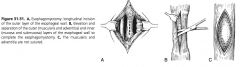
Describe ventral approach:
|
dorsal recumbency, 10 cm incision made ventral midline, sternothyroid, sternohyoid, omohyoid muscles bluntly separated, expose trachea, retract trachea to right, esophagus exposed by conservative dissection loose adventitial tissue
|
|
|
Describe ventrolateral approach:
|
dorsal, RLR, or standing, 5 cm incision made just ventral to jugular vein, sternocephalicus and brachiocephalicus separated, blunt dissection of deep cervical fascia exposes the esophagus
|
|
|
Why is ventrolateral approach advantageous for esophagostomy?
|
better access to the middle and distal cervical access due to increased ventral cervical musculature
|
|
|
Use of thoracotomy approach to the esophagus:
|
vascular ring anomalies, or esophageal lesions in the distal half of the esophagus that do not require penetration of the esophageal lumen
|
|
|
Medical adjuncts to esophageal impaction:
|
atropine (0.02 mg/kg), acepromazine, oxytocin (0.11-0.2 IU/kg), xylazine
|
|
|
Affects of atropine of esophagus:
|
esophageal relaxation, reduction of salivary secretions
|
|
|
Closure after esophagotomy:
|
apposition of the mucosa-submucosa with continuous suture (with knots to the lumen), esophageal musculature in interrupted pattern, place drain, closure of the approach incision in 2-3 layers
|
|
|
Benefits of drain placement:
|
remove dead space fluid, monitor for saliva leakage
|
|
|
Post-operative esophagotomy care:
|
withholding feed for 48 hours, supportive fluid therapy, electrolyte correction
|
|
|
Esophageal rupture management:
|
early establishment of ventral drainage
|
|
|
Complications of esophageal rupture:
|
Feed and saliva accumulation causes severe soft tissue inflammation, infection, mediastinitis, or plueritis
|
|
|
How long should Esophagostomy tubes remain in place?
|
at least 7 to 10 days to allow granulation tissue to form a stoma around the tube
|
|
|
Types of esophageal stricture:
|
mural lesions involving only the adventitia and muscularis, rings that involve only the submucosa and mucosa, and annular stenosis that affect all 4 layers
|
|
|
When do strictures form and resolved?
|
maximally formed by 30 days after esophageal trauma, in most cases return to normal after 60 days
|
|
|
Disadvantages of bougienage or dilators for esophageal stricture:
|
require special equipment and may fail due to the chronicity of the stricture
|
|
|
Optimal surgical intervention for esophageal stricture:
|
delayed for at least 60 days after the original trauma
|
|
|
Surgical options for esophageal stricture:
|
esophagomyotomy, partial or complete resection and anastomosis, or patch grafting
|
|
|
Complications after resection and patch grafting:
|
leakage of luminal contents, reformation of the stricture.
|
|
|
Best approach for mural strictures:
|
esophagomyotomy
|
|
|
Describe esophagomytomy:
|
ventral approach, linear incision made into muscularis, not penetrating mucosa, NGT passed through the lumen, muscularis separated from the mucosa by sharp dissection circumferentially myotomy not closed
|
|
|
Describe partial resection:
|
approached as longitudinal esophagomyotomy, mucosal stricture resected within the muscular tube, mucosal edges apposed without tension, if performed for esophageal ring, myotomy closed, if performed for annular stenosis, myotomy is not closed
|
|
|
When is a complete resection and anastomosis is performed?
|
esophagus has ruptured and the muscularis is not viable
|
|
|
Describe complete resection and anastomosis:
|
esophagus exposed throug ventral approach, lumen occluded using rubber drains or umbilical tape secured circumferentially proximally and distally, entire esophagus transected proximally and distally removing the diseased segment, anastomosis performed by apposing the mucosal-submucosal layer, The muscularis layer is apposed but relief incisions may be required to reduce tension
|
|
|
What can reduce post-operative tension on esophageal resection and anastomosis:
|
standing martingale
|
|
|
Patch grafting:
|
longitudinal incision made through the muscularis layer, extending 3 cm proximal and distal, mucosa incised after passage of NGT, strip of brachio- or sternocephalicus muscle separated from belly of the muscle but not transected proximally or distally, edge of submucosa-mucosa, and muscularis layers are sutured to the graft
|
|
|
Management of esophageal fistulas:
|
establishment of ventral drainage and elevating food and water to limit loss
|
|
|
Types of esophageal diverticula:
|
traction or true, pulsion or false
|
|
|
Development of traction diverticula:
|
contraction of periesophageal scar tissue and outward traction or tenting of all layers of the esophagus
|
|
|
Causes of transction diverticula:
|
esophagostomy sites, wounds allowed to heal by second intension, or after luminal penetration with leakage and secondary inflammation or abscessation
|
|
|
Development of a pulsion or false diverticulum:
|
protrusion of the mucosa-submucosa through a defect in the muscularis layer
|
|
|
Causes of pulsion diverticula:
|
fluctuations in esophageal intraluminal pressure, overstretch damage to esophageal muscle fibers after impaction, and external trauma
|
|
|
Treatment of pulsion diverticula:
|
diverticulectomy (resection of mucosa-submucosal sac) or inversion of the sac followed by reconstruction of layers
|
|
|
When is diverticulectomy chosen?
|
mucosal sac is very large and the neck of the diverticula is very narrow
|
|
|
Megaesophagus:
|
dilation and muscular hypertrophy of the esophagus oral to a constriction
|
|
|
Types of megaesophagus:
|
congenital ectasia (dilation of unknown origin), achalasia (failure of the distal esophagus to relax because of neural dysfunction) and secondary to vascular ring anomalies
|
|
|
Complications of esophageal surgery:
|
dehiscence and stricture, laryngeal hemiplegia, or carotid artery rupture and complication of esophageal disease such as acid-base and electrolyte alterations
|
|
|
Why do acid-base and electrolyte alteration occur?
|
loss of saliva
|
|
|
What acid-base and electrolyte abnormalities usually occur?
|
hyponatremia, hypochloremia, transient metabolic acidosis which progresses to metabolic alkalosis
|
|

|

|
|

|
|
|

|

|
|

|

|
|

|

|