![]()
![]()
![]()
Use LEFT and RIGHT arrow keys to navigate between flashcards;
Use UP and DOWN arrow keys to flip the card;
H to show hint;
A reads text to speech;
76 Cards in this Set
- Front
- Back
|
All of the followingare signs of regeneration on the Diff Quik blood smear except: |
retics. |
|
|
Anisocytosis is bestdescribed as: |
variable size. |
|
|
Assuming these are your values: PCV of 36, Hgb 5.1, RBC of 3.8 and the WBC is 12.3 from the auto cell counter, calculate the MCHC: _____________ |
14.1% or 14.1 g/dl |
|
|
A yellow lab ispresented with a PCV of 58% and a 8.0 g/dl total protein. Pick the mostprobable description of his health status. |
Dehydrated |
|
|
Background counts on cell counting machines are done for the following reason: |
To assure reagents and diluents are not contaminated so as to affect patient cell counts. |
|
|
Best evaluation for aregenerative anemia besides the retic count would be: |
RBC morphology on a Diff Stain |
|
|
Blood samples must bewell mixed before use: |
to insure even distribution of cells. |
|
|
Blood smears: should be made from |
fresh blood. |
|
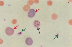
Canine ( ignore the arrows) |
Hypochromic |
|
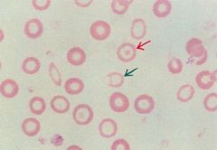
Canine - Red Arrow These RBC's are bestdescribed as: |
Macrocytic, polychromic |
|
|
Crenated RBC's can be caused by: |
excessive use of anticoagulants. cells being put into a hypertonic solution. not drying smear quickly enough. all of the above. |
|
|
Compute the indices for adog with the following values: PCV33%, TP 7 g/dl, WBC 20,000, RBC 4.75, Hgb 11g/dl. |
MCV = 69 MCH = 23 MCHC = 33 |
|
|
Crenation is best described as: |
usually due to technique. |
|
|
Definition, Decreased erythrocyte mass |
Anemia |
|
|
Heinz body is bestdescribed as: |
denatured Hgb. |
|
|
How is anemia noted clinically? |
Pale mucus membranes and Low PCV |
|
|
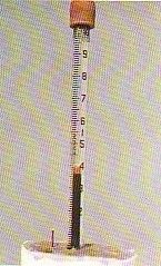
How is arefractometer calibrated? |
Measure a drop of distilled water. Adjust until the specific gravity is 1.000. |
|
|
Howell Jolly Body isbest described as: |
nuclear remnant. |
|
|
Hypochromic is best described as: |
decreased Hgb. |
|

Identify microscope part A |
Ocular |
|
|
If the PCV is 36 whatis the RBC estimate? |
6 |
|
|
Mean Corpuscular Volume isan indices expressed in what kind of units? |
Femtoliters |
|
|
One breed of dog mentioned in the reading may have microcytic RBC's. What is the breed? |
Akitas |
|
PCV 36% The best description of this buffy coat is: |
increased |
|
|
PCV may remain stable6 hours post trauma due to vasoconstriction and concentration |
TRUE |
|
|
Poikilocytosis is/arebest described as: |
varying shape. |
|
|
Polychromatic: |
many colored. |
|
|
RBC's are evaluatedfor three criteria. What are they ? |
size, shape and color |
|
|
Rouleaux is bestdescribed as: |
coin like stacking. |
|
|
Stomatocyte is bestdescribed as: |
mouth cell. |
|
|
Supravital stainssuch as NMB would be used to demonstrate: |
nuclear material. |
|
|
Target cell is bestdescribed as: |
leptocyte. |
|
|
The 100X objective is used to |
Evaluate RBC morphologyPerform a differential WBC countEstimate platelet numbers*All of the above* |
|
|
The best terminologyto describe a young RBC seen on the differential smear is: |
polychromatic and macrocytic. |
|
|
The normal totalprotein average for the dog and cat is: |
6.0-7.5 g/dl t |
|
|
The only informationyou have on a cat is PCV = 37%. What is his MCHC? |
33 g/dl (or %) 20 g/dl (or %) |
|
|
We prefer push smearsover coverslip smears in order to ensure better distribution of cells. |
False |
|
|
What are the units?T.P.: 7.0 (____) |
g/dl |
|
|
What causes or enhances hypersegmentation of neutrophils? |
Cell agingTreatment with steroidsHyperadrenocortisolism |
|
|
What does lipemic plasma indicate? |
post prandial sample poor sampling technique |
|
|
What is the units?Hgb: 5.1 (____) |
g/dl |
|
|
What is the units? PCV: 36 (____) |
% |
|
|
what is the fecal flotation media used in our lab and in most small animal practices? |
Sodium nitrate |
|
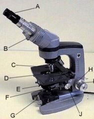
What is the name of the microscope part labeledH ? |
Voltage regulator or course adjustment knob |
|
|
What magnificationare the RBC's morphologically evaluated? |
Oil |
|
|
What objective(magnification) is used to find a good monolayer? |
High |
|
|
What species has RBCthat normally appear like spherocytes making it hard to identify anabnormality? |
Cat |
|
What type of RBC morphology might you expect to see with this type of tumor or DIC? |
schistocytes |
|
|
What would aspherocyte look like? |
Small dense cell with no zone of central pallor |
|
|
With a PCV of 36,what is the hemoglobin estimate? |
12 |
|
|
Which species has adiscocytic normal shape RBC? |
Dog |
|
|
Which is statement isfalse when refering to Dif Quick staining? |
Dif quick is a supra vital stain |
|
|
Which type of feline reticulocyte is a polychromatophil on Romanowsky stained blood films? |
Aggregate |
|
|
Why do we prefercoverslip smears to push smears. ONE answer please. |
even distribution of cells |
|
|
You are able toclearly see your specimen on low power but can't focus on high or oil. What isthe problem? |
Your cytology or blood smear is on the the down side of the slide. |
|
|
Normal PCV Dog : |
Avg. 45% Range: 36-54% |
|
|
Normal PCV cat |
Avg. 37% Range: 25-46% |
|
|
Normal TP Dog: |
6-7.5 g/dl |
|
|
Normal TP Cat: |
5.6-7.4 g/dl |
|
|
RBC estimate = |
1/6 of the PCV mm3 |
|
|
Hgb estimate is |
1/3 PCV g/dl |
|
|
PCV and TP increased= |
Dehydration |
|
|
PCV decreased TPnormal = |
Anemic |
|
|
PCV decreased and TPincreased= |
Anemic with Dehydration |
|
|
PCV normal TPdecreased= |
Protein loss/liver disease/burns |
|
|
Total protein is thefirst to fall with trauma patients |
TRUE |
|
|
MCV (Femtoliters fl )= |
PCV x 10 divided by RBC |
|
|
MCH (Picogramspg)= |
HGB x 10 divided by RBC |
|
|
MCHC (g/dl) = |
HGB x 100 divided by the PCV |
|
|
Which breed naturally has low platelet counts? |
Greyhound |
|
|
Which breeds cells are naturally macrocytic? |
Miniature Poodle |
|
|
The purpose of performing a background count on auto cell counters is to assure the reagents and diluents are free of debris that might interfere with patient cell counts. |
TRUE |
|
|
What does yellow plasma indicate? |
liver disease |
|
|
Given a PCV of 33%. What isthe RBC estimate? |
5.5 x 10^6 mm^3 |
|
|
Generally speaking, the higher the magnification the lower the condensor. |
False |
|
|
These RBC's look fragmented and like they were hung out carelessly on a clothesline! They may indicate DIC or hemangiosarcomas of the liver/spleen. What are they? |
schistocytes |