![]()
![]()
![]()
Use LEFT and RIGHT arrow keys to navigate between flashcards;
Use UP and DOWN arrow keys to flip the card;
H to show hint;
A reads text to speech;
23 Cards in this Set
- Front
- Back
|
Temporal & Spatial Resolutions |
Spatial: amount of information per sample (blocky video game) Temporal: # of samples per unit time |
|
|
Three broad tool types |
Imaging -often trade off spatial for temporal Point tracking -high spatial and temporal (can be limited by points) Others - may have high temporal but no spatial (recordings) |
|
|
Brain Study Methods |
Interruption of normal activity (lesions, brain damage, open brain surgery, TMS) Measuring structure of the brain (mri, CT scan, x-ray, etc) Measuring activity in the brain (measure blood flow, electrical or magnetic fields generated by brain) |
|
|
Hemodynamic methods |
Useful for where, not when activity is occuring Lag in imaging, stimuli need to be presented in blocks Sometimes introduce radioactive isotope into blood Creates photons, picked up by scanner |
|
|
Functional Magnetic Resonance Imaging |
When hemoglobin in blood carrying oxygen, it's diamegnetic. Will create opposite magnetic field. When not carrying oxygen, is paramagnetic: will align itself to magnetic field |
|
|
Electro physiological methods |
Good for when something happens Bad for showing where
EEG (electroencephalography): Brain creates electricity, travels through tissue/bone/scalp and electrodes detect it MEG (magnetoencephalography): Electrical currents created by active neurons generate magnetic field Detected by SQUIDS Better spatial resolution |
|
|
CNS/PNS |
Central nervous system (brain/spine) Peripheral Nervous System (cranial nerves, spinal nerves) |
|
|
Neural Tissue |
Neural pathways transmit signals through the body Contains two types of cells: Neurons (specialized nerve cells that generate and conduct nerve impulses) - motor - sensory Neuroglia Supporting cells that provide physical support, remove debris, or electrical insulation |
|
|
Neuron |
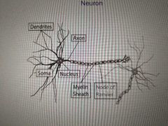
Nucleus Soma Axon (send signals, white matter) Dendrites (receive signals, grey matter) Myelin Sheath Node of Ranvier |
|
|
Gray/White Matter |
CNS: nuclei (grey) tracts (white) PNS: ganglia (grey) nerves (white) |
|
|
Synapse |
Action potential runs through to terminal/button/bouton, neurotransmitters get released into synaptic gap, picked up by Dendrites of next neuron |
|
|
Brain |
Densely packed with neurons Half are in cerebellum Folded for more surface area inside of skull Cerebral cortex, corpus callosum, amygdala, pons, medulla, cerebellum |
|
|
Cerebellum/brainstem (hindbrain) |
Multisensory integration in perception Last stop of timing/coordination of motor output Cerebellum - 10% of brain weight Half of brain neurons Fine tuning motor control, balance, posture, movement, cognition |
|
|
Pons/Medulla |
Pons Breathing/sleeping Medulla Heart rate, breathing, blood pressure |
|
|
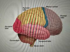
Basic Cortex Structure |
Hemispheres Lobes Gyri (ridges) Sulci (furrows) Fissures (deep Sulci) |
|
|
Corpus Callosum |
Connects hemispheres Communication between the two Eye movement and vision Maintaining balance of arousal and attention Tactile localization |
|
|
Lobes |
Frontal: cognitive function, planning, initiating and inhibiting voluntary motion Parietal: sensory perception Temporal: audition Occipital: vision |
|
|
Sulci |
Sylvian fissure (lateral sulcus) - Heschel's Gyrus (lots of language in this area) Rolandic Fissure (Central sulcus) |
|
|
Neural Impulses (action potential) |
Electrical charges that move through nervous stem Ions (charged atoms, positive or negative) Resting potential Outside neuron: positive (Na+ sodium) Inside neuron: negative (K- potassium) Gates and pumps that facilitate movement. 3 Na out for every 2 K in |
|
|
Action potential |
Na gates open and Na rushes in K channels open and K flows out Refractory period while rebalancing Only happens at nodes, not all along Axon (saltatory conduction) Thicker Axon = faster |
|
|
Language in the brain |
Lateralized, generally left hemisphere Studied injured people (Phineas Gage) Broca: aphasia in producing speech (Frontal lobe) Wernicke: aphasia in understanding speech (temporal lobe) |
|
|
Arcuate Fasciculus |
Tract (white matter) between Brocas area and Wernickes area Not present in non-human primates Damage to this area causes conduction aphasia (speaking, understanding okay, but can't repeat stuff) |
|
|
Other areas of brain related to language |
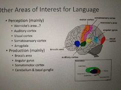
Motor cortex: Frontal lobe, anterior to central sulcus movement/articulatory Somatosensory cortex: posterior to Central sulcus, sensory Amygdala: processes fear/threats (and even swear words) Angular Gyrus: Parietal lobe, high-level speech planning (irony, metaphor) Perisylvian Language Zone Auditory cortex (along sylvian fissure) |