![]()
![]()
![]()
Use LEFT and RIGHT arrow keys to navigate between flashcards;
Use UP and DOWN arrow keys to flip the card;
H to show hint;
A reads text to speech;
245 Cards in this Set
- Front
- Back
|
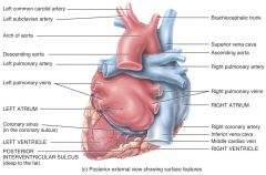
Where is the HEART LOCATED? |
The heart is located at the MEDIASTINUM (so if you think about it, 'media' --> medial which means middle, and 'stinum' --> 'sternum' which is the one holding your ribs. Therefore, the heart is located in the mediastinum -- the middle of your sternum!) |
|
|
What are PLEURAL MEMBRANES? |
Pleural Membranes are serous membranes that cover the lungs and act in the same way as the pericardium does for the heart -- in that it provides protection, and lubrication for the lungs. |
|
|
The Mediastinum has how many compartments? |
The mediastinum contains 3 compartments: the ANTERIOR, the MIDDLE, and the POSTERIOR. The heart is located in the MIDDLE compartment, and the other 2/3 of the heart's mass is slightly to the left of the body's midline! |
|
|
What are the 2 anatomical portions of the Heart? |
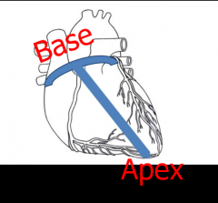
There is the BASE of the heart, which is tipped up medially and posteriorly; and then there's the APEX of the heart, which projects inferiorly and laterally. (Or, in other words, the BASE is the TOP of the heart, and the APEX is the BOTTOM of the heart). |
|
|
What is the FUNCTION of the PERICARDIUM? |
The Pericardium serves in a few ways: - Holds the heart in place - Forms a barrier against infections - Helps keep the heart from over expanding - Fluids within the sacs of the pericardium lubricate the outer wall of the heart and allow it to beat without friction. |
|
|
What is the STRUCTURE of the PERICARDIUM like? |
The pericardium is a tough, fibrous outer layer lined by a delicate serous membrane. |
|
|
What is the FIBROUS PERICARDIUM? |
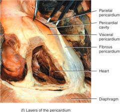
The FIBROUS PERICARDIUM is the dense and non-flexible connective tissue that helps to protect and anchor the heart. |
|
|
What is the INNER SEROUS MEMBRANE? |

The inner serous membrane is subdivided into two layers: - the Parietal Layer which adheres to the outermost fibrous layer - the Visceral Layer which also forms the outer surface of the heart's wall. The INNERMOST layer! |
|
|
What is Pericardial Fluid and what is its importance? |
Pericardial Fluid is the fluid located within the pericardium that acts to reduce friction within the pericardium by lubricating the epicardial surface and allowing the membranes to glide over each other with each heartbeat. |
|
|
What are the 3 Layers of the Heart Wall? |
There are 3 Layers to the Heart Wall: - Epicardium - Myocardium - Endocardium |
|
|
What is the Epicardium? |

The Epicardium is the OUTERMOST wall of the heart, composed of connective tissue covered by epithelium. It is also known as the visceral pericardium and serves as an additional layer of protection for the heart |
|
|
What is the Myocardium? |

The Myocardium is the middle layer of the heart's walls. It is composed of cardiac muscle and serves a few functions: - provides a scaffolding for the heart's chambers - assists with the contraction/relaxation of cardiac muscle - conducts electricity to coordinate contraction/relaxation of cardiac muscle |
|
|
What is the Endocardium? |
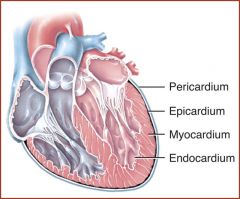
The Endocardium is the innermost layer of the heart's walls, and it lines the chambers of the heart. It provides protection to the valves and the heart chambers and is composed of endothelial cells which help to maintain smooth blood circulation/pumping |
|
|
Cardiac Muscle |
Cardiac Muscle is STRIATED, with short, branched fibers that contain only one centrally located nucleus. These muscle cells connect to and communicate with neighboring cells via GAP JUNCTIONS in INTERCALATED DISCS! |
|
|
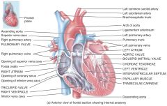
How many chambers does the heart contain? |
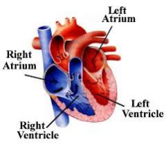
The Heart contains 4 Chambers: - 2 Upper Chambers known as the RIGHT and LEFT Atria - 2 Lower Chambers, known as the RIGHT and LEFT Ventricles **The heart can be thought of as "right and left" pumps or even "top and bottom" pumps. |
|
|
What is the FUNCTION of the TOP part of the Heart? |
The TOP part of the heart consists of the Right and Left Atria. These are rather WEAK pumps which serve to load the ventricles via an 'atrial kick' before ventricular contraction occurs. |
|
|
What is the FUNCTION of the BOTTOM part of the Heart? |
The Bottom part of the heart consists of the Right and Left Ventricles, which are rather STRONG pumps, and serve as the main pump for pulmonary and systemic circulation! **Specifically the LEFT ventricle is the strongest of all 4 chambers! |
|
|
What is the 'Atrial Kick'? |
The Atrial Kick is the last contraction the atrium makes as it sends blood into the ventricles. As this last contraction takes place, the ventricles take over and draws in the blood. It is responsible for about a 20% increase in the amount of blood ejected by the ventricles (and eventually amounts to a flutter as we age) |
|
|
What is Atrial Fibrillation? |
Atrial Fibrillation is when there is no atrial kick present in the processes of the atrium, and can attribute to shortness of breath, heart palpitations and weakness. |
|
|
Which way does blood flow? And why is blood flow important to heart functioning? |
Blood flows from an area of HIGH to LOW pressure! It is dictated strictly by differences in pressure (nothing to do with muscles). The flow of blood operates the valves of the heart! |
|
|
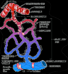
How do our heart valves operate? |
![Valves operate in TWO pairs:
- Atrioventricular Valves (AV) allow blood flow from the atria into the ventricles.
- Outflow (Semilunar Valves [SL]) allow blood flow from the ventricles into the outflow vessels.](https://images.cram.com/images/upload-flashcards/34/45/61/16344561_m.jpg)
Valves operate in TWO pairs: - Atrioventricular Valves (AV) allow blood flow from the atria into the ventricles. - Outflow (Semilunar Valves [SL]) allow blood flow from the ventricles into the outflow vessels. |
|
|
What are the parts of the AV valves? |
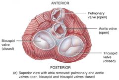
The AV valves are positioned at the entrance of each ventricle, thus meaning there are two of them. - Right AV Valve is AKA the Tricuspid Valve and it opens into the right ventricle - Left AV Valve is AKA the Bicuspid/Mitral Valve and it opens into the left ventricle |
|
|
What is the Chordae Tendineae? |
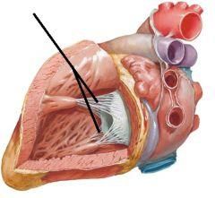
The chordae tendineae are strong, fibrous strings attached to the cusps of the heart on the ventricular side (lower part).They originate from the papillary muscles and serve as the attachment for the AV valves to the walls of the ventricles. |
|
|
What happens when the Papillary Muscles contract? |
The AV valve closure slows to prevent trauma |
|
|
What are the OUTFLOW VALVES? |

These are positioned at the entrance to outflow vessels leading into the pulmonary and systemic circulation. They are the [top] part of the heart, and consist of two parts: - Right Outflow Valve (Pulmonary Valve) opens into the pulmonary trunk - Left Outflow Valve (Aortic Valve) opens into the aortic arch |
|
|
How do Outflow Valves work? |
Well, outflow valves open with the act of ventricular ejection and close when blood in the aorta and pulmonary outflow tract begins to leak back into ventricles. |
|

|
|
|
|
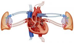
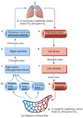
What are the two circuits of blood flow? |
There are TWO circuits of blood flow: - Systemic Circuit - Pulmonary Circuit |
|
|
What does the Systemic Circuit do? How does it work? |
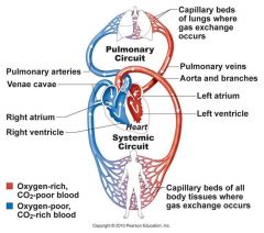
The Systemic Circuit ejects blood into the aorta, systemic arteries, and arterioles. It is powered by the LEFT side of the heart. |
|
|
What does the Pulmonary Circuit do? How does it work? |

The Pulmonary Circuit ejects blood into the pulmonary trunk. It is powered by the RIGHT side of the heart. |
|
|
What is the PULMONARY TRUNK? |
The pulmonary trunk is a major vessel of the human heart that originates from the RIGHT ventricle. It branches into the R&L pulmonary arteries, which lead straight to the lungs. |
|
|
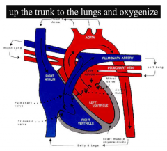
What are the 3 Steps in Blood Flow? |
1.) Venous return to the heart; deoxygenated blood flows into the R atrium from the two vena cava and the coronary sinus 2.) Blood flows through the R heart to the lungs to be oxygenated 3.) Oxygenated blood returns to the L heart to be pumped through the outflow tract of the systemic circulation |
|
|
What are the 3 Major Arteries attaching to the heart? |
- Arch of the Aorta - Pulmonary Trunk - Coronary Arteries |
|
|
What is the Superior Vena Cava? |

The Superior Vena Cava carries deoxygenated blood into the heart from the head, neck and upper limbs. It also dumps blood into the R atrium. |
|
|
What is the Inferior Vena Cava? |

The Inferior Vena Cava carries deoxygenated blood into the heart from the lower body. It also dumps blood into the R atrium. |
|
|
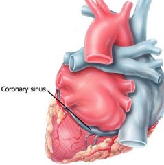
What is the Coronary Sinus? |
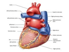
The Coronary Sinus is where the blood is returned to the R atrium. It receives its blood via the Coronary Arteries (which branches from the aorta). |
|
|
What is the Aorta? |
The Aorta is the main artery of the body. It supplies large amounts of oxygenated blood to the rest of the body with a high pressure. It passes over the L ventricle and runs down in front of the backbone. |
|
|
What are the major VEINS attaching to the heart? |
There are THREE major veins that attach to the heart: - Superior and Inferior Vena Cava - the 4 Pulmonary Veins - The Coronary Sinus (which is located on the back of the heart). |
|
|
What do the 4 Pulmonary Veins do? |
They carry oxygenated blood into the L atrium |
|

|
|
|
|
WHat is the direction of blood flow? |
|
|
|
Where are the Coronary Vessels located on the heart? |
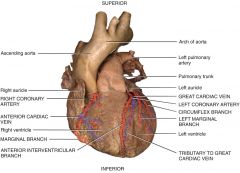
** Only the innermost tissues lining the chambers of the heart can derive oxygen from the blood flowing through those chambers |
|
|
What is the difference in functioning of the coronary veins and the coronary sinus? |
The coronary veins all collect into the coronary sinus at the posterior of the heart. Meanwhile, the coronary sinus empties into the RIGHT atrium. Deoxygenated coronary blood joins oxygen-depleted blood from the rest of the body. |
|
|
Where are the coronary veins located on the heart? |
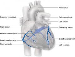
the coronary veins drain blood from the heart wall and empty into the coronary sinus. |
|
|
What is Autorhythmicity? |
Autorhythmicity is the rhythmical electrical activity produced individually by special myocytes within the heart. |
|
|
What is the Cardiac Conduction System? |
The Cardiac Conduction System is a specialized group of myocytes that have the unusual ability to spontaneously depolarize. |
|
|
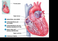
What are the MAIN components of the Cardiac Conduction System? |
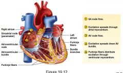
There are 5 MAIN components of the Cardiac Conduction System: * SA Node * AV Node * Bundle of His * Bundle branches * Purkinje fibers |
|
|
What are the 2 important roles of the self-excitable myoctyes that "act like nerves"? |
The self-excitable myocytes that "act like nerves" have two important roles: ~ Conduction System of the heart ~ Pacemaker |
|
|
What is the Sinoatrial (SA) Node? |
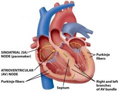
The SA node is located in the RIGHT ATRIAL WALL, below the entry of the superior vena cava. It has TWO main features: ~ To act as the Pacemaker of the heart ~ It has the fastest rate of depolarization |
|
|
How often do the autorhythmic fibers in the SA node spontaneously depolarize? |
They fire about once every 0.6 seconds, or 100 action potentials per minute. This act [of spontaneous depolarization] is modified by the ANS |
|
|
What are the steps of the Cardiac Conduction System? |
|
|
|
What is functional syncytium? |
This is the way that bands of muscles wind around the heart and work together as a unit. This allows the top ad bottom parts to contract in their own unique way. |
|
|
Why does the atrial muscle synctyium contract as a single unit? |
The atrial muscle syncytium contracts as a single unit to force blood down into the ventricles! |
|
|
Where does the syncytium of the ventricular muscle begin contraction at? Why does it start there? |
The syncytium of the ventricular muscle starts contracting at the APEX of the heart, in order to squeeze blood upwards to exit out the outflow tracts |
|
|
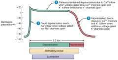
What is an Action Potential (AP)? |
The change in electrical potential associated with the passage of an impulse along the membrane of a muscle or nerve cell. |
|
|
What is the job of the AP within the cardiac muscle? |
The AP of a cardiac muscle is initiated by SA node and travels through the conduction system in order to excite 'working' contractile muscle fibers within the atria and ventricles. |
|
|
What are contractile fibers? |
Contractile fibers are fibers that are made up mostly of proteins that contract when stimulated and relaxes when not stimulated. |
|
|
What does RMP stand for? |
Resting Membrane Potential |
|
|
What is the stable RMP of contractile fibers? |
The stable RMP of contractile fibers is -90mV (unlike autorhythmic fibers) |
|
|
How do AP spread widely throughout the heart? |
AP is able to spread widely throughout the heart by opening and closing the Na+ [sodium] and K+ [potassium] channels |
|
|
Analyze this diagram: |
|
|
|
What is the Refractory Period? |
The refractory period lasts longer than the contraction itself. In fact, another contraction cannot begin until relaxation is underway! (this is where cardiac muscle differs from skeletal muscle) |
|
|
What is Tetanus? |
Tetanus is maintained contraction caused by rapidly repeating stimuli. |
|
|
Why can't Tetanus happen in cardiac muscle? |
Because it is maintained, constant contraction it therefore cannot occur in cardiac muscle, because cardiac muscle MUST have a refractory period in order to leave a sufficient amount of time between contractions for the chambers of the heart to fill with blood! |
|
|
What are the mechanisms of contraction in cardiac muscle? |
* Electrical activity leads to Ca2+ release from the SR * Actin & Myosin filaments go through the contraction cycle * Tension develops as filaments slide past one another |
|
|
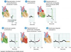
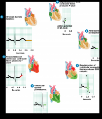
What are the steps of an EKG? |
|
|
|
What is an ECG/EKG? |
An EKG/ECG is a recording of electrical changes on the surface of the body resulting from depolarization and repolarization of the myocardium. |
|
|
What do EKGs measure? |
EKGs measure the presence or absence of certain waveforms (deflections), the size of the waves, and the time of the intervals of the cardiac cycles. *EKGs allows us to quantify and correlate, electrically, the mechanical activities of the heart. |
|
|
What are the 5 Major Waves within an EKG? |
*P-wave *P-Q Interval *QRS Wave *T-wave *S-T Segment |
|
|
What does the P Wave measure? |

The P-wave measures the atrial depolarization |
|
|
What does the P-Q Interval measure? |

The P-Q Interval measures the time it takes for the atrial kick to fill the ventricles |
|
|
What does the QRS Wave measure? |
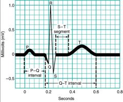
The QRS Wave measures the ventricular depolarization and atrial repolarization. |
|
|
What does the S-T Segment measure? |

The S-T segment measures the time it takes to empty the ventricles before they repolarize |
|
|
What does the T-Wave measure? |

The T-wave measures ventricular repolarization (which occurs as the ventricles relax). |
|
|
KNOW THIS DIAGRAM |
|
|
|
What is the Pressure-Volume Loop? |
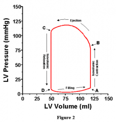
The pressure-volume loop is when the cycle that occurs within the heart when ALL FOUR valves are closed at the same time for a quick second. This then promotes a large level of pressure within the heart, which is what helps the heart to eject its blood. Afterwards, the four valves all close once again very quickly while it refills with blood to start the ejection cycle again. |
|
|
What is Hemodynamics? |
Hemodynamics relates the forces and motion of blood flow and the science concerned with the study of the circulation of blood. It determined oxygen and nutrient delivery, as well as metabolic end product removal. |
|
|
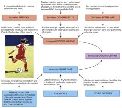
What is Cardiac Output? |
Cardiac Output is the amount of blood ejected by the heart per minute! (it is around 5 L per minute!) |
|
|
What is the formula that goes along with cardiac output? |
Q = SV x HR where Q --> cardiac output SV --> stroke volume HB --> heart beat |
|
|
What is the typical Q in adult males? |
The typical Q in adult males is around 5.25L/min (in formula, it would look like SV = 75mL/beat x 75 bpm) |
|
|
What are the THREE important factors affecting the Stroke Volume? |
* Preload * Myocardial Contractility * Afterload |
|
|
What is Preload? |
Preload is the degree of myocardial fiber stretching at the end of diastole and before contraction. |
|
|
What is diastole? |
Diastole is the phase of the heartbeat when the heart muscles relax and allows the chambers to fill with blood. |
|
|
What is Starlings Law of the Heart? |
With a greater preload on cardiac muscle just before they stretch comes an increase in their force of contraction during stystole |
|
|
Define systole |
Systole is the phase of the heartbeat when the heart muscle contracts and pumps blood from the chambers into the arteries. |
|
|
What is Afterload? |
Afterload is the resistance of flow, which affects how hard the heart must pump in order to push blood out (the pressure of the ventricle must overcome to eject its own volume). |
|
|
What is the peripheral component of the afterload? |
The pressure the heart must overcome to open the aortic valve. This process depends on TWO things: *Aortic compliance *Total systemic vascular resistance (or the blood viscosity and arteriolar constriction). |
|
|
What is Myocardial Contractility? |
Myocardial contractility is the force and velocity with which ventricular ejection occurs INDEPENDANT of the effects of preload/afterload. |
|
|
What is an example of myocardial contractility? |
When catecholamines are released they can increase contractility (in moments of fear, stress, pain). |
|
|
What is myocardial compliance? |
Myocardial compliance is the ventricle's ability to stretch and receive a given volume of blood. Normally, ventricles are very compliant, so large changes in their volumes produce only small changes in pressure. |
|
|
What is LOW COMPLIANCE? (in regards to myocardial compliance) |
LOW COMPLIANCE is when small changes in volume result in large changes in pressure within the ventricle. This occurs when the ventricle cannot stretch, which thus limits its ability to increase cardiac output with an increased preload. |
|
|
What is Ejection Fraction? |
Ejection Fraction (EF) is the percentage of blood that is pumped out of the ventricles with each heartbeat. |
|
|
What does the EF formula look like? |
EF = SV / EDV where EF --> Ejection Fraction SV --> stroke volume EDV --> end diastolic volume |
|
|
What happens in decreased systolic function? |
A decrease in contractility, and valvular diseases |
|
|
What happens in increased systolic function? |
Hypertrofic cardiomyopathy |
|
|
What are the normal values of Ejection Fraction (EF)? |
Greater than or equal to 50-55% EF is known as an increased EF and other inotropic actions Less than 40% indicates systolic dysfunction. |
|
|
Why is the Frank-Starling Law of the Heart important? |
It equalizes the output of R & L ventricles; keeps same volume of blood flowing to both systemic and pulmonary circuits). |
|
|
What are two agents that affect Myocardial contractility? |
1) Positive inotropic agents INCREASE contractility as well as stroke volume 2) Negative inotropic agents DECREASE contractility as well as stroke volume. |
|
|
What is cardiac reserve? |
Cardiac Reserve is the difference between Q at rest and the max Q. |
|
|
What is the average Cardiac Reserve? |
The average cardiac reserve is about 4-5x resting values **exercise draws upon cardiac reserve to meet the body's increased physiological demands and maintain homeostasis. |
|
|

|
|
|
Why do we measure blood pressure instead of blood flow? |
Blood flow is hard to measure, but blood pressure is easier and it is related to blood flow so we measure that instead! |
|
|
What is Ohms Law? |
Ohms Law is the equation that draws out the relationship between blood flow, blood pressure, and peripheral resistance. It looks like this: BP = Flow x Resistance |
|
|
What is Blood Flow? |
Blood flow is the amount of blood that actually reaches the end organs |
|
|
What is Blood Pressure? |
Blood Pressure (BP) is a measure of the force (in mmHg) exerted in the lumen (cavity) of blood vessels. |
|
|
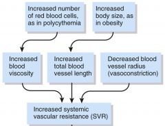
What is Resistance? |
Resistance is the sum of many factors which oppose the flow of blood! |
|
|
What sort of factors aid in the resistance of blood? |
|
|
|
What are the TWO types of venous return? |
~~ Skeletal Muscle Pump ~~ Respiratory Pump |
|
|
What does the Skeletal Muscle Pump do? |
The Skeletal Muscle Pump uses the action of muscles to milk blood in one direction (dude to the valves). |
|
|
What does the Respiratory Pump do? |
The Respiratory Pump uses the negative pressures in the thoracic and abdominal cavities generated during inspiration to pull venous blood towards the heart |
|
|
What are the names of the two valves utilized in the skeletal muscle pump? |
The proximal valve and the distal valve |
|
|
What are the factors affecting BP?
|
|
|
|
What are Baroreceptors? |
Baroreceptors are pressure-sensitive sensory receptors located in the aorta, internal cortid arteries, and other lage arteries located in the neck/chest. |
|
|
What type of changes occur in HR during parasympathetic and sympathetic reactions? |
Para --> DECREASE HR Symp --> INCREASED HR |
|
|
What are chemoreceptors? |
Chemoreceptors are sensory receptors that monitor chemical composition of blood. They are located in the carotid and aortic bodies and detect changes in O2, CO2, and H+ with their input being sent to the cardiovascular and cardiorespiratory centers. |
|
|
BP: Hormonal Regulation |
|
|
|
How can one measure peripheral circulation? |
By checking the PULSE! |
|
|
What is a pulse? |
The pulse is a result of the alternate expansion and recoil of elastic arteries after each systole. They are strongest in arteries closest to the heart, and get weaker the further they get away. |
|
|
What is the relationship between pulse and HR? |
Normally, the pulse is the same as the heart rate |
|
|
Tachycardia |
... |
|
|
Bradycardia |
... |
|
|
What do Arteries do? |
Arteries carry blood AWAY from the heart. They are normally oxygenated blood, but carry deoxygenated blood in the pulmonary circuit |
|
|
What are the three types of arteries? |
--Large elastic arteries (>1cm) --Medium muscular arteries (0.1 - 10mm) --Arterioles (<0.1 mm) |
|
|
What are capillaries? |
Capillaries are the site of nutrient and gas exchange! |
|
|
What are do veins do? |
Vein carry blood TOWARDS the heart. Usually oxygenated only in the pulmonary circuit and deoxygenated in the systemic circuit |
|
|
What are venules? |
Small veins (<0.1 mm) |
|
|
What are the 3 basic layers of blood vessels? |
*Tunica Intima **Tunica Media ***Tunica Externa (adventitia) |
|
|
What is the Tunica Intima? |

The Tunica Intima is the endothelium lining the cavity (lumen) of all vessels |
|
|
What is Tunica Media? |

Tunica Media is the muscular and connective layer that is responsible for vasodilation and vasoconstriction |
|
|
What is the Tunica Externa? |

The Tunica Externa anchors the vessels to their surrounding tissues. They contain elastic and collagen fibers, nerve endings, and small blood vessels. |
|
|
What is VASA VASORUM? |
The vasa vasorum is a network of small blood vessels that supply the walls of large blood vessels such as elastic arteries and large veins. They are related to the tunica externa |
|
|
Label the passage of blood through the arteries in order from first to last place visited. |
Elastic arteries --> Muscular arteries --> Arterioles |
|
|
What exactly are Elastic (conducting) Arteries? |
Elastic arteries are the largest arteries, about the size of a garden hose. Their walls are thin compared to their overall size, and they serve to store mechanical energy during ventricular systole as a sort of pressure reservoir. They also are in charge of transmitting that energy to keep blood moving after aortic and pulmonary valves close. |
|
|
What are Muscular (disturbing) Arteries? |
These are the medium sized arteries, found in two places of the body: brachial artery in the arm and the radial artery in the forearm. They have even more reserves of smooth muscle in their tunica media. Their main function is to help maintain proper vascular tone to ensure efficient blood flow to distal tissue beds. |
|
|
What are Anastomoses? |
Anastomoses is a union of vessels supplying blood to the same body tissue. |
|
|
Why are anastomoses beneficial? |
They provide collateral circulation (an alternative route) for blood to reach a tissue in instances of occlusion /blockage. |
|
|
What are Arterioles? |
These are abundant microscopic vessels with a thin tunica interna and tunica media. They act to regulate blood flow into capillaries and influence the greatest collective influence on both the local blood flow and on overall blood pressure. |
|
|
What are Metarteriole? |
Metarterioles are the termical ends of an arteriole that tapers towards the capillary junction |
|
|
What is a Precapillary Sphincter? |

A precapillary sphincter is formed by most distal muscle at the metarteriole capillary junction. It monitors and regulates blood flow into the capillary bed. |
|
|
What is the structure of capillaries like? |
Capillaries have a single layer of endothelial cells and a thin basemement membrane. Meaning they lack tunica media and tunica externa. The way they are formed allows them to be freely permeable to many susbtances. |
|
|
What is the function of capillaries? |
to exchange gases, fluids, and small ionic molecules |
|
|
In relation to pressure, how do veins operate compared to arteries? |
Because veins have thinner walls, less muscle and elastic tissue they operate at LOWER PRESSURES than arteries. |
|
|
What are the intravenous pressures? |
Venules have a pressure of 16mmHg Large Veins have a pressure of 1-2 mmHg Arterioles have a pressure of 35 mmHg |
|
|
Why are valves required? |
Because veins have lower pressures, so there is a low intravenous pressure in the body circulation system. |
|
|
What are varicose veins? |
Varicose veins are when veins are exposed to higher than normal pressure, and they become incompetent because of it. |
|
|
How many divisions does the respiratory system consist of? |
The respiratory system is divided into 2 zones: the -- Conducting Zone -- Respiratory Zone |
|
|
What are the duties of the CONDUCTING ZONE? |
The conducting zone is in charge of filtering, warming, and moistening the air as well as delivering it to the site of respiration in the lungs. |
|
|
What does the conducting zone consist of? |
The conducting zone consists of the nose, pharynx, larynx, trachea, bronchi, bronchioles, and terminal bronchioles. |
|
|
What are the duties of the RESPIRATORY ZONE? |
The respiratory zone is the MAIN SITE OF GAS EXCHANGE between air and blood. |
|
|
What does the respiratory zone consist of? |
The respiratory zone consists of the respiratory bronchioles, alveolar ducts, alveolar sacs, and alveoli |
|
|
What are the functions of the respiratory system? |
~ Provides gas exchange (intake O2, removal of CO2) ~ Helps regulate pH ~ Contain receptors for smell, filters inspired air, produces vocal sounds, and excretes small amounts of water and heat. |
|
|
What is the order of air passage through the Upper respiratory tract? |
Nose --> nasal cavity --> pharynx |
|
|
What is the order of air passage through the Lower respiratory tract? |
Larynx --> Trachea --> Primary (1) bronchi --> Secondary (2) bronchi --> Tertiary (3) bronchi --> Bronchioles --> Alveoli |
|
|
What is the NASAL CAVITY? |
The nasal cavity is inferior to the nasal bone and superior to the oral cavity, divided by the nasal septum into the right and left nares. It is important in: ~ ducts from paranasal sinus (mucus) and nasolacrimal ducts drain into the nasal cavity ~ Olfactory receptors ~ contains nasal conchae (which protrude from each lateral wall). |
|
|
What does the nasal cavity do? |
The nasal cavity warms, moistens, and filters incoming air. It also detects olfactory stimuli. |
|
|
What is the PHARYNX? |
The passageway to the pharynx begins posteriorly to internal nares and descends to opening of larynx in the neck. |
|
|
What are the three anatomical regions of the pharynx? |
~ Nasopharynx ~ Oropharynx ~ Laryngopharynx |
|
|
What are the duties of the pharynx? |
the functions of the pharynx are to: ~serve as a passageway for air/food ~resonating chamber ~housing for tonsils |
|
|
What is the Nasopharynx and what does it do? |

The Nasopharynx is the first (top) location of the pharynx. It contains the opening of the Eustachian Tubes (auditory). It also contain the pharyngeal tonsils (adenoids) and internal nares. |
|
|
What is he Oropharynx and what does it do? |

The oropharynx is the middle region of the pharynx. It serves both the respiratory and digestive functions. It's where the opening of the mouth is located as well as the main palatine tonsils & small lingual tonsil. |
|
|
What is the Laryngopharynx and what does it do? |
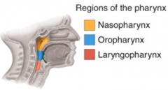
It is the bottom region of the pharynx. It also serves both respiratory and digestive functions. It open into the larynx and the esophagus. |
|
|
What is the larynx?
|
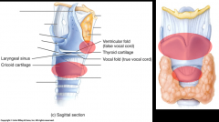
The larynx connects the laryngopharynx with the trachea. It's composed of 9 pieces of cartilage that altogether form a short passageway. |
|
|
What is the Thyroid Cartilage? |
The "Adam's Apple" found in the larynx |
|
|
What is the importance of knowing where the cricoid cartilage is? |
The cricoid cartilage is found in the larynx and it's the serves as the landmark for performing a cricothyrotomy (putting straw through neck to allow person to breathe). |
|
|
What is the trachea? What is its function? |
The trachea is anterior to the esophagus and is around 12 cm in length. It's a semi-rigid pipe of semi-circular incomplete circular incomplete cartilaginous rings. |
|
|
What are special about the branches from the trachea? |
All branches from the trachea to the terminal branchioles are part of the CONDUCTING AIRWAYS and DO NOT participate in gas exchange!! |
|
|
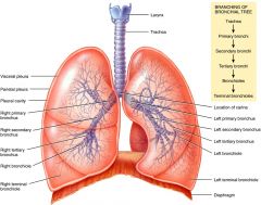
What are the PRIMARY BRONCHI? |
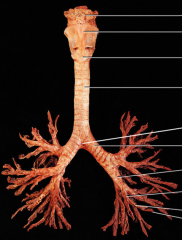
The primary bronchi emerge from the inferior trachea and split into RIGHT and LEFT main bronchus. They then also divide into SECONDARY (lobar) BRONCHI, which each supply one lobe: 3 on the right and 2 on the left. From there, the lobar bronchi breaks into TERTIARY (segmental) bronchi which also continues dividing down another 23 times. They then continue on into the bronchioles (tubes smaller than 1 mm) and then onto the terminal bronchioles. |
|
|
What is the CARINA? |
The carina is an internal ridge located at the junction of the two main stem bronchi - which is a very sensitive area for triggering the cough reflex. |
|
|
What is the RESPIRATORY ZONE? |
This is the end-point of the respiratory tree where the terminal bronchioles divide in respiratory bronchioles that lead into alveolar ducts that then open unto alveolar sacs that contain alveoli |
|
|
What are alveoli? |
Air exchange chambers |
|
|
What provides a large area for gas exchange? |
millions of alveoli provide a large area for gas exchange (close to around 1000 sq ft). |
|
|
Describe alveoli. |
Alveoli are surrounded by fine elastic fibres and are interconnected with the alveolar pores. |
|
|
What are alveolar macrophages? |
free floating 'dust cells' |
|
|
What type(s) of cells are present in alveoli? |
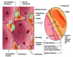
Type I and Type II cells are present in alveoli. Type I cells are epithelial cells and granular pneumocytes. Type II cells are also epithelial cells that secrete surfactant. |
|
|
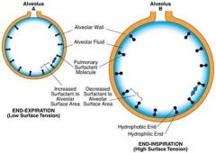
What is surfactant? |
Surfactant is a detergent-like substance secreted in fluid coating alveolar surfaces (alveolar fluid) that acts to decrease tension. It is secreted by TYPE II cuboidal epithelial cells (which are scattered within the alveolar walls). |
|
|
What happens without surfactant? |
Without surfactant alveolar walls would stick together during exhalation. |
|
|
Describe the anatomy/structure of the R lung
|
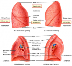
The R lung is divided by the oblique and horizontal fissures into 3 lobes: 1) Superior Lobe 2) Middle Lobe 3) Inferior Lobe |
|
|
Describe the anatomy/structure of the L lung |

The L left is divided by the oblique fissure into 2 lobes: 1) Superior Lobe 2) Inferior Lobe |
|
|
Where is the BASE of the lung? |

Actually located at the base - the bottom |
|
|
Where is the APEX of the lung located? |
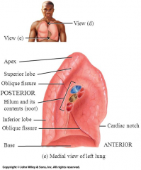
The apex is located at the top of the lung |
|
|
What is the cardiac notch? |

it is located in the L lung and makes the left lung 10% smaller than the right lung. It is the indentation for the heart. |
|
|
What is the structure of the thoracic cavity? |
|
|
|
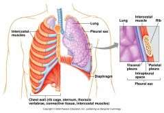
What is the PLEURA? |
A double-layered serous membrane that encapsulates the lungs. Split into 2 layers: ~ Parietal pleura ~ Visceral pleura |
|
|
What is the parietal pleura? |

The parietal pleura lines the walls of the thoracic cavity |
|
|
What is the visceral pleura? |

The visceral pleura adheres tightly to the surface of the lungs themselves. |
|
|
What is the intrapleural space? |
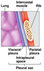
The intrapleural space is a slit-like space filled with pleural fluid. This is crucial to the mechanism of breathing. |
|
|
What is pleural fluid? |
Pleural fluid prevents friction of lungs against the rib cage and maintains lung expansion. |
|
|
What is pulmonary ventilation? |
The movement of air between the atmosphere and the alveoli |
|
|
What is Boyle's Law? |
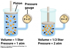
The pressure of a gas in a closed container is said to be inversely proportional to the volume of the container. |
|
|
Why does pulmonary ventilation occur? |
Pulmonary ventilation occurs due to implications of Boyle's Law. By changing the VOLUME of the thoracic cavity and lungs, the PRESSURE in the lungs will also change. |
|
|
What 2 things does pulmonary ventilation consist of? |
Inhalation and Exhalation |
|
|
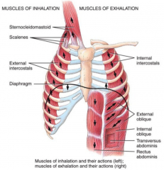
What are the muscles of inhalation/exhalation? |
|
|
|
What happens during the contraction of the diaphragm? |
The diaphragm and the external intercostal (rib) muscles increase the size of the thorax and decreases intrapleural pressure, which results in inhalation. |
|
|
What happens during the relaxation of the diaphragm? |
With or without the contraction of the interal intercostalis, the size of the thorax decreases and the air pressure increases, resulting in exhalation. |
|
|
What is the difference between quiet (normal) and forced (labored) respiration? |
During normal quiet inhalation the diaphragm and the ext. intercostalis contract. However, during forced, labored inhalation the sternocleidomastoid, scalenes, and pectoralis minor also contract. |
|
|
What are the characteristics of inspiration? |
during inspiration: ~Chest walls expand due to muscle contraction ~Pressure in alveoli decreases ~Air moves towards the alveoli |
|
|
What are the characteristics of exhalation? |
during exhalation: ~Passive process, so the muscles relax. ~Chest wall returns to resting state ~Alveoli become compressed ~Alveolar pressure increases ~Air moves outwards. |
|
|
What is Tidal Volume? |
Tidal volume is the volume of air inhaled or exhaled with each breath during quiet breathing, |
|
|
What is respiratory frequency? |
the number of breaths per minute |
|
|
What is minute ventilation? |
the total volume of inhaled/exhaled air oer minute |
|
|
How many breaths per minute does a healthy adult take at rest? |
12 breaths per minute |
|
|
Approximately how much air is moved in/out of the lungs per breath? |
~500mL of air is moved in/out of the lungs per breath. |
|
|
What is inspiratory reserve volume? |
the max volume f inhaled air beyond the end-inhaled level |
|
|
What is expiratory reserve volume? |
the max volume of exhaled air beyond the end-expiratory level. |
|
|
What is the residual volume of males/females? |
Males = 1200mL and females = 1100mL |
|
|
What factors effect the airflow? |
~Pressure ~Surface Tension ~Compliance ~Airway Resistance |
|
|
What is surface tensions? |
Surfactant decreases surface tension and prevents the complete collapse of alveoli |
|
|
What is compliance? |
The indicator of expandability. It is the change in volume when subjected to an applied force. There are 3 factors that affect compliance: ~Connective tissue structure of the lungs ~Level of surfactant production ~Mobility of the thoracic cage |
|
|
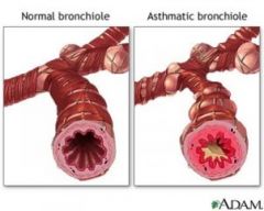
What is airway resistance? |
Airway resistance is when bronchodilation causes a drop in resistance. Airway diamter is regulated by smooth muscle tone, which is dependent on the sympathetic and parasympathetic tone. |
|
|
Define/describe central chemoreceptors |
Central chemoreceptors are located on the medullary rhythmicity area. It is sensitive to carbon dioxide and pH in the CSF. |
|
|
Define/describe the dorsal respiratory group |
the dorsal respiratory group is always functioning during normal breathing. It is sensitive to blood, CO2 and pH levels. Its also located on medulla |
|
|
Define/describe the ventral respiratory group |
It is always functioning during forceful breathing. Also located on the medulla (controls the accessory muscles). |
|
|
Define/describe the peripheral chemoreceptors |
Peripheral chemoreceptors detect variations of CO2, O2, and the pH of blood |
|
|
Define/describe pneumotaxic center |
the pneumotaxic center recieves inputs from higher brain centers and peripheral receptors and fine tunes the breathing rhytm during speaking/sleeping/exercising. |
|
|
Define/describe apneustic center |
apneustic centers sends inhibitory signals to the medulla, reducing duration of inhalation impulse and thus causing shorter cycles which increases the ventilation rate. |
|
|
What other brain areas are involved in the control of respiration? |
-cortex has voluntary control of breathing -stretch receptor sense over-inflation and arrests breathing temporarily (herring-breuer reflex) -emotions w/in limbic system affect breathing -the hypothalamus senses fever/moderate pain and increases breathing |
|
|
What stimulates central chemoreceptors? |
An increase in the pressure of CO2 (via a decrease in pH levels) |
|
|
what stimulates the peripheral chemoreceptors? |
it follows the aortic arch and carotid bodies and it's stimulated by an increase in the pressure of CO2 (via a decrease in pH levels), a decrease in the pressure of O2 and a decrease in the pH levels. |
|
|
Define respiration |
the exchange of gas |
|
|
What is Dalton's Law? |
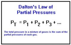
Dalton's Law is the total pressure of a mixture f gases is equal to the sum of the partial pressures of the individual gases in the mixture |
|
|
What is partial pressure |
the hypothetical pressure of a gas if it alone occupies the volume of the mixture at the same temperature. |
|
|
What is total pressure? |
the sum of the partial pressure of all gasses in a mixture |
|
|
What 4 gasses compose the atmosphere? |
-nitrogen (N2) -oxygen (O2) -carbon dioxide (CO2) -water vapor |
|
|
What does the respiratory system depend on? |
the medium of the earth's atmosphere to extract the oxygen necessary for life |
|
|
WHat What are the 5 effficiencies of gas exchange? |
1) Substantial differences in partial pressure across the respiratory membrane 2) short distance for gas exchange 3) O2 and CO2 are lipid soluble 4) total surface area if large 5) coordinated blood flow and airflow |
|
|
What is respiration? |
The exchange of gases |
|
|
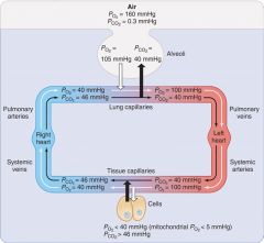
What is external respiration (pulmonary)? |
The gas exchange between alveoli and blood |
|
|
What is internal respiration (tissue)? |
gas exchange between the systemic capillaries and the tissues of the body |
|
|
What is cellular respiration? |
ATP production in the tissue cells |
|
|
How many molecules of O2 can each molecule of hemoglobin (Hb) carry? |
Each Hb molecule can carry 1,2,3, or 4 molecules of O2. |
|
|
What is the most important factor when determining how much O2 binds to Hb? |
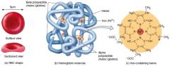
PO2 is the most important factor that determines how much O2 binds to Hb |
|
|
What does the Oxyhemoglobin Dissociation Curve show? |
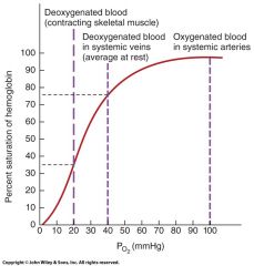
It illustrates the relationship between the % of saturation of Hb and PO2. |
|
|
What does oxyhemoglobin mean? |
hemoglobin that's fully saturated |
|
|
What is SaO2 |
The amount of Hb saturated with O2 |
|
|
oxyhemoglobin has a % level of _____SaO2? |
95-98% |
|
|
When blood returns, it still has 3/4 O2 binding sites, making the % level of SaO2 at _____? |
75% |
|
|
What is the average PO2 of tissue at rest? |
40 mmHg (that's when Hb is 75% saturated) |
|
|
What is the PO2 when Hb is 90% saturated with O2? |
The PO2 60-100 mmHg |
|
|
When PO2 is at 40-20 mmHg, what is the Hb? |
Hb becomes 35% saturated with O2 |
|
|

|
|
|
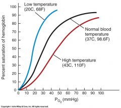
What is the BOHR EFFECT?
|
increasing blood acidity towards tissue (pH decreases), shifts the O2-Hb saturation curve "to the right", thus enhancing the unloading of O2 |
|
|
What is the HALDANE EFFECT? |
The removal of O2 from Hb increases Hb affinity for CO2 and thus allows CO2 to "ride" on the empty Hb. |
|
|
What is Henry's Law? |
the quantity of a gas that will dissolve in a liquid is proportional to the partial pressures of the gas and its solubility. |
|
|
What's partial pressure in relation to henry's law? |
a higher partial pressure of a gas (O2) over a liquid (blood) causes more of the gas to stay in the solution |
|
|
What is solubility in relation to henry's law? |
because CO2 is 24x more soluble in blood than O2, much more CO2 is dissolved in blood plasma. |