Reading...
![]()
Play button
![]()
Play button
![]()
Use LEFT and RIGHT arrow keys to navigate between flashcards;
Use UP and DOWN arrow keys to flip the card;
H to show hint;
A reads text to speech;
211 Cards in this Set
- Front
- Back
- 3rd side (hint)
ID #3
|
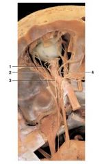
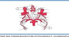
superior sagittal sinus; venous dural sinus located at attachment of falx cerebri with skull; receives blood from superior cerebral hemispheres
|
|
|
ID #3
|
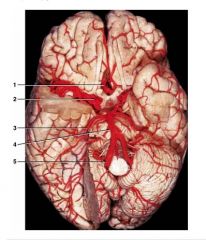
internal carotid artery; supplies anterior two-thirds of cerebral circulation via its branches, anterior and middle cerebral arteries
|
|
|

ID #5
|
vertebral artery; branches supply medulla, spinal cord, and part of cerebellum
|
|
|

ID # 4
|
basilar artery; branches supply most of rostral brainstem and cerebellum
|
|
|

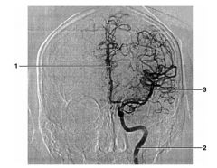
ID #1
|
anterior cerebral artery; branches supply anterior two-thirds of medial surface of cerebral hemisphere
|
|
|
ID #2
|
middle cerebral artery;
branches supply lateral surface of cerebral hemisphere |
|
|

ID #3
|
posterior cerebral artery; branches supply much of inferior and posterior cerebral hemisphere; occlusion may produce homonymous hemianopia
|
|
|
ID #4
|
ophthalmic artery;
branch of internal carotid that enters orbit within optic nerve dural sheath |
|
|

ID #1
|
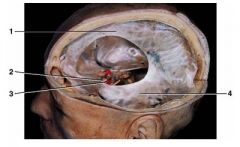
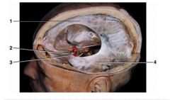
falx cerebri;
sickle-shaped dural fold located between two cerebral hemispheres |
|
|
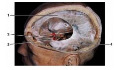
ID #4
|
tentorium cerebelli;
dural fold that separates cerebellum from temporal and occipital lobes |
|
|
ID #3
|
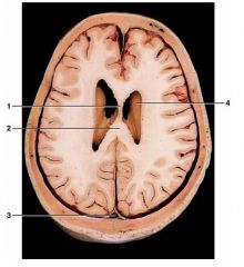
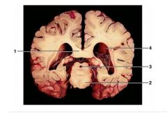
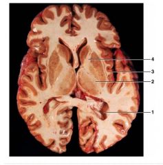
posterior horn of lateral ventricle
|
|
|

ID #4
|
anterior horn of lateral ventricle
|
|
|

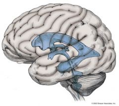
ID #1
|
lateral ventricle
|
|
|

ID #2
|
third ventricle
|
|
|

ID #3
|
fourth ventricle
|
|
|
ID #4
|
cerebral aqueduct
|
|
|
ID #3
|
cerebral aqueduct;
narrowest part of ventricular system; frequent point of blockage in ventricular system that results in hydrocephalus |
|
|

ID #4
|
lateral ventricle;
largest part of brain's ventricular system |
|
|
ID #1
|
posterior horn of lateral ventricle
|
|
|

ID #1
|
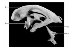
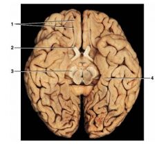
CN 1
olfactory bulb and tract; olfactory nerve terminates at olfactory bulb |
brain exit: olfactory bulb
Skull exit: cribiform plate w/ no companion nerve |
|
ID #2
|
optic nerve (II);
relays visual information from retina to central nervous system |
brain exit: thalamus
skull exit: optic canal |
|
ID #2
|
optic nerve (II);
relays visual information from retina to central nervous system |
brain exit: thalamus
skull exit: optic canal |
|

ID #4
|
oculomotor nerve (III); motor axons innervate many muscles controlling eye movement and preganglionic parasympathetic axons important for control of pupil diameter
|
brain exit: interpeduncular fossa
skull exit: supraorbital fissure companion nerves: 3,4,6, ophthalmic 5 motor or sensory tracts near brainstem: corticospinal (bulbar) |
|
ID #1
|
facial (VII) and vestibulocochlear (VIII) nerves;
facial nerve innervates muscles of facial expression; vestibulocochlear nerve conducts information from organs of hearing and balance |
facial:
brain exit: pontomeduallary junction skull exit: internal auditory meatus, stylomastoid foramen companion nerve: 8 vestibulocochlear/auditory: brain exit: pontomeduallary junction skull exit: internal auditory meatus companion: 7 BOTH: motor or sensory tracts near brainstem course: anterior lateral fasciculus spinal trigeminal |
|

ID #2
|
abducens nerve (VI); innervates lateral rectus muscle of eye
|
brain exit: pontomeduallary junction
skull exit: supraorbital fissure companion nerves: 3,4, ophthalmic 5 |
|

ID #3
|
trigeminal nerve (V);
contains sensory fibers from face and motor axons that control muscles of mastication |
brain exit: middle pons, middle cerebellar peduncle
skull exit: foramen rotundum (V2), foramen ovale (V3), supraorbital fissure (V1) companion nerves: 3,4,6 for only V1 |
|
ID #3
|
hypoglossal nerve (XII);
motor innervation of tongue; damage produces deviation of tongue toward side of damage |
brain exit: preolivary sulcus
skull exit: hypoglossal canal NO companion nerves motor or sensory tracts near brainstem course: corticospinal |
|
ID #3
|
trochlear nerve (IV); innervates superior oblique
|
brain exit: caudal to inferior colliculus
skull exit: supraorbital fissure companion nerves: 3,6, ophthalmic 5 |
|
ID #3
|
vagus (X); emerges from jugular foramen and traverses neck in carotid sheath
|
brain exit: postolivary sulcus
skull exit: jugular foramen companion nerves: 9, 11 motor or sensory tracts near brainstem course: anterior lateral fasciculus spinocerebellar |
|
ID #3
|
foramen rotundum
for V2 |
|
|
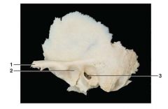
ID #3
|
external acoustic/auditory meatus
conducts sound to tympanic membrane |
|
|
ID #2
|
internal carotid artery
supplies anterior two-thirds of cerebral circulation via its branches, anterior and middle cerebral arteries |
|
|
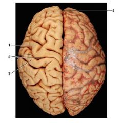
ID #1
|
precentral gyrus; location of primary motor cortex
|
|
|

ID # 2
|
central sulcus; separates frontal lobe from parietal lobe
|
|
|

ID #3
|
postcentral gyrus; location of primary somatosensory cortex
|
|
|

ID #4
|
longitudinal fissure
|
|
|

ID # 1
|
precentral gyrus; location of primary motor cortex
|
|
|

ID #2
|
central sulcus; separates frontal lobe from parietal lobe
|
|
|

ID #3
|
lateral sulcus; separates frontal and parietal lobes from temporal lobe
|
|
|
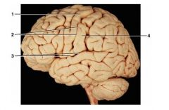
ID #4
|
postcentral gyrus; location of primary somatosensory cortex
|
|
|

ID #1
|
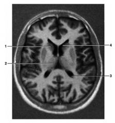
cingulate gyrus; part of limbic lobe
|
|
|
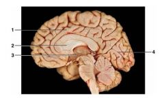
ID #2
|
septum pellucidum; separates two lateral ventricles
|
|
|

ID # 3
|
hypothalamus; important area for autonomic and endocrine control
|
|
|

ID #4
|
calcarine sulcus; primary visual cortex located along banks of this sulcus
|
|
|
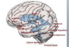
ID:
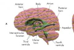
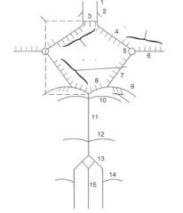
Body of lateral ventricles Anterior horn Foramen of monro/ IV foramen 3rd ventricle inferior horn lateral aperature 4th ventricle posterior horn cerebral aqueduct medial aperature central canal |
|
|
|
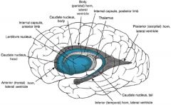
ID:
anterior horn body atrium posterior horn aqueduct 4th ventricle inferior horn third ventricle interventricular formamen |
|
|
|
|
|
|
|

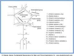
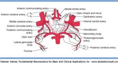
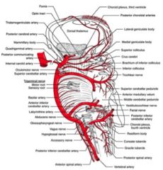
ID
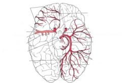
Internal carotid a. Vertebral a. Basilar a. Anterior cerebral a. (ACA) Middle cerebral a. (MCA) Posterior cerebral a. (PCA) Ophthalmic a. Posterior communicating a. Anterior communicating a. Pontine a. Anterior choroidal a. Anterior inferior cerebellar a. (AICA) Posterior inferior cerebellar a. (PICA) Superior cerebellar a. (SuCA) Posterior spinal a. Anterior spinal a. Central branch of the anterior spinal a. |
|
|
|
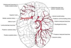
ID
Internal carotid a. Basilar a. Anterior cerebral a. (ACA) Middle cerebral a. (MCA) Posterior cerebral a. (PCA) Ophthalmic a. Posterior communicating a. Anterior communicating a. Anterior choroidal a. |
|
|
|
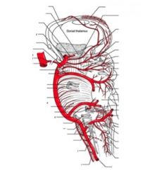
ID
Internal carotid a. Vertebral a. Basilar a. Posterior cerebral a. (PCA) Posterior communicating a. Pontine a. Posterior inferior cerebellar a. (PICA) Superior cerebellar a. (SuCA) Posterior spinal a. Anterior spinal a. Central branch of the anterior spinal a. |
|
|
|
ID
Internal carotid a. Anterior cerebral a. (ACA) Middle cerebral a. (MCA) Posterior cerebral a. (PCA) Posterior communicating a. |
|
|
|
|
Spinal cord segments named based on
|
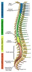
the intervertebral foramen the nerve leaves through, not location of nearby vertebra
|
Lumbar cord deep to T11-T12
Sacral at Conus medullaris Cauda Equina |
|
|
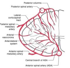
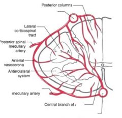
2 Posterior spinal
Off PICA 1 anterior spinal Off vertebral Segmental a. Artery of Adamkiewicz (L2) (not pictured |
|
|
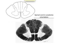
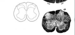
what level is this
|
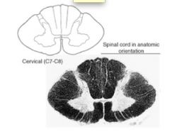
cervical
|
|
|
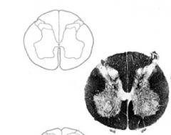
what level is this
|
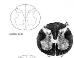
lumbar
|
|
|
what level is this
|
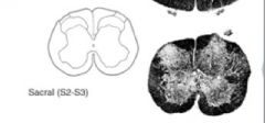
sacral
|
|
|
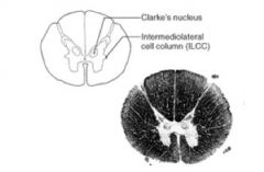
what level is this
|
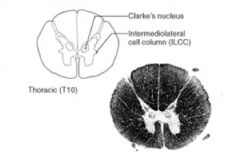
thoracic
|
|
|

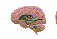
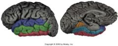
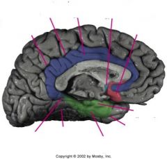
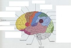
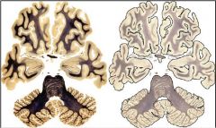
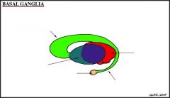
name the lobes by color
|
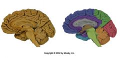
blue: frontal
Green: parietal aqua:temporal red:occipital light green: pons/medulla oblongota/ brainstem Orange: cerebellum Purple:cingulate gyrus Grey: corpus callosum Brown: thalamus and hypothalamus |
|
|
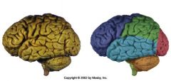
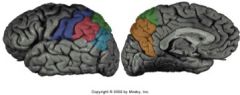
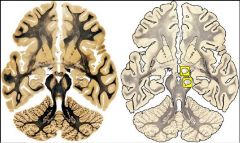
label the different sections
|

blue: frontal
Green: parietal aqua:temporal red:occipital light green: pons/medulla oblongota/ brainstem Orange: cerebellum |
|
|
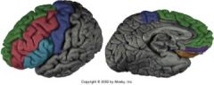
label the different gyri
|

blue: precentral gyrus
green: superior frontal gyrus Red: middle frontal gyrus aqua: inferior frontal gyrus |
|
|

label the different gyri
|
blue: superior temporal gyrus
green: middle temporal gyrus red: inferior temporal gyrus orange: lingual? gyrus aqua: medial occipitotemporal gyrus |
|
|

label the gyri
|
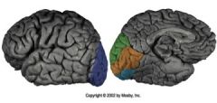
blue: occipital pole
green: orange: aqua: |
|
|

label the gyri
|
blue: post central gyrus
red:supramarginal gyrus green:? aqua: angular gyrus |
|
|
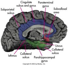
ID the following:
Isthmus Collateral sulcus subparietal sulcus parahippocampal gyrus cingulate sulcus cingulate gyrus collateral sulcus uncus paraterminal gyrus subcallosal area |
|
|
|
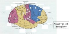
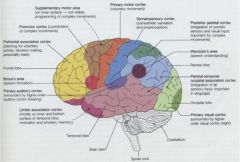
ID the following sensory/motor areas:
visual association primary visual somesthetic association primary somesthetic primary motor premotor frontal eye broca's primary gustatory primary auditory auditory association |
|
|
|
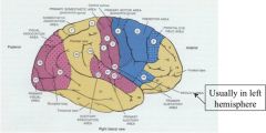
label the following areas of the cerebral cortex:
prefrontal association broca's primary auditory limbic association premontor supplememtary motor area primary motor somatosensory posterior parietal wernicke's parietal-temporal-occipital association primary visual |
|
|
|
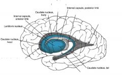
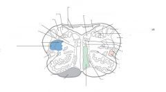
label the following:
anterior frontal horn of the lateral ventricle body (parietal) horn of lateral ventricle thalamus posterior (occipital) horn of the lateral ventricle inferior (temporal) horn of lateral ventricle |
|
|
|
ID the following
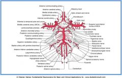
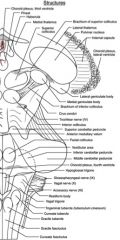
olfactory nerve optic nerve oculomotor trochlear trigeminal abducens facial vestibulocochlear glossopharyngeal vagus accessory hypoglossal pyramidal decussation postolivary sulcus olive preolivary sulcus pyramid |
|
|
|
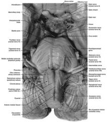
ID the following:
olfactory tract optic nerve optic chiasm oculomotor abducens facial vestibulocochlear glossopharyngeal vagus hypoglosseal acessory decussation of pyramids choroid plexus olive olivary eminence middle cerebellar peduncle trigeminal trochlear basilar pons interpeduncular fossa mammillary body infundibulum |
|
|
|
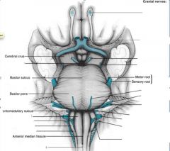
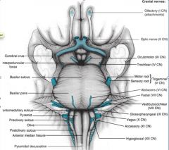
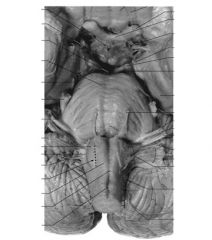
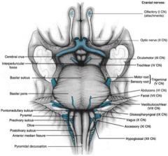
ID:
all cranial nerves pyramidal decussation anterior median fissure postolivary sulcus olive preolivary sulcus pyramid pontomedulary sulcus basilar pons basilar sulcus interpeduncular fossa cerebral crus |

|
|
|

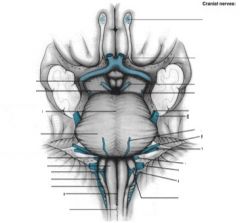
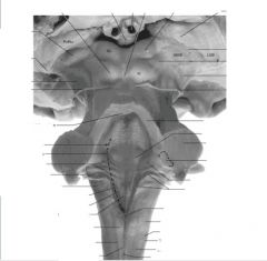
ID:
all cranial nerves pyramidal decussation anterior median fissure postolivary sulcus olive preolivary sulcus pyramid pontomedulary sulcus basilar pons basilar sulcus interpeduncular fossa cerebral crus |
|
|
|

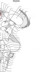
ID:
all cranial nerves pyramidal decussation anterior median fissure postolivary sulcus olive preolivary sulcus pyramid pontomedulary sulcus basilar pons basilar sulcus interpeduncular fossa cerebral crus |

|
|
|
ID:
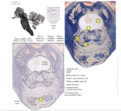
internal cerebral vein pineal superior and inferior colliculi trochlear nerve superior cerebellar peduncle middle cerebellur peduncle inferior cerebellar peduncle vestibular area tuberculum cuneatum cunate fasciculus posterior median sulcus gracille fasciculus posterior intermediate sulcus tuberculum gracile |
|
|
|
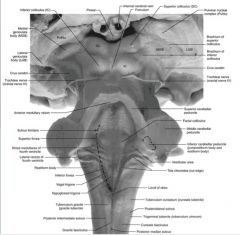
ID:
choroid plexus, third ventricle pineal medial thalamus superior colliculus lateral thalamus internal capsule choroid plexus, lateral ventricle brachium of inferior and superior colliculi crus cerebri trochlear nerve inferior colliculus superior cerebellar peduncle facial colliculus inferior cerebellar peduncle choroid plexus, 4th ventricle glossopharyngeal nerve vagus nerve acessory nerve cuneate tubercle gracile tubercle gracile fasciculus cunate fasciculus |
|
|
|
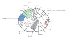
what level is this?
ID the following: gracile fasciculus gracile nucleus cuneate fasciculus cuneate nucleus spinal trigeminal: nucleus and tract reticulospinal/vestibulospinal fibers pyramidal (motor)decussation medial longitudinal fasciculus accessory nucleus anterior spinocerebellar tract anterolateral system posterior spinocerebellar tract rubrospinal tract hypoglossal nucleus dorsal motor vagal nucleus central gray central canal |
motor decussation level
|
|
|
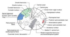
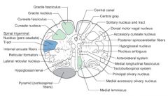
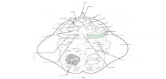
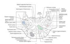
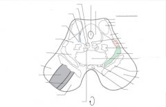
what level of medulla is this?
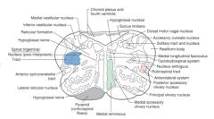
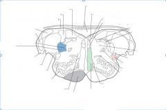
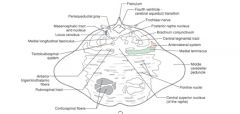
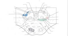
ID the following: chorid plexus and 4th ventricle hypoglossal nucleus sulcus limitans dorsal motor vagal nucleus acessory cuneate nucleus solitary tract and nucleus restiform body medial longitudinal fasciculus tectobulbospinal system nucleus ambiguus rubrospinal tract anterolateral system posterior accessory olivary nucleus principal olivary nucleus medial accessory olivary nucleus medial lemniscus pyramid (corticospinal fibers) hypoglossal nerve lateral reticular nucleus anterior spinocerebellar tract spinal trigeminal: nucleus and tract hypoglossal nerve reticular formation inferior vestibular nucleus medial vestibular nucleus |
caudal open
|
|
|
what level of medulla is this?
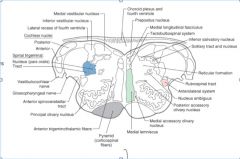
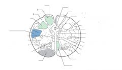
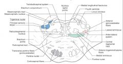
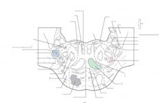
ID the following choroid plexus and 4th ventricle prepositus nucleus medial longitudinal fasciculus tectobulbospinal system inferior salivatory nucleus solitary tract and nucleus reticular formation rubrospinal tract anterolateral system nucleus ambiguus posterior accessory olivary nucleus medial accessory olivary nucleus medial lemniscus pyramid anterior trigeminotalmic fibers principal olivary nucleus anterior spinocerebellar tract glossopharyngeal nerve vestibulocochlear nerve spinal trigeminal (nucleus and tract) cochlear nuclei (posterior/anterior) lateral recess of fourth ventricle inferior vestibular nucleus medial vestibular nucleus |
rostral open
|
|
|
what level of medulla is this?
ID the following: gracile fasciculus gracile nucleus cuneate fasciculus cuneate ucleus spinal trigeminal: nucleus/tract internal arcuate fibers reticular formation lateral reticular nucleus hypoglossal nerve pyramid central canal central gray solitary nucleus and tract dorsal motor vagal nucleus accessory cuneate nucleus posterior spinocerebellar fibers hypoglossal nucleus nucleus ambiguus anterolateral system medial longitudinal fasciculus tectobulbospinal system principal olivary nucleus medial accessory olivary nucleus medial lemniscus |
sensory decussation
|
|
|
ID the following tracts through the pons:
corticospinal-pyramidal system trigeminal nuclei posterior column - medial lemniscus system anterolateral system |
|
|
|
ID the following in this upper level slice of the pons:
4th ventricle/cerebral aqueduct transition trochlear nerve anterolateral system medial lemniscus middle cerebellar peduncle pontine nuclei central superior nuclues of the raphe corticospinal fibers retrospinal tract anterior trigeminothalmic fibers medial longitudinal fasciculus periaquaductal gray |
|
|
|
ID the following on this slice through the middle of the pons:
tectobulbospinal system trigeminal nuclei (sensory/motor) transveres pontine fibers pontine nuclei (multiple) corticospinal fibers medial lemniscus anterior trigeminothalmic fibers anterolateral system lateral lemniscus anterior spinocerebellar fibers 4th ventricle medial longitudinal fasciculus |
|
|
|
ID the following from this inferior slice of the pons:
medial longitudinal fasciculus internal genu of facial nerve superior salivatory nucleus spinal trigeminal (nucleus/tract) facial nucleus facial nerve superior olivary nucleus transverse pontine fibers corticospinal fibers pontine nuclei abducens nerve medial lemniscus anterior trigeminothalmic tract anterolateral system solitary tract and nucleus vestibular nuclei (sup/med/lat) abducens nucleus facial colliculus inferior cerebellar peduncle containing: juxtarestiform body and restiform body |
|
|
|
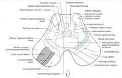
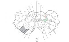
ID the following in this caudal section of the midbrain:
trochlear nucleus medial longitudinal fasciculus mesencephalic tract and nucleus reticular formation anterior trigeminothalmic fibers porieto, occipto and temporopontine fibers corticocobulbar fibers interpeduncular nucleus substantia nigra decussation of the superior cerebellar peduncle medial lemniscus anterolateral system lateral lemniscus inferior colliculus including: pericentral, external and central nuclei periaqueductal gray cerebral aqueduct |
|
|
|
ID the following in the rostral slice of the mid-brain:
cerebral aqueduct periaqueductal gray medial longitudinal fasciculus superior colliculus anterolateral system medial geniculate nucleus brachium of inferior colliculus medial lemniscus cerebellothalmic fibers substantia nigra (pars reticulat and pars compacta) red nucleus oculomotor nerve corticospinal fibers parieto, occipito, temporopontine fibers optic tract lateral geniculat nucleus posterior and anterior trigeminothalmic tracts brachium of superior colliculus mesencephalic tract and nucleus oculomotor nucleus edinger-westphal nucleus |
|
|
|
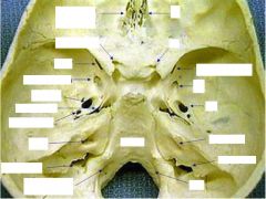
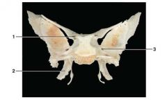
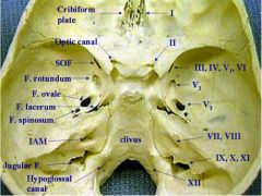
ID the following:
cribiform plate optic canal superior orbital fissure foramen rotundum foramen ovale foramen lacerum foramen spinosum IAM Jugular foramen hypoglossal canal clivus where the following CN exit: I, II, III, IV, V1,V2,V3, VII, VIII, IX, X, XI, XII |
|
|
|
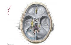
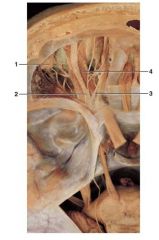
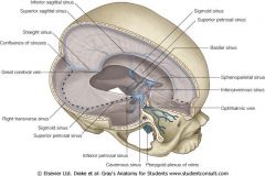
ID the following:
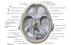
olfactory bulb/tract trigeminal 1,2,3 basilar artery oculomotor nerve trigeminal ganglian optic nerve trochlear nerve infundibulum middle meningeal artery facial nerve abducens vertebral artery olfactory nerve vestibulocochlear nerve hypoglossal nerve superior sagittal sinus (ant/post) glossopharyngeal spinal accessory sigmoid sinus vagus nerve cerebral falx cerebellar tentorium onferior sagittal sinus spinal accessory nerve transverse sinus |
|
|
|

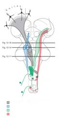
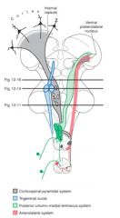
ID the following:
Corticospinal Posterior column Anterolateral (spinothalamic) Trigeminal nuclei |

Corticospinal (grey, movment)
Posterior column – medial lemniscus (green- fine touch, conscious proprioception from neck and below, NOT from face) Anterolateral (spinothalamic)- (temp and pain in red from neck and down into cerebrum so you can be consciously aware of it) Trigeminal nuclei- (sensory info from face, touch, prprioception, pain, etc all from face, in blue) |
|
|
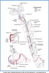
anterolateral system
|

Sits between anterior column and lateral column
Black is descending analgesic pathway Contains multiple tracts: spinothalmic (localization, intensity) and spinoreticular (collaterals into reticular formation) Spinothelamic pathway comes in on spinal cord in dorsal horn, synapses in dorsal horn and cross to contralateral side of spinal cord and then ascends. Damage to spinal cord on right side lose pain on left side. Is a fully contralateral pathway |
|
|
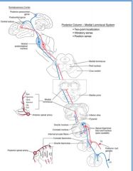
posterior column - medial leminiscus pathway
|

responsible for fine touch and 2 point discrimination, stereognosis, and conscious proprioception
White matter in posterior column Legs go most medial Upper extremity is more lateral Fasiculus gracilis- lower extremety Fascilulus cuneatus – upper extremity Synapses in medulla on 2 tubercules (cuneate and gracile) after you synapse you cross over and ascend contralaterally in medial lemniscus and then synapse in thalmus Medial lemniscus- have a pole you have 2 dancers next to the poles standing on the pyramids- the best girls slide down the pole and do a flip |
|
|
|
trace pathway of posterior column medial leminiscus
|

dorsal root
fasiculus gracillus (leg) and fasiculus cuneatus (arm) nucleus gracilis/cuneatus internal arcuate fibers medial lemniscus thalamus cerebrum (primary sensory cortex/parietal lobe) |
|
|
|
trace pathway of anterolateral system- spinothalmic divison
|

doral root
lissauer's tract anterior white commissure contralateral anterolateral funiculus anterolateral pathway thalamus internal capsule S1 |
|
|
|
where are the spinomesencephalic synapses and spinoreticular synapses of anterolateral system
|

spinomesencephalic synapses in periaqueductal gray
spinotreticular synapses in reticular formation |
|
|

what is spinal trigeminal tract responsible for?
trace it's path |

blue path: responsible for pain from face
CN V spinal trigeminal tract spinal V nucleus contralateral anterior trigeminal tract thalamus S1 |
|
|

what is trigeminal tract responsible for?
trace it's path |

blue: responsible for touch from face
trigeminal ganglion principal sensory nucleus medial lemniscus thalamus S1 |
|
|
trace the corticospinal tract
|

grey tract in picture
cerebrum internal capsule cerebral peduncle pyramid pyramidal decussation contralateral lateral funiculus (column) anterior horn |
|
|
|
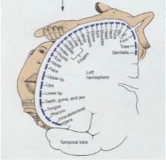
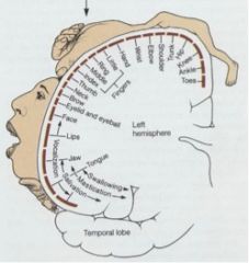
what is the general scheme of the somatotopic map for SOMATOSENSORY cortex (homonculus view)
|
|
|
|
|
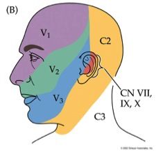
know the nerves responsible for general sensation from the head
|
|
|
|
|
trace path of trigeminal pain/temp
|
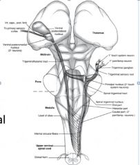
Sensory info descends down into medulla and spinal cord once it enters, synapses between medulla and spinal cord and ascends contralaterally up to thalamus
|
|
|
|
trace the trigeminal - touch pathway
|
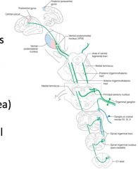
Trigeminal nerve touch comes in synapses in principal nuclus, crosses over and ascends up to VPM
‘pain takes a painful pathway’ aka it goes down to go up |
|
|
|
what provides afferent limb of corneal blink reflex
|

trigeminal - touch division
|
|
|
|
what is the general scheme for somatotopic map for primary MOTOR cortex
|
|
|
|
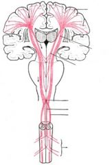
what is the course of the corticospinal tract
|
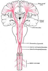
Out of cerebral cortex into corona radiata and the posterior limb of the Internal (the white matter mass where the pink lines all seem to bunch first -supplied by MCA), travel down in midbrain (past cerebral peduncles/ crus cerebri) then down through basilar pons (potbelly of pons) then into medulla (pyramids) and at lowest possible level they cross (decussation) and then they descend in lateral column of spinal cord and synapse in anterior horn
All this is considered an upper motor neuron |
|
|
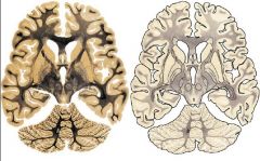
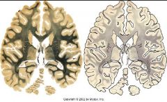
ID: lateral ventricles, 3rd ventricle, thalmus makes walls of 3rd ventricle, caudate nucleus (part of basal ganglia) projects into lateral wall of lateral ventricles, the rest of the basal ganglia are the little slice of pie looking things , the tract between caudate nucleus and rest of basal ganglia is the internal capsule (black stained)
Uncus is internal above inferior temporal gyrus |

|
|
|
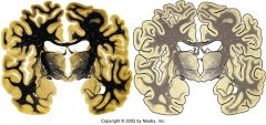
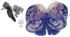
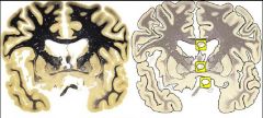
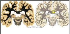
where are we?
ID: superior colliculi peduncles aquaduct periductal grey matter oculomotor nucleus corticospinal tract posterior medial lemniscus ALS red nucleus superior cerebellar |

We’re in the tentorium/midbrain! See bumps superior colliculi on the back and can see peduncles (one is atrophied due to person having a stroke) and can see aquaduct, around aquaduct is periaquaductal grey matter
Oculomotor nucleus is below aquaduct, deep in there is anglo-westinfall Corticospinal tract is in the peduncles Posterior medial lemniscus is the little c shaped dark shadow Just superior to that is the ALS (slightly darker) The mustache is from the red neucleus and superior cerebellar What is the little greyish junk below the peduncle? is CN3 White spot up above and lateral is CN 2 |
|
|
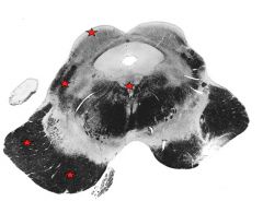
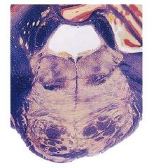
where are we?
ID: CS tract medial lemniscus ALS superior cerebellar peduncle 4th ventricle |

This is the pons
Think: Potbelly pons CS tract is broken up in the potbelly (dark) (VENTRAL) Medial lemniscus is on the ventral side before the potpelly- 2 stars ALS- lateral to ML with star marked on left Can see superior cerebellar peduncle (red star on right) Can see aqueduct has expanded to become 4th ventricle |
|
|

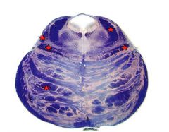
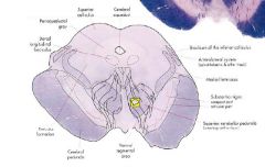
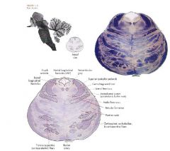
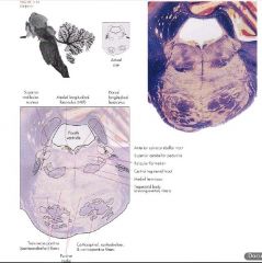
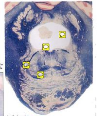
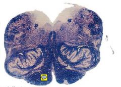
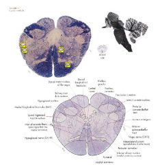
where are we?
ID: pyramids medial lemniscus anteriorlateral tract spinal trigeminal solitary tract |
This is the medulla
Can see pyramids – dark areas ventral (starred on right) Medial lemniscus (pole girl) sits right above the pyramid Anteriorlateral tract isn’t well defined more lateral than anterior (marked with star on left) pain for the body Spinal trigeminal is above ALS (pain for the face) marked with star on left Solitary tract is surrounded by it’s nucleus like little eyes, marked with red star on right – has taste for tongue |
|
|
|
|
|
|
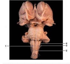
ID the following:
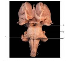
optic chiasm gyrus rectus orbital gyri infundibular stalk olfactory bulb/tract optic nerve optic tract mammillary body uncus CN 4 CN 3 basal pons CN 5 CN 7 CN 6 CN 8 CN 9 CN 10 CN 11 CN 12 cerebellar hemisphere pyramid |
|
|
|
ID
superior colliculus periaqueductal gray dorsal longitudinal fasciculus reticular formation cerebral peduncle cerebral aqueduct brachium of inferior colliculus anterolateral system (ALS) medial lemniscus substantia nigra superior ceregellar peduncle CN 3 |
THIS IS ROSTRAL MIDBRAIN- when you see peduncles you know you're in midbrain
CN3 are the little dark lines going down over superior cerebellur peducle from oculomotor nucleus (heart shaped) |
|
|
what section is this?
ID: periaqueductal gray cerebral aqueduct inferior colliculus lateral lemniscus anterolateral system central tegmental tract medial lemniscus pontine nuclei decussation of the superior cerebellar peduncles cerebral peduncle reticular formation dorsal longituidnal fasciculus |
this is caudal midbrain
|
|
|
what level is this?
ID: 4th ventricle medial longitudinal fasciculus periventricular gray superior cerebellar peduncle central tegmental tract lateral lemniscus anterolateral system medial lemniscus reticular formation pontine nuclei corticospinal, corticebulbar, corticopontine fibers basilar artery transverse pontine fibers doral longitudinal fasciculus |
rostral pons
|
|
|
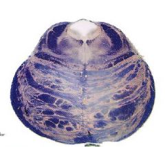
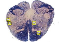
what level is this?
ID: superior vestibular nucleus medial longitudinal fasciculus dorsal longitudinal fasciculus anterior spinocerebellar tract superior cerebellar peduncle reticular formation central tegmental tract medial lemniscus trapezoid body pontine nuclei corticospinal,corticobulbar, corticopontine fibers transverse pontine fibers |
midpons
|
|
|
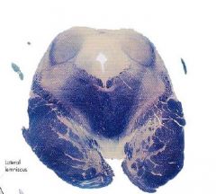
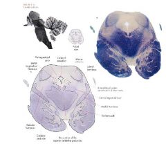
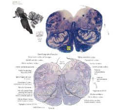
what level is this?
ID: dorsal longitudinal fasciculus superior cerebellar peduncle inferior cerebellar peduncle solitary tract/nucleus anterior spinocerebellar tract middle cerebellar peduncle lateral lemniscus trapezoid body transverese pontine pontine nuclei fourth ventricle |
caudal pons
top yellow sticky: 4th ventricle 2nd yellow sticky: bump in 4th ventricle are the facial colliqui 6 is dark line in the center around colliculi and 7 is dark line outside colliculi third yellow sticky: ALS is around here (feet of the stripper) 4th sticky: ML is now laying down |
|
|
what level is this?
if you damage area where yellow sticky is what happens? ID: dorsal longitudinal fasiculus solitary tract hypoglossal nucleus lateral cunate nucleus posterior spinocerebellar tract anterior spinocerebellar tract vagus nucleus ambiguus inferior olivary nucleus pyramid medial lemniscus hypoglossal reticular formation spinal trigeminal tract medial longituidinal fasciculus |
rostral medulla level
if we damage CST in the BRAIN you lose motor control contralateraly. if you damage this right pyramid you lose control on left side. |
|
|
what level is this?
ID: nucleus cunectus nucleus gracilis central canal dorsal longitudinal fasciculus solitary tract hypoglossal nucleus medial longitudinal fasciculus spinal trigeminal internal arcuate fibers hypoglossal medial lemniscus pyramid inferior olivary nucleus reticular formation anterolateral system vagus nerve anterior spinocerebellar tract nucleus ambiguus posteior spinocerebellar tract lateral cuneate nucleus fasciculus cuneatus |
caudal medulla level
|
|
|
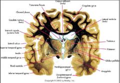
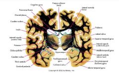
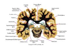
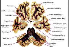
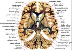
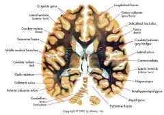
ID:
longituidinal fissure corpus callosum cingulate gyrus lateral ventricle choroid plexus thalamus insula superior temporal gyrus middle temporal gyrus inferior temporal gyrus parahippocampal gyrus anterior commissure middle cerebral branches globus pallidus putamen lateral sulcus inferior frontal gyrus internal capsule caudate nucleus middle frontal gyrus superior frontal gyrus |
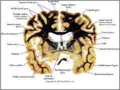
yellow stickies top to bottom:
septum pellucidum is thin seperation. lower is fornix- also fornix is white matter so it'll stain dark 2. anterior commissure 3. optic chiasm |
|
|

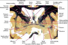
ID the following:
fornix septum pellucidum septal nuclei caudate nucleus internal capsule globus pallidus (internal/external) putamen optic chiasm optic nerve olfactory tract thalamus |

|
|
|

ID the following:
fornix septum pellucidum septal nuclei caudate nucleus internal capsule globus pallidus (internal/external) putamen optic chiasm optic nerve olfactory tract thalamus |
|
|
|

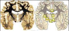
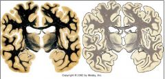
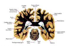
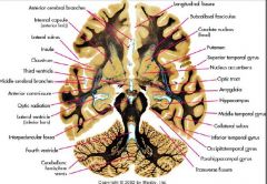
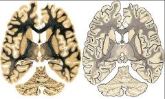
ID:
corpus calosum (body) cingulate gyrus lateral ventricle (body) thalamus putamen globus pallidus amygdala parahippocampal gyrus occipitotemporal gyrus inferior temporal gyrus third ventricle middle temporal gyrus insula superior temporal gyrus lateral sulcus internal capsule caudate nucleus choroid plexus transverse fissure |
yellow sticky's L to R:
1. putamen on outside of pie 2. globus pallidus - has internal and extrnal, is slightly darker area of pie 3. CN 2 4. corpus callosum 5. 3rd ventricle remember walls of 3rd ventricle is thalamus 6. hypothalamus |
|
|
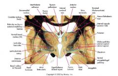
ID:
septum pellucidum internal capsule putamen globus pallidus claustrum amygdala fornix hypothalamus anterior commissure reticular nuculus caudate nucleus |
|
|
|
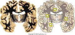
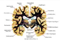
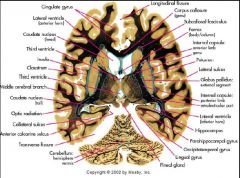
ID:
corpus callosum lateral ventricle (body) thalamus lateral sulcus superior temporal gyrus internal capsule middle temporal gyrus lateral ventricle (inferior horn) occipitotemporal gyrus hippocampus cerebral peduncle third ventricle caudate nucleus globus pallidus putamen insula caudate nucleus choroid plexus transverse fissure cingulate gyrus |
yellow sticky's L - R:
1. insular cortex- cortex on the inside when you go in through the lateral fissure aka insular gyrus, aka insular lobe 2. amygdala- more anterior than posterior uncus is the surface structure and amygdala is the nuclei inside 3. cingulate gyrus 4. putamen - light crust of pie |
|
|
ID the following:
corpus callosum cingulate gyrus caudate nucleus lateral sulcus superior temporal gyrus middle temporal gyrus inferior temporal gyrus hippocampus cerebral peduncle lateral ventricle (inferior horn) third ventricle thalamus choroid plexus lateral ventricle (body) transverse fissure |

|
|
|

ID the following:
corpus callosum cingulate gyrus caudate nucleus lateral sulcus superior temporal gyrus middle temporal gyrus inferior temporal gyrus hippocampus cerebral peduncle lateral ventricle (inferior horn) third ventricle thalamus choroid plexus lateral ventricle (body) transverse fissure |
|
|
|

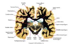
ID:
corpus callosum (body) cingulate gyrus lateral ventricle (body and inferior horn) insula lateral sulcus superior temporal gyrus middle temporal gyrus inferior temporal gyrus basal pons cerebral peduncle hippocampus caudate nucleus x 2 thalamus choroid plexus transverse fissure |
|
|
|
ID:
cingulate gyrus corpus callosum (splenium) lateral ventricle (body and inferior horn) superior cistern choroid plexus hippocampus thalamus cerebral peduncle basal pons inferior temporal gyrus lateral sulcus caudate nucleus middle temporal gyrus superior temporal gyrus lateral ventricle |
|
|
|
ID:
corpus callosum lateral ventricle (body/inferior horn) choroid plexus thalamus basal pons hippocampus caudate nucleus superior cistern cingulate gyrus |
|
|
|
ID the following on this AXIAL cross section:
longitudinal fissure superior temporal gyrus middle temporal gyrus inferior temporal gyrus parahippocampal gyrus cerebellum: hemisphere/vermis 4th ventricle basal pons hippocampus lateral ventricle amygdala lateral sulcus optic chiasm |
|
|
|
ID the following on these axial slices:
lateral sulcus internal capsule longitudinal fissure insula caudate nucleus 3rd ventricle optic tract amygdala hippocampus anterior commissure superior temporal gyrus putamen lateral ventricle middle temporal gyrus inferior temporal gyrus 4th ventricle transverese fissure cerebellum |
yellow stickies top to bottom:
1. mammillary bodies 2. cerebellar peduncles of midbrain |
|
|
ID the following on this axial slice:
3rd ventricle caudate nucleus longitudinal fissure corpus callosum (genu/rostrum) globus pallidus cerebral aqueduct cerebellum transverese fissure lateral ventricle (inferior /anterior horn) hippocampus internal capsule fornix insula putamen anterior commissure |
|
|
|
ID the following:
cingulate gyrus longitudinal fissure corpus callosum (genu) putamen fornix lateral sulcus cerebellum (hemisphere/vermis) internal capsule insula cingulate gyrus transverse fissure third ventricle globus pallidus pineal gland caudate nucleus lateral ventricle (inferior/anterior horn) hippocampus |
|
|
|
ID the following:
cingulate gyrus longitudinal fissure lateral sulcus corpus callosum (genu/body) lateral ventricle (inferior/anterior horn) caudate nucleus fornix caudate/putamen gray bridges transverse fissure cerebellum (veris/hemisphere) hippocampus |
|
|
|

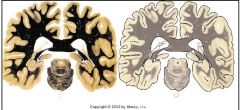
ID:
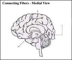
tentorium cerebelli falx cerebri |
falx cerebri: descends vertically in the longitudinal fissure between the cerebral hemispheres
tentorium cerebelli: an extension of the dura mater that separates the cerebellum from the inferior portion of the occipital lobes |
|
|
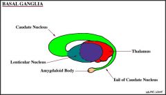
ID:
caudate nucleus thalamus amydaloid body |
|
|
|
ID:
corpus callosum posterior commissure anterior commissure |

|
|
|
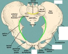
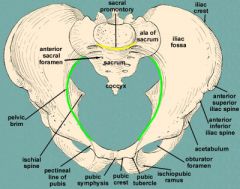
ID:
ASIS Pubic crest |
|
|
|
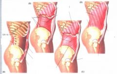
ID the following:
Rectus Abdominis (very anterior) Transverse abdominis Internal Oblique External oblique |

Rectus means running with the longitudinal axis of the body
Rectus abdominis – trunk flexion. Covered by rectus sheath in front and back, made up of apneurosis (broad flat tendon) of all three other muscles BCD are all flank muscles and end around the semilunar line Ext oblique's (pic D) go down and in from ribs to iliac crest Int obliques run down and out, come off iliac crest and also goes to throaco lumbar fascia to lineal alba – in general they rotate the trunk Ext oblique- your right rotates you left- contralateral rotations Int oblique – your left rotates you left- ipsilateral rotation Transverse abdominis – from thoracolumbar fascia runs anteriorly in transverse plane so it will compress your abdominal content, is more important for stabilizing the low back |
|
|
|
External oblique's
orgin insertion innervation main action |

orgin: external surfaces of rib 5-12
Insertion: linea alba, pubic tubercle, anterior half of iliac crest Innervation: nerves from between T7 and L1 main action: contralateral rotation |
|
|
|
internal oblique
orgin insertion inneration main action |

Origin: thracolumbar fascia, anterior 2/3 of iliac crest, CT deep to lateral 1/3 of inguinal ligament
Insertion: interior borders of rib 10-12, linea alba, and pecten pubis innervation: nerves from between T7 and L1 main action: ipsilateral rotation |
|
|
|
transverse abdominis:
orgin insertion innervation main action |

orgin: 7-12 costal cartilage, throacolumbar fascia, iliac crest, CT deep to lateral 1/3 of inguinal ligament
Insertion: linea alba w/ aponeurosis of internal oblique, pubic crest, pecten pubis Innervation: nerves between T7 and L1 main action: compress and support abdominal viscera |
|
|
|
rectus abdominis
orgin insertion innervation main action |
orgin: pubic symphysis and pubic crest
insertion: xiphoid process and 5-7 costal cartilages innervation: nerves between T7 and L1 action: flexes trunk, compresses abdominal viscera, stabilizes/tilts pelvis |
|
|

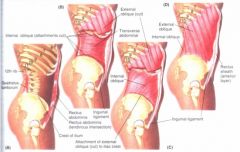
ID:
inguinal ligament inguinal canal deep ring superficial ring internal oblique transverse abdominis external oblique describe function of each |
Inguinal Ligament
Folds of ext. oblique, create a thickening Connects ASIS to pubic tubercle Inguinal canal Consists of 3 arcades traversed by spermatic cord or round ligament of the uterus and vessels Deep ring – superior to middle inguinal lig. Lateral to infreior epigastric a. Superficial ring – split in ext. oblique apoonerurosis – superolateral to pubic tubercle. Contraction of int. oblique and TA makes roof of canal descend. |
|
|
|
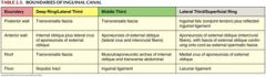
describe the boundaries of the inguinal canal
|
|
|
|

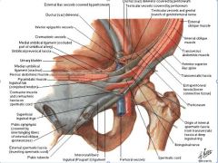
ID:
transversalis fascia peritoneum median umbilical fold medial umbilical fold lateral umbilical fold deep inguinal ring medial inguinal triangle lateral inguinal fossa Describe the different types of hernia (direct/indirect) |
Transversalis fascia
Peritoneum Median umbilical fold (midline) Covers median umbilical ligmaent Medial umbilical folds(red) Covers remnants of umbilical a. Lateral umbilical folds (blue) Covers inferior epigastric vessels Deep inguinal ring (yellow circle) Medial Inguinal (Hesselbach’s) triangle (yellow triangle) Direct hernia: new opening Lateral inguinal Fossa Indirect hernia: through canal, more common |
|
|
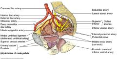
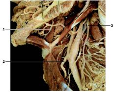
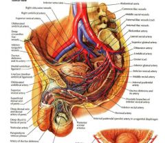
ID:
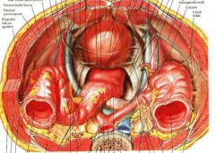
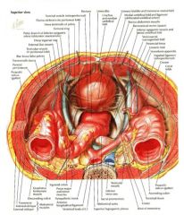
internal iliac artery common iliac artery superior/inferior gluteal iliolumbar artery lateral sacral artery inferior vesical artery internal pudendal artery/nerve middle rectal artery prostatic branch of inferior vesicle artery superior vesical arteries inferior epigastric artery deep circumflex iliac artery obturator artery external iliac artery |
review table 3.4 from Moore to see what each supplies
|
|
|
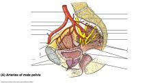
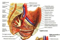
ID:
abdominal aorta common iliac vessels external iliac vessels superior gluteal a. obturator a. umbilical a. inferior gluteal a. inferior vena cava |
|
|
|
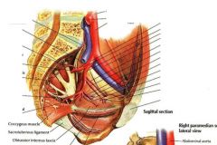
ID:
abdominal aorta R/L common iliac a. internal iliac a. obturator a. umbilical a. inferior gluteal a. superior gluteal a. |
|
|
|
|
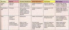
Describe the muscles that make up the boundaries of the pelvic floor including their proximal and distal attachment and action
|
|
|
|
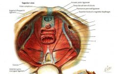
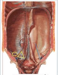
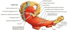
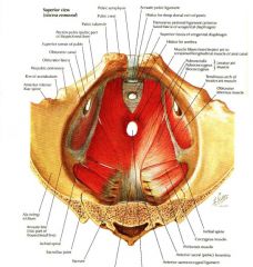
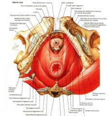
ID:
urethra vagina obturator canal obturator internus coccygeus levator ani rectum piriformis |
|
|
|
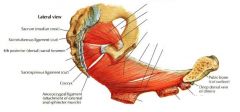
ID:
urethra vagina rectum piriformis levator ani coccygeus obturator internus |
|
|
|
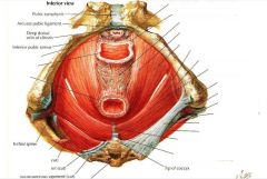
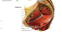
ID:
piriformis rectum vagina urethra coccygeus |
|
|
|
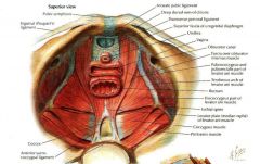
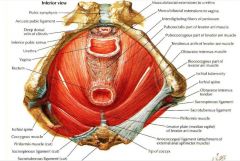
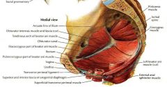
ID:
piriformis obturator canal coccygeus levator ani rectum vagina urethra obturator internus |
|
|
|
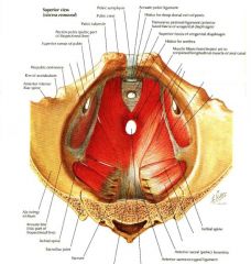
ID:
levator ani obturator internus coccygeus piriformis |
|
|
|
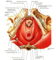
ID:
levator ani obturator internus coccygeus prostate urethra rectum |
|
|
|
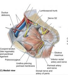
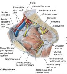
ID:
ureter internal iliac a. obturator nerve piriformis coccygeus obturator internus inferior epigastric a/v external iliac a/v |
|
|
|
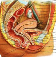
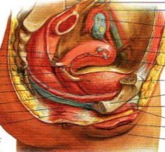
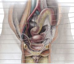
ID:
rectum vagina uterus bladder urethra ovary external iliac a/v levator ani |
|
|
|
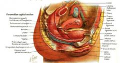
ID:
uterus bladder ovary fallopian tube rectum ureter levator ani |
|
|
|
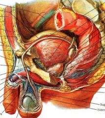
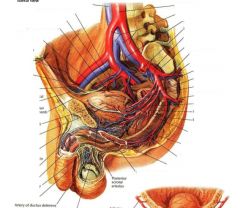
ID:
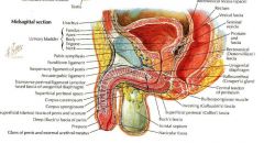
vas deferens bladder seminal vesicle rectum prostate testis external iliac a/v peritoneum rectus abdominis corpus cavernosum corpus spongiosum |
|
|
|
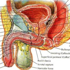
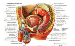
ID:
urinary bladder corpus cavernosum corpus spongiosum scrotal septum prostate seminal vesicle rectum |
|
|
|
ID:
bladder linea alba rectum seminal vesicle femoral ring deep inguinal ring external iliac vessels transverse abdominals internal/external oblique sigmoid colon inferior vena cava abdominal aorta ureter terminal ileum ascending colon cecum rectus abdominis |
|
|
|
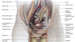
ID:
aorta sigmoid colon uterine tube ovary broad ligament of uterus fundus of uterus round ligament of uterus trigone of urinary bladder obturator externus vestibule inferior vena cava ureter internal/external iliac a. crura of clitoris |
|
|
|
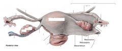
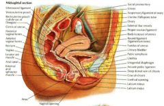
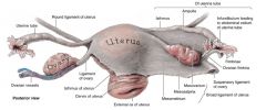
ID:
uterus external os cervix isthmus of uterus isthmus of uterine tube ampulla of uterine tube infundibulum of uterine tube fimbriae suspensory ligament of ovary broad ligament of uterus mesometrium ovary |
|
|
|
|
mesentery
|
dbl layer peritoneal formation that anchors structures to the abdoinal wall
2 parts: proper (small intestine) and mesocolon (colon) |
|
|
|
omentum
|
double fold peirtoneal fomation that extends from stomach:
greater- from greater curveratue of stomach lesser - from lesser curveratue of stomach |
|
|
|
peritoneal ligaments
|

double peritoneum connects organs to organ/ walls:
falciform (liver) phrenicolic (diaphragm to colon), coronary lig (liver to diaphragm), gastrophrenic (stomach to diaphragm), gastrosplenic (stomach to spleen) |
|
|
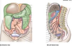
ID:
omental bursa (lesser sac) supracolic compartment(greater sac) infracolic compartment (greater sac) |
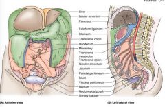
-greater sac is divided by transverse mesocolon
-supra/infracolic communicate via lateral Paracolic gutters |
|
|
|
omental foramen
aka foramen of winslow |
connects greater and lesser sac
is made of posterior to gallpladder and free edge of lesser omentum |
|
|
|
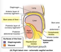
where does fluid accumulate when a person is supine
|
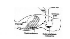
morrison's pouch (hepatorenal)
and rectovesicular/retrouterine pouch of Douglas |
|
|
|
describe the path of food
|

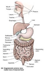
mouth
pharynx esopagus stomach and pyloric sphincter small intestine (duodenum, jejunum, ileum) Large intestine (cecum, ascending, transverse, descending, sigmoid) rectum anus |
|
|

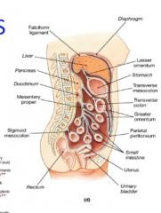
ID the following:
mouth pharynx esopagus stomach and pyloric sphincter small intestine (duodenum, jejunum, ileum) Large intestine (cecum, ascending, transverse, descending, sigmoid) appendix rectum anus |
|
|
|
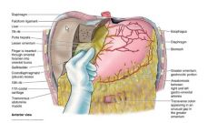
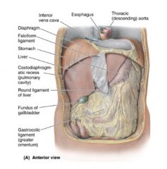
ID the following:
IVC esophagus descending aorta falciform ligament liver diaphragm stomach costodiaphragmatic recess (pulm cavity) fundus of gallbladder round ligament of liver gastrocolic ligament (greater omentum) |
|
|
|
|
where are surface landmarks of stomach
|
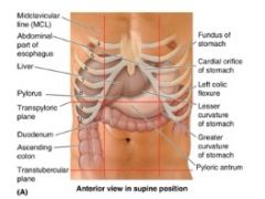
esopgastric junction at Z line (T11/tip of xiphoid @ L 6th costal cartilage)
distends 50mL - 3L |
|
|
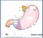
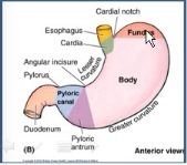
ID the following:
cardial notch fundus body lesser curverature greater omentum pyloric antrum pyloric canal pylorus angular incisure duodenum cardia pyloric antrum |
|
|
|
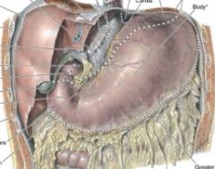
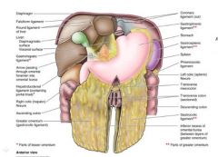
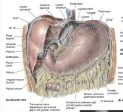
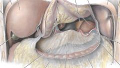
ID the following
lesser / greater curverature liver falciform ligament porta hepatis omental foramen cardial notch cardia fundus diaphragm body greater omentum transverse colon angular incisure pyloric canal pylorus omental foramen pyloric antrum |
|
|
|
|
what are rugae
|
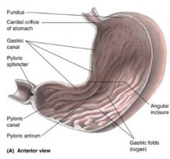
gastric folds inside the stomach
|
|
|
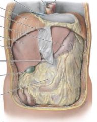
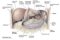
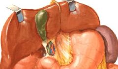
ID the following:
liver stomach (reflected) gallbladder diaphragm portal triad left kidney/adrenal spleen splenic vein/artery body/tail of pancreas transverse colon transverese mesocolon greater omentum |
|
|
|
|
4 parts of pancreas
|
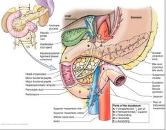
head (duodenum wraps around this)
neck (narrowing that goes over superior mesentaric artery and under pylorus) body (over aorta and L2) tail (anterior to L kidney and hilum of spleen) All structures are retroperitoneal |
|
|
|
where is jejunum located
|
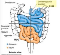
intraperitoneal
LUQ, infracolic |
|
|
|
where is ileum
|

ends at ileocecal junction in pelvis
RLQ |
|
|
|
what anchors the small intestines
|
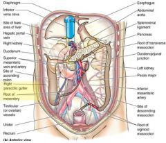
mesentery root directed obliquiely, inferior and right
duodenojejunal junction to ileocolic junction (more transparent area in picture) |
|
|
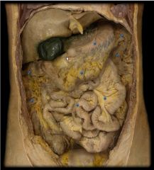
be able to ID:
liver diaphragm gallbladder stomach small intestines mesentery illeum/cecum |
you don't need a guide you know this :)
|
|
|
|
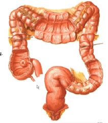
large intestine parts include
|

4-5' long, 2.5 in diameter
includes cecum, ascending, right hepatic flexure (9/10 rib) transverese splenic flexure (slightly higher) descending colon sigmoid colon (iliac fossa to s3) rectum |
|
|
|
at what levels are the following structures:
right hepatic flexure sigmoid colon |
RHF: 9th/ 10th ribs
SC: iliac fossa to S3 |
|
|
|
at what levels are the following structures:
right hepatic flexure sigmoid colon |

RHF: 9th/ 10th ribs
SC: iliac fossa to S3 |
|
|
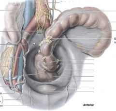
thinking intra/retro/subperitoneal where is the sigmoid colon? the rectum?
ID: IVC internal iliac artery ureter tenia coli sigmoid colon sigmoid mesocolon rectum medial umbilical ligament external iliac artery/vein |
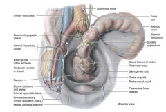
sigmoid colon = intraperitoneal
rectum = retroperitoneal and subperitoneal |
|
|
|
appendix is usually off the cecum in what direction?
how large is it? |
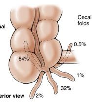
posterior medial
usually 6-10cm long mcBurney's point is 1/3 of the way up spinoumbilical line and is referred pain from appendix |
|
|
|
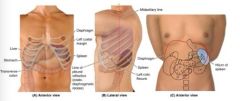
what surface structures are used to find the liver
|
it's deep to 7-11 ribs, moves with diaphragm
|
|
|
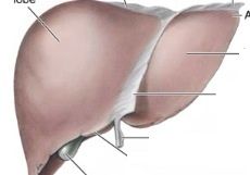
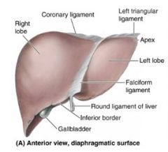
ID:
right lobe coronary ligament left triangular ligament apex left lobe falciform ligament round ligament inferior border gallbladder |
|
|
|
|
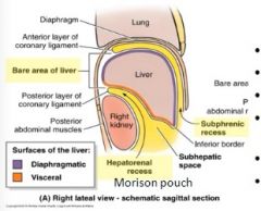
-what divides the subphrenic recess of the liver?
-where is the bare area? -where is the coronary ligament? |
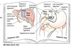
-subphrenic recess divided by falciform ligament
-bare area is behind coronary ligament and is where liver contacts diaphragm - coronary ligament is a peitoneal fold from diaphragm to liver |
|
|
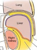
ID:
bare area hepatorenal recess subphrenic recess |
|
|
|
|
portal triad includes:
|
common bile duct
hepatic artery portal vein |
|
|
|
hepatopancreatic ampulla
is the combo of what 2 structures |
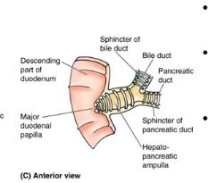
bile duct + pancreatic duct
|
|
|
|
bile duct is the mergine of one two structures
|

cystic duct (from gallbladder)
common hepatic duct (from liver) |
|
|
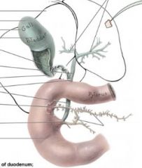
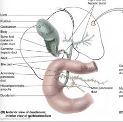
ID:
cystic duct common hepatic duct bile duct hepatopancreatic ampulla duodenum main pancreatic duct hepatic duct |
|
|
|
|
where is the spleen in relation to other structures?
what is the spleen's function? |
in LUQ behind ribs 9-11
posterior to stomach but superior to L colic flexure function: lymphocyte proliferation and immune surveillance, hematopoiesis prenatally and in severe stress |
|
|
|
where is the hilum and the renal pelvis of the kidneys?
|
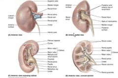
hilum is concave medial margin
renal pelvis is funnel shaped expansion of end of ureter |
|
|
|
kidneys are deep to what structures
|
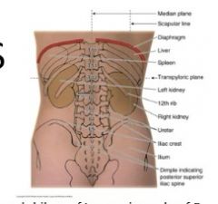
11th and 12th rib
|
|
|
|
ureters are how long?
trace their path |
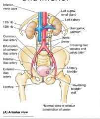
25-30cm
starts at L2, goes over pelvic brim (common area of damage), enter bladder posterior and inferior (to avoid dripping and splashing into the bladder which would cause use to feel like we need to pee all the time) |
|
|
|
bladder is located where?
how does it move as it fills? how is it held in place? |
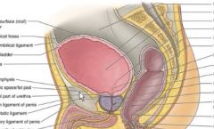
retropubic space btw pelvis and bladder (behind pubic bone)
ascends as it fills up to umbilicus neck of bladder is held in place by lateral ligaments |
|
|

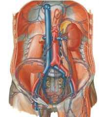
ID:
inferior phrenic lumbar arteries (1-4) median sacral suprarenal renal gonadal celiac trunk superior mesenteric inferior mesenteric |
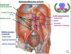
these go to the body wall (segmental vessels): inferior phrenic, lumbar arteries and median sacral
these go midline to organs and are unpaired: celiac trunk, superior mesenteric, inferior mesenteric these are paired and run to UG organs: suprarenal, renal, gonadal |
|
|

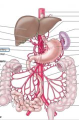
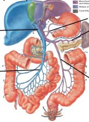
what does celiac trunk supply
|
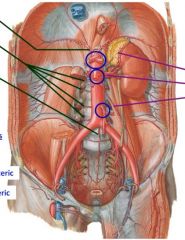
foregut and spleen: abdominal esophagus, stomach, proximal 1/2 of duodenum, liver, gallbladder and pancreas
(top circled blue) |
|
|

what does superior mesenteric artery (SMA) supply
|

midgut: distal 1/2 duodenum, jejenum, ileum, cecum, appendix, ascending colon, proximal 2/3 of transverse colon
think S to S- SMA to superior intestines (second blue circle) |
|
|
|
describe the blood supply to the spleen
|
splenic artery off the celiac artery
drains by splenic vein off hepatic portal vein |
IF IT CAME OFF AN UNPAIRED ARTERY THEN IT GOES INTO HEPATIC PORTAL:
aka celiac trunk, sma, ima supplies then it goes into hepatic portal |
|
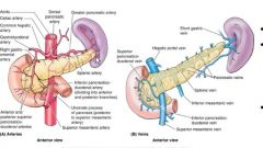
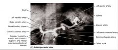
ID:
Liver left/right hepatic artery gastroduodenal artery spleen celiac trunk splenic artery left gastro-omental common hepatic |
|
|
|
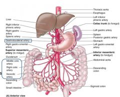
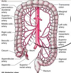
ID:
superior mesenteric artery middle/right colic artery ileocolic artery celiac trunk left/right inferior phrenic artery right/left gastric artery splenic artery left/right gastro-omental a. inferior mesenteric artery arterial arcades/loops |
|
|
|
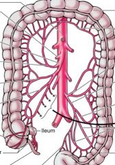
ID:
inferior mesenteric a. left colic a. sigmoid branches superior rectal artery superior mesenteric artery marginal artery |
|
|
|
where is the vena cava in relation to aorta?
where is superior mesenteric in relation to the renal vein? what is nutcracker effect? |

if pressure to abdomen where pancreas is you can crush either mesenteric artery or the vein
|
|
|
|
what is the drainage of body wall posteriorly?
|
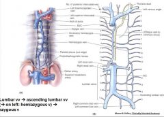
Posteriorly
Intercostal/lumbar veins drain: -directly to IVC -via ascending lumbar vv to azygous system to IVC -to epidural venous plexus, to azygous to IVC |
|
|
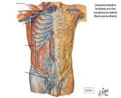
what is drainage of anterior body wall?
|
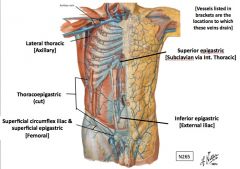
Anteriorly: Numerous pathways, all of which could communicate with each other
-Superior epigastric > internal thoracic > subclavian -inferior epigastric> external iliacs -Superficial circumflex iliac & superficial epigastric to femoral -Thoracoepigastric to lateral thoracic to axillary |
|
|
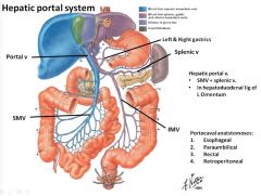
hepatic portal system
ID: portal vein SMV IMV splenic |
|
|
|
|
What is the parasympathetic innervation of the abdomen
|
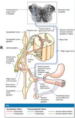
Vagus
S2 - S4 provides excitation |
|
|
|
what is the sympathetic innervation of the abdomen
|

sympathetic trunk from T2-T12
splanchnic nerve function is inhibition |
|