![]()
![]()
![]()
Use LEFT and RIGHT arrow keys to navigate between flashcards;
Use UP and DOWN arrow keys to flip the card;
H to show hint;
A reads text to speech;
24 Cards in this Set
- Front
- Back
|
2. Blood supply and blood vessels of the kidney. |
Abdominal Aorta > Renal Artery > Segmental Artery > Interlobar Artery > Arcuate Artery > Interlobular Artery > Affarent Artery > Glomerular Capillaries > Efferent Arteriole > Peritubular Capillaries > Interlobular veins > Arcuate Veins > Interlobar Veins > Segmental Vein > Renal Vein > Inferior Vena Cava |
|
|
10. Normal and abnormal constituents of urine. |
Urine is an aqueous solution of greater than 95% water. Other constituents include urea, chloride, sodium, potassium, creatinine and other dissolved ions, and inorganic and organic compounds. Urea is a non-toxic molecule made of toxic ammonia and carbon dioxide. Abnormal constituents of urine: red blood cells, large or medium proteins (constituents normally too large to pass into tubules) |
|
|
11. Regulation of sodium and potassium secretion/excretion in the kidneys by hormones. |
If there is no Aldosterone, Sodium will not be reabsorbed which will cause sodium excretion? The presence of aldosterone causes potassium secretion? |
|
|
1. Knowthe regions and the structures of the kidney – Cortex |
•Renal Cortex–Outerlayer The renal cortex is the outer portion of the kidneybetween the renal capsule and the renal medulla. In the adult, it forms a continuous smooth outer zone with a number of projections (cortical columns) that extend down between the pyramids. |
|
|
1. Know the regions and the structures of the kidney – Medulla. |
•Renal Medulla –Innerlayer –Renal Pyramids •Whereurine is formed •Renal papillae –Tipof pyramid –Renal Columns |
|
|
3. The functional units of the kidney – Nephron – know parts and functions. |
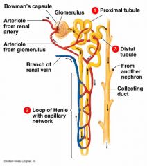
•Over1 million nephrons in each kidney •Sitesof urine formation •Extendfrom cortex to medulla –Tubulesform pyramids Consists of the 1. Bowman's Capsule 2. Proximal Convoluted tubule 3. Loop of Henle 4. Distal Convoluted Tubule 5. The Collecting Duct |
|
|
6. Know the three processes/steps and sites in urine formation. |
•Three processes are involved in the formationof urine in the nephron. 1. Filtration –Mostof the blood and its solutes move from the capillaries into the nephron 2. Reabsorption –Nutrients,ions and water are reabsorbed back into the blood. 3. Secretion –Wastesnot absorbed during filtration are actively moved into the nephron and out withthe urine. |
|
|
4. Components of the renal corpuscle: a. Glomerulus |
•Madeup of 3 layers of filtration membranes•Outercovering –Podocytes•Createslit membrane•Layersallow plasma and most dissolved particles to pass into capsule•Redcells and most proteins remain behind•Afferent arteriole–Largervessel–Bringsblood in•Efferent arteriole–Carriesblood out |
|
|
4. Components of the renal corpuscle: b. Bowman’s capsule. |
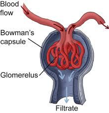
a capsule-shaped membranous structure surrounding the glomerulus of each nephron in the kidneys of mammals that extracts wastes, excess salts, and water from the blood. |
|
|
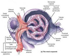
5. Juxtaglomerular apparatus – components and function of JG cell, Macula Densa |
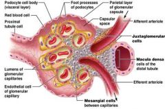
•Regulatesblood pressure and osmolarity•Locatedat contact point between afferent arteriole and distal tubule –Macula densa •Distaltubule •Highfiltrate osmolartiy –Juxtaglomerular cells •Afferentarteriole •Lowblood pressure |
|
|
Where the Juxtaglomerular Apparatus is located with respect to the Glomerulus/Bowman's Capsule |
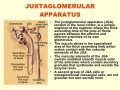
The JG apparatus is located where the ascending limb of the loop of Henli passes between the affarent artery and the effarent arteriole. |
|
|
Function of the juxtaglomerular cells |

The JG Cells Surround the affarent artery. smooth myoepithelioid cells lining the glomerular end of the afferent arterioles in the kidney that are in opposition to the macula densa region of the early distal tubule. These cells synthesize and store renin and release it in response to decreased renal perfusion pressure, increased sympathetic nerve stimulation of the kidneys, or decreased sodium concentration in fluid in the distal tubule. |
|
|
Function of the Macula Densa Cells |
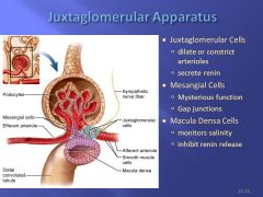
In the kidney, the macula densa is an area of closely packed specialized cells lining the wall of the cortical thick ascending limb of the loop of Henle, at the transition to the distal convoluted tubule. The cells of the macula densa are sensitive to the concentration of sodium chloride in the late thick ascending limb. A decrease in sodium chloride concentration initiates a signal from the macula densa that has two effects: (1) it decreases resistance to blood flow in the afferent arterioles, which raises glomerular hydrostatic pressure and helps return GFR toward normal, and (2) it increases renin release from the juxtaglomerular cells of the afferent and efferent arterioles, which are the major storage sites for renin. |
|
|
7. Functions/actions of the following hormones in the kidney: a. Aldosterone |
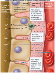
•RegulatesNa+excretion •Mineralocorticoid hormone produced in AdrenalCortex •StimulatesNa+reabsorption in –Distal tubule –Collecting tubule •MostNa+already reabsorbed by the time it reaches distal tubule •2%left, around 30 grams (still a lot) •Ifmore Na+is reabsorbed moreH20 is reabsorbed •Thegoal –regulatebloodpressure •Stimulates Na+ reabsorptionfromdistal tubule –Water follows •Stimulates K+ secretion in urine |
|
|
7. Functions/actions of the following hormones in the kidney: b. ADH |
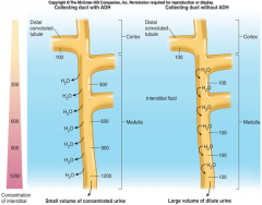
•Theamount of waterreabsorbed from the collectingduct is regulated by ADH activated Channels •Secretedby the posterior pituitary When Antidiuretichormone is present •Distaltubule + collecting tubule become permeableto H20causing •More water reabsorption causing –Concentrated (hypertonic)urine –Increased blood pressure When antidiuretichormone is absent •TheDistal tubule + Collecting tubules become impermeableto water causing •Less water absorption causing –Dilute (hypotonic) urine –Decreased blood pressure Factors that affect ADH Stimulation •Dehydration •Reducedblood volume •Pain Inhibition •Alcohol,caffeine •Ingestionof water |
|
|
7. Functions/actions of the following hormones in the kidney: c. Calcitonin |
High concentrations of calcitonin may be able to increase urinary excretion of calcium and phosphate, via actions on the kidney tubules.[12] However, this is a minor effect with no physiological significance in humans. It is also a short-lived effect because the kidneys become resistant to calcitonin, as demonstrated by the kidney's unaffected excretion of calcium in patients with thyroid tumors that secrete excessive calcitonin. |
|
|
7. Functions/actions of the following hormones in the kidney: d. Atrial natriuretic peptide (ANP) |
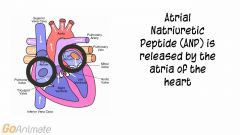
•Fromatria of heart in response to high blood pressure •Fouractions 1. Dilatesafferent arteriole, Constricts efferent arteriole - Increases GFR 2. Inhibitsrenin and aldosterone secretion 3. Inhibitssecretion of ADH 4. InhibitsNaClreabsorption by collecting duct •Results 1. Excretionof more salt and water in the urine 2. Reduced blood volume and pressure |
|
|
7. Functions/actions of the following hormones in the kidney: e. Angiotensin II |

Drop in Blood Pressure > Renin Angiotensinogen (Plasma Protein) > Renin> Angiotensin I Angiotensin I > Angiotensin Converting Enzyme (ACE) (In lungs and Kidneys) > Angoitensin II (Active Hormone) RESULT: ELEVATED BLOOD PRESSURE. |
|
|
Angiotensin II Effects |
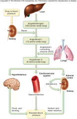
1.Vasoconstriction •Afferentand efferent arterioles •IncreasesGFR –Arterioles throughout body 2. Aldosteronesecretion •IncreasedNa+ andH2O reabsorption from nephron to blood 3. AntidiureticHormonesecretion •IncreasedH2O reabsorption from nephron to blood 4. It stimulates the sense of thirst and encourages water intake. Overall result: IncreasedBP by: a)Reducing water loss b)Encouraging water intake c)Constricting blood vessels |
|
|
8. Distribution of body fluid, intracellular vs extracellular fluid compartments. |
Intracellular fluid – 65% |
|
|
8. Distribution of body fluid, intracellular vs extracellular fluid compartments. |
– Interstitial fluid –25% – Plasma and lymphatic fluid –8% – Transcellular fluid-2% • Where is this found? (everywhere) |
|
|
Fluid Compartments |
Fluid Exchanges Fluid compartments are separated by membranes • Cell membranes • Vessel walls • Nephron etc.Water + solutes constantly move between compartments |
|
|
9. Calculate the Net Filtration Pressure given the blood hydrostatic pressure, capsular pressure and osmotic pressure. |
FILTRATION PRESSURES Moving fluid OUT of glomerulus 1. Glomerular blood pressure- +60mm Hg Moving fluid INTO glomerulus 1.Blood Osmotic pressure- -32mm Hg 2. Capsular Hydrostatic pressure- -18mm Hg Net filtration pressure +10mm Hg |
|
|
Filtration Pressures Diagram |
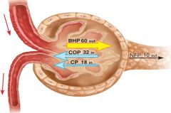
See Flashcard 23 for breakdown of Filtration Pressures |