Reading...
![]()
Play button
![]()
Play button
![]()
Use LEFT and RIGHT arrow keys to navigate between flashcards;
Use UP and DOWN arrow keys to flip the card;
H to show hint;
A reads text to speech;
66 Cards in this Set
- Front
- Back
|
What is the antecubital fossa?
|

The dip on the medial side of the elbow region
|
|
|
What is the antebrachial?
|

The forearm
|
|
|
What separates muscles and limbs into discrete compartments?
|

Intermuscular septa that arise from deep fascia
|
|
|
What do muscles within the same compartment share?
|
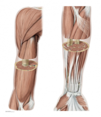
Function, innervation, and blood supply.
Muscles of the anterior arm are common flexors whereas posterior muscles are extensors. |
|
|
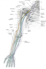
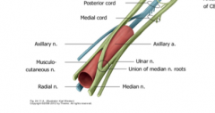
Name the nerves (overall excluding exception) that supply the different parts of the upper limb:
Anterior Arm Posterior Arm Anterior Forearm Posterior Forearm Intrinsic Hand |

Musculocutaneous
Radial Median Radial Ulnar |
|
|
What is the name for the shoulder joint?
|

Glenohumeral joint
|
|
|
What are the neurovascular vessels that are compromised with a dislocated glenohumeral joint
|

The axillary nerve and the posterior circumflex humeral artery that run around the surgical neck of the humerus.
|
|
|
What is the major long bone of the arm (brachium)?
|
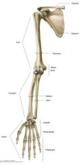
Humerus
|
|
|
What are the major long bones of the forearm?
|

Medial Ulna and Lateral Radius
|
|
|
What are the main bones of the hand?
|

8 carpals, 5 metacarpals, and 14 phalanges
|
|
|
What type of nerves compose the brachial plexus?
|
Ventral Primary Rami/Spinal nerves/Mixed Nerves
|
|
|
What is the difference between dermatomes and myotomes?
|
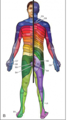
Dermatomes are areas of skin supplied by the cutaneous (sensory) branches from a single spinal nerve. Myotomes are muscle sections supplied by a single spinal nerve. The somites from embryonic development drag their nerve supply with them out to either the motor or sensory areas they innervate
|
|
|
What nerves supplies the skin of the anterolateral aspect of the forearm?
|

Musculocutaneous nerve
|
|
|
What nerve supplies the palmar surface of the lateral 3.5 digits?
|

Median nerve
|
|
|
What nerve supplies the palmar surface of the medial 1.5 digits?
|
Ulnar nerve
|
|
|
What nerve supplies the skin of the posterior and inferolateral arm, posterior of the forearm, and dorsum of the hand?
|

Radial nerve
|
|
|
What is a dermatome sensory area to test if the axillary nerve is damaged?
|
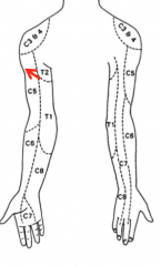
A patch on the lateral upper arm
|
|
|
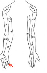
What is a dermatome sensory area to test if the median nerve is damaged?
|
The lateral finger-distal phalanx (pointer finger)
|
|
|
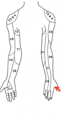
What is a dermatome sensory area to test if the ulnar nerve is damaged?
|
Medial surface of the proximal finger-distal phalanx (pinky finger)
|
|
|
What is a dermatome sensory area to test if the radial nerve is damaged?
|
The first dorsal web space of the hand (between thumb and pointer finger)
|
|
|
Which artery supplies structures in the neck, part of the thoracic wall and entire upper limb?
|
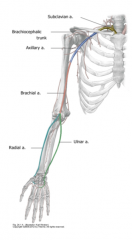
The subclavian artery
|
|
|
Which artery begins at the lateral boarder of the first rib?
|

The axillary
|
|
|
Which artery begins at the lateral, lower boarder of the teres major?
|

Brachial
|
|
|
What two vessels are branches of the brachial artery and what compartment do they supply?
|

The radial and ulnar artery are branches of the brachial artery starting at the cubital fossa supplying blood to the forearm.
|
|
|
What is the meaning of venae comitantes?
|
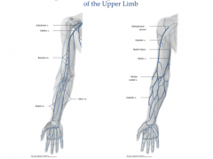
The deep veins which "accompany" larger arteries. These include the subclavian vein and axillary vein.
|
|
|
What are the superficial veins?
|

They do not run with arteries and are subcutaneous and drain into the deep venous system. These include the cephalic vein, basilic vein, and median cubital vein (blood draw vein)
|
|
|
What is the breast simplistically?
|

It is a derivative of the epidermis and a modified sweat gland.
|
|
|
What forms the areola and nipple?
|
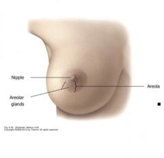
The areola is a dark pigmentation of skin and the nipple is a result of circular and longitudinal muscle
|
|
|
What factors influence the size and shape of breasts?
|

Race, Genetics, and Diet
|
|
|
What area of skin is covered by the breast typically?
|

The lateral boarder of the sternum to the mid-axillary line from ribs 2-6.
|
|
|
Where is breast tissue located?
|
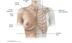
In the superficial fascia.
|
|
|
Which quadrant of the breast contains the axillary tail?
|
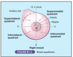
The superolateral quadrant or the upper quadrant contains the tail of Spence.
|
|
|
When does breast development start in utero?
|
In the fourth week of development.
|
|
|
What is the technical term for supernumerary breasts and nipples
|
Polymastia and polythelia respectively
|
|
|
What artery supplies the medial side of the breast?
|

The internal thoracic artery (a branch of the subclavian artery also known as the internal mammillary artery) This has perforating branches then medial mammary branches to supply the entire medial side
|
|
|
What artery supplies the lateral side of the breast?
|
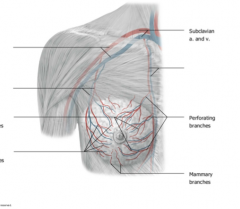
The lateral thoracic artery (a branch of the axillary artery) with perforating and mammary branches.
|
|
|
What is the main venous drainage of the breast?
|

The axillary and internal thoracic veins.
|
|
|
Which nerves supply the breast?
|

Anterior and lateral cutaneous branches of the 4-6th intercostal nerves.
|
|
|
What do nerve branches convey to the breast?
|
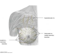
Sensory fibers to the skin
Sympathetic fibers to the blood vessels in the breast Fibers to smooth muscle in the overlying skin and nipples |
|
|
What muscles are just deep to the breast?
|
Subclavious
Pectoralis Minor Pectoralis Major (superficial to minor) |
|
|
What separates the breast from the underlying muscle?
|

The retromammary space
|
|
|
What two types of tissue compose the breast?
|
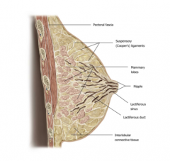
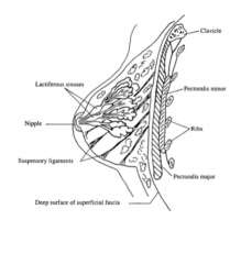
Glandular Tissue (Parenchyma)
-Lactiferous glands: mammary lobes (15-20) with lobules that give rise to lactiferous ducts which emerge to form lactiferous sinuses which exit the nipple Connective tissue (stroma) -Connective tissue ligaments -Fat |
|
|
What are Coopers ligaments?
|

Suspensory ligaments that hold up the breast and provide support. Part of the connective tissue.
|
|
|
What is the leading cause of cancerous death in women worldwide?
|

Breast cancer
|
|
|
What percentage of breast cancer occurs in postmenopausal women?
|

2/3
|
|
|
What percent of cancers develop in the upper outer quadrant?
|

60%
|
|
|
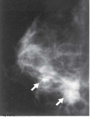
What are common signs of breast cancer?
|
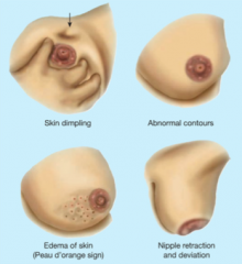
Lump, skin dimpling, peau d'orange, and nipple retraction
|
|
|
How can breast cancer metastasize ot the vertebrae, brain, and cranium?
|
Through venous communication with the azygos system and the vertebral venous plexus
|
|
|
What are the functions of the lymphatic system?
|
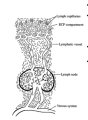
-Drains excess ECF from the body and returns it to the circulatory system
-Mounts immune responses in the body -Transports fat and large protein molecules that are too big for venous transport |
|
|
What are normal and abnormal symptoms of the lymph node?
|

A normal lymph node is : small, mobile, and pain-free/non-tender
Infected are swollen and soft (tender) cancerous nodes can be swollen and hard (non-tender) |
|
|
What is lymphedema?
|
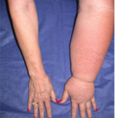
Accumulation of interstitial fluid such as when cancerous lymph nodes are removed
|
|
|
What is drained by the right lymphatic duct?
|
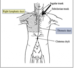
The right side of the head, neck and upper limb
|
|
|
What is drained by the thoracic duct?
|

Everything else not drained by the right lymphatic duct. This drains at the intersection between the left internal jugular and left subclavian veins.
|
|
|
What is the cisterna chyli?
|

Thin-walled sac at the inferior end of the thoracic duct dilated because this is a convergent point of the right and left lumbar lymphatic trunks, intestinal lymph trunks and descending thoracic lymph trunks.
|
|
|
What is the convergence point of lower lymph vessels and where they enter the thoracic duct?
|

The Cisterna Chyli
|
|
|
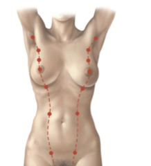
What are the two groups of lymph nodes for draining the breast?
|
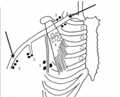
Axillary (75%) and Parasternal (25%) lymph node groups
|
|
|
What are the sections of the axillary lymph node group?
|

Pectoral (anterior) nodes
Subscapular (posterior) nodes Brachial (lateral) nodes Central nodes Apical nodes |
|
|
Which vein are the parasternal lymph node group likely to drain into?
|

The internal thoracic vein
|
|
|
What are the proximal attachments of the pectoralis major?
|
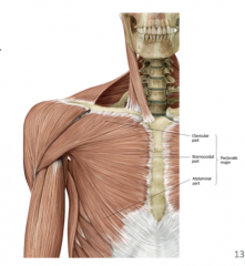
Clavicular part
Sternocostal part Abdominal part |
|
|
What are the primary actions of the pectoralis major?
|

Adduction of the arm
Medially rotate the arm |
|
|
What is the main function of the sternocostal head of the pec major?
|

Extend the arm that is already flexed
|
|
|
What is the main function of the clavicular head of the pec major
|

Arm flexion (zombie walk)
|
|
|
What are the proximal and distal attachments of the pectoralis minor?
|
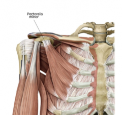
Ribs 3-5 and the coracoid process of the scapula respectively
|
|
|
What are the main functions of the pectoralis minor?
|

-Stabilizes the scapula
-Depresses the shoulder |
|
|
What are the innervations of the pectoralis major and minor?
|
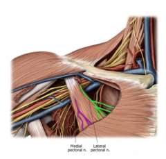
Pectoralis major: Lateral and Medial pectoral nerve
Pectoralis minor: The medial pectoral nerve |
|
|
The names for the medial and lateral pectoral nerves come from where?
|

The brachial plexus cords from which they arise.
|