Reading...
![]()
Play button
![]()
Play button
![]()
Use LEFT and RIGHT arrow keys to navigate between flashcards;
Use UP and DOWN arrow keys to flip the card;
H to show hint;
A reads text to speech;
49 Cards in this Set
- Front
- Back
|
|
|
|
|
|
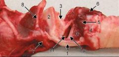
Left Side of Larynx Removed
|
1) Thyroid Cartilage
2) Epiglottic Cartilage 3) Arytenoid Cartilage 4) Cricoid Cartilage 5) Cricoarytenoideus Lateralis m. 6) Cricoarytenoideus Dorsalis m. 7) Basihyoid bone 8) Thyrohyoid bone 9) Vocal lig. 10) Vocalis m. 11) Laryngeal Ventricle |
|
|
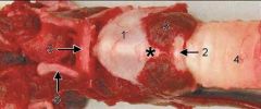
1) Thyroid Cartilage
2) Cricoid Cartilage 3) Cricothyroideus m. 4) Trachea 5) Basihyoid bone 6) Ceratohyoid bone * Cricothyroid lig. |
|
|
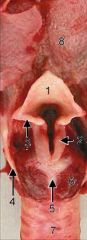
1) Epiglottic cartilages
2) Arytenoid cartilages 3) Aryepiglottic fold 4) Thyroid cartilage 5) Cricoid cartilage 6) Cricoarytenoideus m. 7) Trachea 8) Root of the Tongue |
|
|
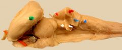
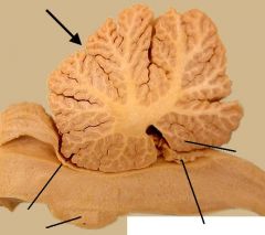
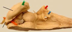
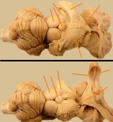
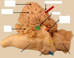
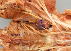
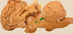
Dorsal view of the right side of an equine brainstem, plus hippocampus. The cerebellum has been removed by cutting three cerebellar peduncles. The rostral cerebellar peduncle (white pick) continues rostrally toward the midbrain. It borders (white asterisk) the fourth ventricle. The middle cerebellar peduncle (red pick) comes from the pons. It is positioned laterally (red asterisk). Fibers of the caudal cerebellar peduncle (blue pick) ascend abruptly from the dorsal edge (blue asterisk) of the medulla oblongata.
The green pick is in the hippocampus. The orange pick passes through the right interventricular foramen. |
|
|
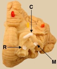
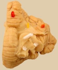
Ventral view of the right half of an equine cerebellum. The three cerebellar peduncles (white picks) were cut as they become confluent. They are: caudal cerebellar peduncle (C); middle cerebellar peduncle (M); and rostral cerebellar peduncle (R).
Also labeled are: nodulus (red pick); flocculonodular tract (yellow pick); and flocculus (orange pick). |
|
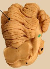
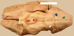
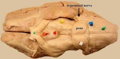
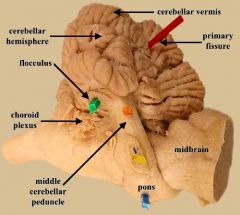
Rostrolateral view of the pons and cerebellum.
|
The green pick is in the middle cerebellar peduncle, which is formed by transverse pontine fibers (asterisk), which are axons of contralateral pontine nuclei. The trigeminal nerve (V) exits the brainstem by passing through the middle peduncle. The arrow points to the primary fissure that divides the cerebellum into rostral and caudal lobes.
|
|
|
The cerebellum has been dissected to reveal the confluence of cerebellar peduncles (medial view). The caudal cerebellar peduncle (white pick) abruptly turns dorsally. The rostral cerebellar peduncle (red pick) extends rostrally and forms a wall of the fourth ventricle. The middle cerebellar peduncle (blue pick) enters the cerebellum laterally.
|
|
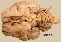
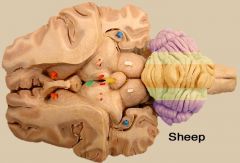
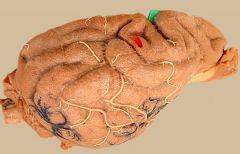
Dorsal view of a dissected sheep brain.
|
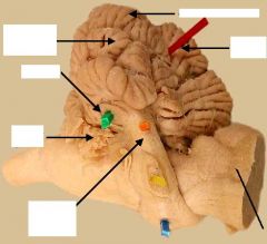
The yellow arrow points to the primary fissure which divides the cerebellum into rostral and caudal lobes. The black arrows point to sulci, which separate folia. The central region of the cerebellum is called cerebellar vermis; it is flanked by cerebellar hemispheres.
Colored pics mark the following structures: caudate nucleus (orange); third ventricle (green); habenular nuclei (red); thalamus (pink); hippocampus (blue); pineal body (yellow); and rostral colliculus (white). |
|
|
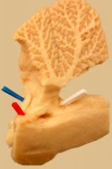
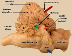
Median view of the vermis of the cerebellum attached to the brainstem. Fissures divide the cerebellar vermis into about ten lobules (to view the lobules, click here). The most caudoventral lobule is the nodulus (N). The arrow marks the location of the primary fissure, which divides the cerebellum into rostral lobe and caudal lobe. The primary fissure is the first to develop embryologically; it is often but not always the deepest fissure. Other labels are: ventral pons (vp); choroid plexus (cp); and rostral medullary vellum forming the roof of the fourth ventricle (v).
|
|
Dorsal view of a dissected sheep brain.
|
(DONT CARE) Bilaterally, colors are used to demonstrate, approximately, the three longitudinal cerebellar zones. From medial to lateral, notice vermal (orange), paravermal (green), and hemispheric (purple) zones. To see the zones in transection, click here.
The zones also have different functional roles: the vermis deals with muscle tone; the paravermis corrects movement based on proprioceptive feedback; and the hemispheric (lateral) zone deals with the cerebral motor cortex. (Colored picks mark the following structures: caudate nucleus (orange); third ventricle (green); habenular nuclei (red); thalamus (pink); hippocampus (blue); pineal body (yellow); and rostral colliculus (white)). |
|
Ventral view of the right half of an equine cerebellum (caudal is toward the top).
|
The nodulus (red pick) of the vermis is connected to the flocculus (orange pick) of each hemisphere by a flocculonodular tract (yellow pick). Collectively these structures constitute the flocculonodular lobe, which functions like vestibular nuclei.
Three cerebellar peduncles have been cut (white picks). |
|
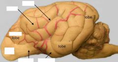
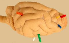
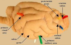
Dog Brain — Lateral View
|
3 lobes of the cerebral hemisphere are labeled: frontal lobe, occipital lobe, and temporal lobe. The parietal lobe, midway between frontal and occipital lob, is not well defined in the canine brain. The surface of the cerebrum features sulci (grooves) which delineate gyri (ridges). To view specific sulci, click here.
Components of the rhinencephalon include: olfactory bulb (olfact bulb), lateral olfactory tract (olfact tr), and piriform lobe (piriform). |
|
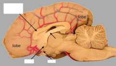
Medial view of a canine brain.
|
The frontal lobe and occipital lobe are rostral and caudal, respectively. The cingulate gyrus (C) is dorsal to the corpus callosum. The fornix, rostral commissure (rc), and lamina terminalis (lam) are also labeled
|
|
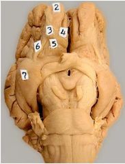
Ventral view of a sheep brain.
|
Various surface components of the rhinencephalon are labeled on the left side. Identify the olfactory bulb (2), piriform lobe (7), and lateral olfactory tract (6). Also, the olfactory peduncle (3) and the medial olfactory tract (4) are labeled. Label 5 indicates an area that has numerous openings indicating a rich blood supply.
The rhinencephalon (nose brain) is concerned with olfaction and emotion. |
|
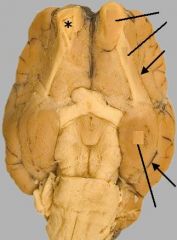
Ventral view of a sheep brain.
|
The arrows indicates the lateral rhinal sulcus, which separates the phylogenetically older rhinencephalon from the newer portion of the cerebral hemisphere that is covered by neocortex. The rhinencephalon (nose brain) is concerned with olfaction and emotion. Identify the following three components of the rhinencephalon on the sheep brain shown on the right: olfactory bulb (O), piriform lobe (P), and lateral olfactory tract (t). The left olfactory bulb is cut open to reveal the lumen of the olfactory bulb.
|
|
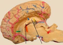
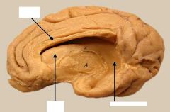
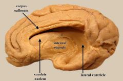
Medial view of an equine half brain. The ventromedial telencephalon has been removed
|
to expose the lateral ventricle. The caudate nucleus (green pic) forms the lateral wall of the ventricle. The floor of the ventricle is formed by the hippocampus (blue; h), which curves caudoventrally. Axons from the hippocampus form the fornix (fo). (The fimbria is not evident because it is positioned lateral to the hippocampus.)
|
|
|
The curved hippocampus (h; green) is preserved in situ on an equine half brainstem. Axons from the hippocampus course rostrally as the fornix (fo). (The orange pic marks the location of the interventricular foramen, between third and lateral ventricles).
|
|
|
A sheep brain was dissected to expose the lateral ventricle and the hippocampus (h; yellow) bilaterally. The fimbria (fi; green) appears as a white matter ledge along the lateral margin of the hippocampus. Fibers of the fimbria are continued rostrally as the fornix (fo; orange
|
|
|
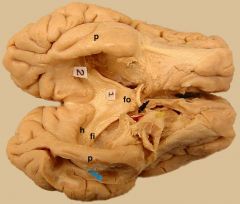
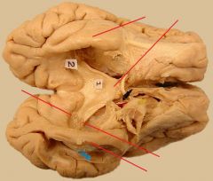
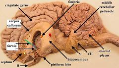
The hippocampus (h; 1, 2) is exposed in this ventral view of an isolated cerebrum, from a sheep. Nerve fibers from the hippocampus emerge laterally in the fimbria (fi) and then run rostrally as the fornix (fo). The fornix passes close to the rostral commissure (black thread). The hippocampus begins deep to the piriform lobe (p).
|
|
Top.
|
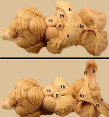
Most of the telencephalon has been removed, but the deeply positioned hippocampus (f) remains. Fibers of the hippocampus extend rostrally as the fornix (fo). Bottom. Same specimen as top, but the hippocampus has been reflected rostrally to produce a ventral view. Notice the hippocampus (h), the fimbria (fi), and the fornix (fo). Also labeled are: pineal body (p); thalamus (th); and rostral colliculus (rc).
|
|
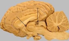
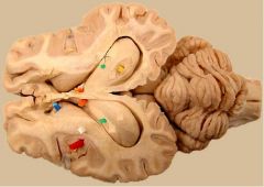
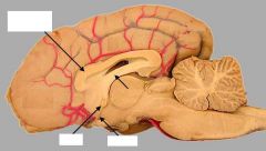
Medial view of an equine half brain.
|
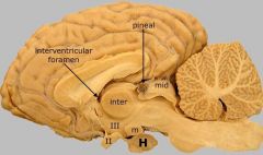
Components of the diencephalon are labeled. The interthalamic adhesion (inter) is surrounded by third ventricle (III). The third ventricle communicates with each lateral ventricle through an interventricular foramen. The floor of the hypothalamus features the optic chiasm (II), the infundibulum, to which the hypophysis (H) is attached, and a mamillary body (m). (Note: the terms "hypophysis" and "pituitary gland" are synonymous.) The equine pineal body is prominent and often pigmented, as is the case here. For a lateral view of the diencephalon, click here.
The rostral colliculus (mid) of the midbrain is labeled. |
|
|
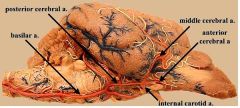
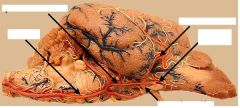
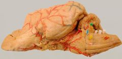
The basilar artery runs rostrally from the spinal cord and bifurcates into posterior cerebral arteries, which complete the arterial circle formed by trifurcation of bilateral internal carotid arteries. Each internal carotid artery gives rise to: an anterior cerebral a., a middle cerebral a., and a posterior communicating branch.
|
|
|
The hippocampus and fornix have been removed in this isolated cerebral hemisphere to show the lateral wall of the lateral ventricle. The caudate nucleus can be seen rostrally in the wall of the ventricle.
|
|
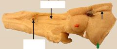
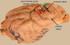
dorsal view of an equine brainstem,
|
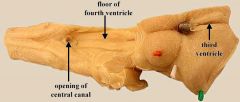
notice the floor of the fourth ventricle (rhomboid fossa) and opening of the central canal of the spinal cord. Rostrally, the third ventricle is evident anterior to the pigmented pineal body
|
|
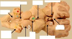
dorsal view of a dissected equine brain.
|
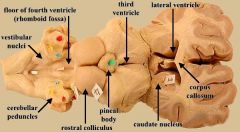
The lateral ventricle is the space medial to the caudate nucleus (1). The medial wall of the ventricle is thin between the corpus callosum and fornix. The third ventricle is evident anterior to the pineal body (green pic). The floor of the flourth ventricle (rhomboid fossa) is marked by white pics
|
|
|
The pons forms a prominent bridge (white pic) on the ventral surface of this sheep brain.The trigeminal nerve emerges laterally from the pons. Other labeled features are: optic chiasm (green pic), infundibulum (orange), mamillary bodies (red), crus cerebri (yellow), trapezoid body (blue), and pyramid (green).
|
|
The cerebellum is seen from a lateral view.
|
The red pic is in the primary fissure (the first fissure to develop embryologically); it divides the cerebellum into cranial and caudal lobes. The green pic indicates the flocculus of the cerebellar hemisphere. The middle cerebellar peduncle is composed of transverse pontine fibers entering the cerebellum. Notice the choroid plexus between the cerebellum and medulla oblongata
|
|
The cerebellum is seen from a lateral view.
|
The red pic is in the primary fissure which divides the cerebellum into cranial and caudal lobes. The green pic indicates the flocculus of the cerebellar hemisphere. The middle cerebellar peduncle (orange) is composed of transverse pontine fibers (blue) entering the cerebellum. The choroid plexus is evident between the cerebellum and medulla oblongata.
|
|
Medial view of a canine brain.
|
The frontal lobe and occipital lobe are rostral and caudal, respectively. The cingulate gyrus (C) is dorsal to the corpus callosum. The fornix, rostral commissure (rc), and lamina terminalis (lam) are also labeled
|
|
|
|
|
|
|
|
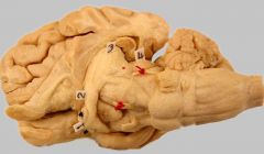
Ventrolateral view of sheep brain following removal of the caudal portion of the left cerebral hemisphere.
|
The oculomotor nerve (bottom) and trochlear nerve (top) are encircled by red thread. (The trochlear nerve emerges from the dorsal surface of the brainstem.) The rostral colliculus of the midbrain is labeled 4.
Some diencephalic structures are labeled: optic chiasm (1), optic tract (2), and lateral geniculate body (3). Also, the medical geniculate body is marked with a small red square. |
|

Lateral view of a sheep brain with the internal capsule transected.
|
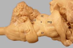
The rostral colliculus (rc) and the caudal colliculus (cc) form the tectum (roof) of the midbrain. Axons travel from the caudal colliculus to the medial geniculate (mg) through the brachium of the caudal colliculus (asterisk). The crus cerebri (cr) is evident on the ventral surface of the midbrain.
|
|
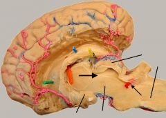
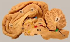
Median view of an equine dissected half-brain (medial components of the rhinencephalon have been removed, as has the cerebellum).
|
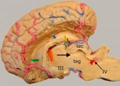
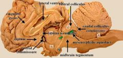
The tectum (tec) of the midbrain is dorsal to the mesencephalic aqueduct (large arrow). The midbrain tegmentum (teg) is ventral to the aqueduct. Labeled telencephalic structures are: caudate nucleus (green), fornix (blue) and interventricular foramen (orange). Labeled diencephalic structures are: pineal body (yellow) and third ventricle (III). A red pick is covered by rostral medullary vellum in the fourth ventricle (IV).
|
|
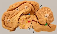
Median view of a sheep half-brain.
|
The rostral colliculus (rc) is a component of the tectum (tec) of the midbrain. The mesencephalic aqueduct has a red pic inserted into it. The midbrain tegmentum (teg) is ventral to the aqueduct. Labeled telencephalic structures are: cingulate gyrus (4), corpus callosum (1 & 2) and rostral commissure (black thread). Labeled diencephalic structures are: pineal body (blue), third ventricle (yellow), and interthalamic adhesion (pink). A green pick is in the floor of the fourth ventricle (IV). The cerebellum is labeled 3.
|
|
Ventral view of a sheep brain.
|
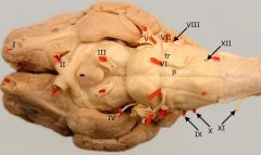
Cranial nerves are marked with red pics and numerical labels. The myelencephalon, located between the pons and the spinal cord, gives rise to seven of the twelve cranial nerves (VI through XIII). The pyramid (p) and the trapezoid body (tr) are also labeled.
|
|
Ventrolateral view of a dog half-brain.
|

The pyramids (pyr) emerge form the pons and run beside the midline along the ventral surface of the myelencephalon. The trapezoid body (yellow pic) is indicated. Cranial nerve locations are marked with colored pics: abducent (pink), facial (blue), vestibulocochlear (green), glossopharyngeal, vagus, and accessory (white), and hypoglossal (orange). The trigeminal nerve (red) emerges from the metencephalon.
|
|
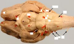
Dorsal view of a sheep brainstem.
|
The fasciculus gracilis (fg) terminates in the nucleus gracilis (nuc gr). The fasciculus cuneatus (fc) coveys axons to the medial cuneate nucleus (mc nuc). The floor of the fourth ventricle (IV) is marked white & pink pics. Also marked: cerebellar peduncles (rostral = blue; middle = red; caudal = yellow), rostral colliculus (4), and pineal body (green).
|
|
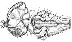
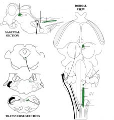
Artist rendition of a dorsal view of the canine brainstem.
|
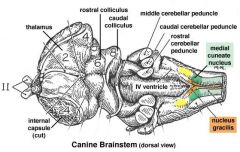
The nucleus gracilis (orange) is located beside the midline at the caudal end of the medulla oblongata (myelencephalon). The nucleus receives touch and kinesthesia from the pelvic limb via the fasciculus gracilis.
The fasciculus cuneatus conveys touch and proprioception from the thoracic limb to the medial cuneate nucleus (green) The nucleus relays the information to the thalamus for conscious perception. (The fasciculus cuneatus also conveys subconscious proprioception to the lateral cuneate nucleus (yellow), for relay to the cerebellum.) |
|
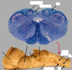
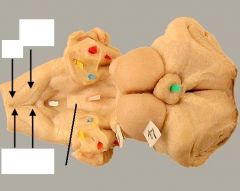
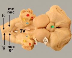
Top: Transection through the the brainstem of a dog at the juncture with the spinal cord.
Bottom: Dorsal view of a half brainstem from a horse. |
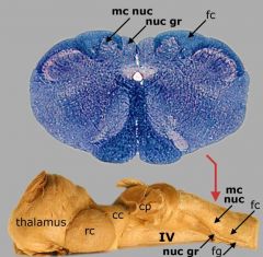
Top:
The nucleus gracilis (nuc gr), medial cuneate nucleus (mc nuc), and fasciculus cuneatus (fc) are labeled. Bottom: The red arrow shows the level of transection for the top image. Locations of the nucleus gracilis (nuc gr), fasciculus gracilis (fg), medial cuneate nucleus (mc nuc), fasciculus cuneatus (fc), and floor of the fourth ventricle (IV) are visible. Also labeled are: cerebellar peduncles (cp), caudal colliculus (cc), rostral colliculus (rc), and thalamus. |
|
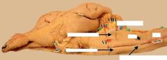
Ventrolateral view of a sheep brainstem.
|
The pyramids (orange) emerge form the pons (white) and run along the ventral surface of the myelencephalon, beside the midline. Because they decussate (cross sides), pyramids disappear from the surface at the juncture with the spinal cord. The green pic is stuck in the trapezoid body. The facial (VII) and vestibulocochlear (VIII) cranial nerves are labeled. The trigeminal nerve (V; red pic) exits through the pons (white). The crus cerebri (blue) is evident on the ventral surface of the midbrain.
|
|
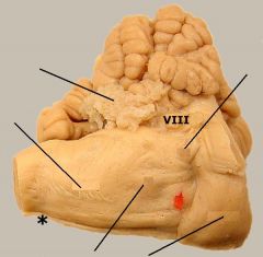
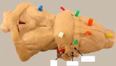
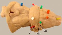
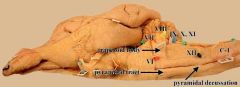
Ventrolateral view of an equine hindbrain.
|
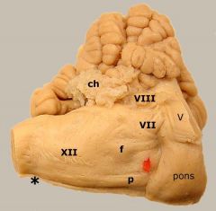
A pyramid (p) emerges form the pons and run along the ventral surface of the myelencephalon. Pyramids disappear from the surface at the juncture with the spinal cord (asterisk), because they turn dorsal and decussate. The red pic is in the trapezoid body. A swelling produced by the motor nucleus of the facial nerve (f) is evident in this specimen. The facial (VII), vestibulocochlear (VIII), and hypoglossal (XII) cranial nerves are labeled. Notice choroid plexus (cp) protruding from the lateral aperture of the fourth ventricle.
The trigeminal nerve (V) exits through the pons (white). |
|
|
|
|

|
|
|
|
|
|
|
|
|
|
|