Reading...
![]()
Play button
![]()
Play button
![]()
Use LEFT and RIGHT arrow keys to navigate between flashcards;
Use UP and DOWN arrow keys to flip the card;
H to show hint;
A reads text to speech;
91 Cards in this Set
- Front
- Back
- 3rd side (hint)
|
Confocal Microscopy
|
Confocal microscopy is now a valuable tool for obtaining high resolution images and 3-D reconstructions of a variety of biological specimens.
|
|
|
|
How does fixation work?
|
Crosslinking of proteins (typically not the sugars or lipids)
|
|
|
|
How does the fixation work in electron microscopy?
|
the most useful fixative agent is glutaraldehyde, its terminal aldehyde groups react with free amine groups of amino acids
|
|
|
|
Immunocytochemistry
|
the most useful fixative agent is glutaraldehyde, its terminal aldehyde groups react with free amine groups of amino acids
|
|
|
|
Histochemistry
|
Some enzymes retain their function after they are immobilized with a fixative, and in such cases, the preserved enzymatic activity can be localized by having it act on a soluble substrate.
|
|
|
|
Freeze Fracture
|
Cell membranes are phospholipid bilayers in which integral membrane proteins are embedded, and the larger of these integral membrane proteins can be visualized in the electron microscope by the freeze-fracture technique.
|
|
|
|
Scanning Electron Microscope
|
Cell membranes are phospholipid bilayers in which integral membrane proteins are embedded, and the larger of these integral membrane proteins can be visualized in the electron microscope by the freeze-fracture technique.
|
|
|
|
Autoradiography
|
Autoradiography is used to identify intracellular biosynthetic or transport pathways. An initial, brief exposure to radioactive substances is brief is followed by washout with excess nonradioactive substance (pulse-chase). Tissue specimens are then prepared at progressively longer time intervals.
|
|
|
|
Where are cisternae located?
|
rER...the stacks
|
|
|
|
What does the r refer to in rER?
|
Rough...polyribosomes
|
|
|
|
Smooth Endoplasmic Reticulum
|
recognized as a network of fine, smooth tubules. It is found in cells that synthesize steroid hormones or are involved in the degradation of noxious substances (e.g. alcohol and barbiturates). Unlike RER, there is no association with polyribosomes
|
|
|
|
Golgi Apparatus
|
consists of arrays of flattened, crescent-shaped membranes and smooth surfaced vesicles. The Golgi acts to concentrate proteins, to complete post-translational modification of newly synthesized protein, and to "address" products that have been synthesized. In cells that actively secrete protein (e.g. exocrine cells), the proteins are packaged and stored as membrane-bound secretory granules.
|
|
|
|
Lysosomes
|
sites of intracellular digestion
work via low pH in a necrotic cell, the lysosomal contents may damage surrounding tissue leading to a substantial inflammatory event |
|
|
|
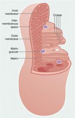
Where are cristae located?
|
Mitochondria mother fuckers!!
|
|
|
|
Mitochondria
|
are characterized by the presence of a double limiting membrane, internal cristae(invaginations of the inner limiting membrane) and a finely granular matrix filling the space between cristae. ______________ produce much of the energy of the cell, in the form of ATP, through the process of oxidative phosphorylation.
|
|
|
|
(3) Cytoplasmic structures
|
microtubules, microfilaments and intermediate filaments that comprise the cell's cytoskeleton
|
|
|
|
Microtubules
|
(~ 25 nm)
_____________________ play a significant role in the maintenance of cell form and symetry, and they have also been implicated in the intracellular transport of organelles and vesicles. ____________________ also provide the structural basis for more complex components such as centrioles, cilia and flagella. |
|
|
|
Microfilaments
|
Actin, Myosin
6-8nm Contractile activity, cell mobility and cell-cell or cell-matrix adhesion are associated with a variety of ________________ |
|
|
|
Intermediate Filaments
|
(10-15 nm).
They are cell type specific. For example, cytokeratins are present in all ecto- and endodermally-derived epithelial cells and desmin is found is muscle cells. |
|
|
|
Centrioles
|
recognized by nine triplets of short microtubules that are arranged in a cylinder, and they are involved in the organization and orientation of microtubules, cilia and flagella.
|
|
|
|
Cellular Inclusions
|
In addition to organelles, the cytoplasm of most cells contain "inclusions", which are usually a storage form of protein, lipid or carbohydrate.
Gylcogen is stored as small electron-dense rosettes Proteins (e.g. granules in eosinophils) are needle-shaped or polygonal lattices Lipids are stored as circular droplets Pigment (e.g. melanin) is found as dense granules or irregularly-shape bodies With normal aging, and under various pathological conditions, the types and amounts of cellular inclusion may increase dramatic. For example, lipofuchsin is an insoluble pigment that doesn't interfere with cell function, but is considered a telltale sign of free radical injury and lipid peroxidation. In the classical "storage diseases", a normal are abnormal endogenous substance accumulate because of genetic or acquired defects in metabolism, packaging, transport or secretion. |
|
|
|
Heterochromatin vs. Euchromatin
|
The dense nuclear aggregates are called heterochromatin and the less dense nuclear matrix is called euchromatin. Euchromatin represents areas where the genetic material is being transcribed into RNA.
|
|
|
|
Nucleolus
|
A very dense, centrally-located structure, the nucleolus, is the center for ribosomal RNA synthesis.
|
|
|
|
How is DNA and RNA transcription regulated?
|
very tightly regulated process that requires the presence of transcription factors, promoters and other factors. There is an ongoing low level of RNA synthesis (basal transcription) in all cells that can be both up-regulated and down-regulated through a variety of mechanisms.
Differentiation can be thought of, therefore, as the selective switching-on and switching-off of the transcription of specific genes. |
|
|
|
proteins have the ability to 'float' and thereby move around in the membrane, this model of the plasma membrane is referred to as the _____________.
|
Fluid Mosaic Model
|
|
|
|
The major functions of plasma membrane:
|
-To maintain the structural integrity of the cell.
-To act as a selective barrier that regulates the passage molecules in and out of the cell, and therefore, to maintain a constant intracellular environment, which is different from that outside of the cell. -To facilitate the transport of specific molecules, e.g. sugars, ion and proteins. -To recognize specific signaling molecules in the external environment, e.g. hormones, neurotransmitters, growth factors. -To regulate cell-cell interactions. |
|
|
|
What are the two major events in the cell cycle?
|
Mitosis - a short period of time when the cell divides.
Interphase - a longer period when the cell increases in size and replicates its DNA |
|
|
|
What are the five distint phases of Mitosis?
|
Prophase - The chromosomes condense and become visible microscopic structures.
Prometaphase -The nuclear envelope disappears and the chromosomes are randomly arranged through out the cytoplasm. Metaphase - Chromosomal condensation is completed and the chromosomes are aligned at the equator of the mitotic spindle. Anaphase - The sister chromatids pull apart and begin to migrate to the opposite poles of the cell. Telophase - The nuclear envelope is reconstituted and the chromosomes uncoil. This is followed by cytokinesis. |
|
|
|
What are the four phases of Interphase?
|
G1 Phase - During this stage the cells resynthesize RNA, regulatory proteins essential to DNA replication and the enzymes to carry out these processes.
S Phase - The entire complement of DNA is replicated. The fidelity of this replication is monitored, and regulatory factors such as the tumor-suppressor gene p53 can arrest the cell cycle until repairs are made, or if repair is not possible lead to programmed cell death (apoptosis). G2 Phase - During this phase the RNA and proteins essential to cell division are synthesized. G0 Phase - Cells that have left the cell cycle permanently (terminally differentiated) or temporarily are said to be in a resting stage. |
|
|
|
Apoptosis
|
Under normal physiological conditions cells often become senescent or damaged, and as a result the cells will commit suicide through the activation of a regulated process called "programmed cell death".
|
|
|
|
Necrosis
|
pathological event that occurs after acute injury. Necrotic cells undergo lysis, and the release of cellular contents induces an inflammatory response. In contrast, cells that undergo apoptosis break down into small apoptotic bodies that are phagocytosed by macrophages and inflammation does not occur.
|
|
|
|
What is the structure-function link?
|
All structures reflect the function of the organ or tissue
|
|
|
|
What is a nucleus bounded by?
|
Nuclear envelope
|
|
|
|
How and where are transmembrane proteins manufactured?
|
on the surface of the rER, pass to the membrane for insertion.
|
|
|
|
Where do proteins fold into their secondary structure?
|
In the walls of the rER due to similar hydrophilic/hydrophobic interactions
|
|
|
|
What proteins are manufactured in the rER?
|
Proteins destined for export and lysosomal proteins
|
|
|
|
What proteins are manufactured via free ribosomes?
|
cytoplasm proteins
mitochondrial proteins nucleus proteins |
|
|
|
What is smooth ER associated with?
|
synthesis of steriod hormones, degradation of barbituates and alcohol.
Main: Lipid biosynthesis, membrane synthesis |
|
|
|
In the liver, the smooth ER contains what special protein?
|
cytochrome P450 and plays a major role in metabolism of glycogen and alcohol degradation
|
|
|
|
Where in the cell are secretory products packaged?
|
golgi apparatus
|
|
|
|
Which surface do proteins arrive to the golgi?
|
Convex side, face, cis
|
|
|
|
What occurs within the golgi?
|
Post translational modification:
glycosylation, phosphorylation, or sulfaction. |
|
|
|
How are secretory granuales created?
|
*rER makes proteins
*Transfer from the rER to the Golgi through transfer vesicles, which fuse with the cis face of the Golgi network *Once these proteins are modified, and pass to the trans face, they bud to form secretory granules, that migrate to the cell surface *fuse with the cell membrane, and release the secretory proteins by exocytosis |
|
|
|
How are coated secretory vessiles created?
|
Coated vesicles form when coat proteins bind to an area of membrane associated with the material to be transported, and form a membrane bud, supported by the coat proteins, which then breaks free to become a coated vesicle containing materials within the membrane or lumen of the vesicle. This vesicle moves to its destination where the protein coat is shed before the membrane fuses with the destination membrane.
|
|
|
|
How does receptor mediated endocytosis work?
|
Receptors for ligands are located in areas with coated pits, which are coated with clathrin protein. When the receptors bind to particles, the coated pit buds off and becomes a coated vesicle, which quickly loses its clathrin coat. These then fuse with sorting endosomes, which dissociate the receptor from the ligand, and the receptors are recycled by recycling endosomes. The remaining part of the sorting endosome becomes a multivesicular body which is moved towards the Golgi, and fuse with lysosomes, which digest the protein components and free cholesterol, for example, for incorporation into membranes. Cells bring in lipoproteins, cholesterol, growth factors by this method.
|
|
|
|
What is receptro mediated endocytosis?
|
Receptor-mediated endocytosis: is used for the uptake of ligands which bind to the cell surface receptors.
|
|
|
|
What is phagocytosis?
|
Phagocytosis: occurs when bacteria are taken up by specialized phagocytic cells, such as neutrophils and macrophages.
|
|
|
|
How does phagocytosis work?
|
. The bacterium binds to the cell surface receptors, triggering the formation of pseudopodia which extend around the organism and fuse to form an engulfed bacterium enclosed in a phagosome within the cytoplasm. Lysosomes then fuse, and these enzymes break down the components of the bacteria, which are then expelled by exocytosis.
|
|
|
|
What is the endocytosis pathway important for?
|
The endocytosis pathway is important for cell protection and metabolism.
|
|
|
|
What can happen if cell mediated endocytosis becomes astray?
|
Excess invagination of extra cellular components, proteins, etc.
They build up within the cell and cause significant problems |
|
|
|
What would happen if a protein was incorrectly made?
|
The exporting process would not occur and the lysosome would recognize it as foreign and destroy it.
This can lead to a lack of an entire class of proteins due to the missense. |
|
|
|
What would happen if a lysosomal protein was incorrectly made?
|
The proteins the lysosome was meant to degrade build up.
Example: Tay-Sachs disease |
|
|
|
What would happen to particles a cell ingests that a lysosome was never designed to metabolize?
|
The lysosome would build up with the material
coal miners lung, asbestos poinoning |
|
|
|
What is the basic idea of how mitochondria create usable energy?
|
Breakdown of cellular organic molecules via cellular respiration.
Energy released is ATP. Byproduct is H2O and CO2. |
|
|
|
Are mitochondria stationary?
|
No, they can move around.
|
|
|
|
What type of 'inclusions' may be present in a mitochondria?
|
Matrix Granule (small dots)
|
|
|
|
Within the liver, what type of cellular inclusions are present?
|
Glycogen rosettes
|
|
|
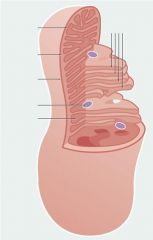
Name the structures:
|
Good job bitch.
|
|
|
|
What is the pore forming protein in mitochondria?
|
Porin, allows for free passage of molecules.
|
|
|
|
What is the purpose of the cristae?
|
For the purpose of increasing surface area of the inner membrane within the mitochondria
|
You know this.....
|
|
|
What does the matrix and inner membrane of the mitochondria contain?
|
The matrix contains enzymes involved in the oxidation of fatty acids and the Krebs cycle.
The inner membrane contains the cytochroms, which are carrier molecules of the electron transport chain, and the enzymes involved in ATP production. |
|
|
|
Are glycogen rosettes and other similar cellular inclusions membrane bound?
|
Often times they are not membrane bound, simply agregated.
|
|
|
|
What do ganglion cells store?
|
brown pigment granules representing lipid-containing residues of lysosomal digestion
|
|
|
|
Describe microfilaments:
|
extremely fine strands of actin protein. Two strings of beadlike subunits twisted together.
|
|
|
|
Intermediate filaments:
|
purely structural function and consist of filaments of protein which self-assemble into larger filaments and bind intracellular structures to each other and to plasma membrane proteins
|
|
|
|
Describe microtubules:
|
are the largest, and are made up of globular protein subunits which can readily be assembled and disassembled. Microtubules originate from a microtubule organizing center called the centriole, found in the centrosome
|
|
|
|
What does the cytoskeleton provide for the cell?
|
*Structural support for the plasma membrane, cellular organelles and some cytosol enzyme systems.
*A means for movement of intracellular organelles, the plasma membrane, and other cytosol constituents necessary for the routine functions of the cell and for cell division. *The locomotor mechanism for amoeboid movements and specialized motile structures such as cilia and flagella. *It is responsible for the property of contractility in muscles. |
|
|
|
What cytoskeletal structure may have striations?
|
Microfilaments of actin can have striations
|
|
|
|
What cytoskeletal structure is responsible for cell motility, intracellular transport, and cell matrix adhesion?
|
associated with a variety of microfilaments (~6-8 nm), e.g. actin and myosin
|
|
|
|
What cell types are characteristic of cytokeratin intermediate filaments?
|
characteristic of epithelial cells where they form a supporting network within the cytoplasm and are anchored to the plasma membrane at intercellular junctions. Certain types of blistering diseases result in a defect in the intermediate filament, the cytokeratins. The epithelial sheet no longer maintains its integrity and a blister forms.
pemphigoid class |
|
|
|
Cytokeratin is the protein for intermediate filaments in epethelial cells. What is the characteristic protein for IF in connective tissues, muscle cells, and nerve cells?
|
Vimentin: is found in cells of mesodermal origin (connective tissues)
Desmin: in muscle cells Neurofilament proteins: in nerve cells |
|
|
|
What cytoskeletal element is important in mitosis?
|
Microtubules are important in mitosis, when the cell divides in forming the spindle apparatus.
|
|
|
|
What cytoskeletal element creates the spindal apparatus?
|
Microtubules
|
|
|
|
What are centrioles used for?
|
are involved in the organization and orientation of microtubules, cilia and flagella
|
|
|
|
In what arrangement are centrioles made?
|
Are recognized by nine triplets of short microtubules that are arranged in a cylinder
|
|
|
|
In a cell the produces a large amount of secretory proteins, what characteristics would be present?
|
Large rER
Many Mitochondria Larger Nucleus Many secretory vessicles |
|
|
|
What goes down in 'Club Nucleoli'?
|
____________________ are dense structures which are the sites of ribosomal RNA synthesis and ribosome assembly. Ribosomal RNA and proteins, which are imported from the cytoplasm, are assembled into subunits which are then passed back to the cytoplasm and aggregate into complete ribosomes
|
|
|
|
What does amphipathic mean?
|
Molecule has a hydrophilic and hydrophobic region
|
|
|
|
How is the fluidity of a cell membrane controlled?
|
By varying the level of cholesterol
|
|
|
|
What is an intrinsic protein? What is an extrinsic protein?
|
Intrinsic - Within the cell membrane
Extrinsic - Transmembrane |
|
|
|
What is the glycocalyx?
|
Glycoproteins and glycolipids project from the surface of the bilayer, forming the glycocalyx. This glycocalyx is involved in cell-recognition, in the formation of intercellular adhesions, and in the adsorption of molecules to the cell surface.
|
|
|
|
What are the three types of transport across a membrane?
|
Passive Diffusion
Facilitated Diffusion Active Transport Bulk Transport |
|
|
|
Describe passive diffusion:
|
dependent on a concentration gradient
|
|
|
|
Describe facilitated diffusion:
|
also concentration dependent, and involves the movement of larger hydrophilic molecules with a protein carrier molecule that binds reversibly
|
|
|
|
Describe Active transport:
|
dependent on concentration gradients, and often operates against them by converting ATP to ADP
|
|
|
|
Describe bulk transport:
|
transport of large molecules or small particles by endocytosis (phagocytosis and receptor mediated endocytosis)
|
|
|
|
What are the (5) main functions of the plasma membrane?
|
*To maintain the structural integrity of the cell
*To act as a selective barrier that regulates the passage molecules in and out of the cell *To facilitate the transport of specific molecules *To recognize specific signaling molecules in the external environment *To regulate cell-cell and cell-matrix interactions |
|
|
|
Name some continuously dividing cells in the human body:
|
epithelium
male sperm cells stem cells for blood |
|
|
|
What is terminally differentiated cell?
|
once they reach a certain stage, they will no longer divide
|
|
|
|
What is the facultative divider?
|
Period in which cell growth appears to be stagnant (almost terminally differentiated), however, can go back info the cycle if needed.
liver cells can regenerate, or when wounded, blood vessels form (angiogenesis) |
|
|
|
What is resolution?
|
The capacity of an optical system to reveal detail in a specimen
|
|