![]()
![]()
![]()
Use LEFT and RIGHT arrow keys to navigate between flashcards;
Use UP and DOWN arrow keys to flip the card;
H to show hint;
A reads text to speech;
65 Cards in this Set
- Front
- Back
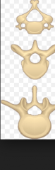
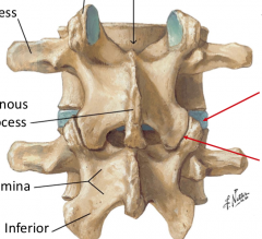
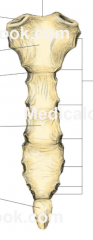
vertebrae number and characteristics |
C7: 1 is atlas 2 is axis. high herniations in C due to high movement. split spinous process. transverse foramen for vertebral artery. T12: heart body, circle foramen. has costal facets for ribs. long spinous process. L5: large kidney shaped body with triangle foramen. built for support. high rate of herniation at l4/l5 and L5/S1 bc of high weight. S1 C1 |
|
|
spinal curves |
Thoracic and saccryl are primary from fetal position. exaggeration is kyphosis. cervical and lumbar are secondary from lifting head and crawling/standing upright. exaggeration in lordosis. scoliosis is curved spine in coronal/frontal plane. |
|
|
vertebral ligaments and epidural/spinal tap importance |
anterior longitudinal ligament posterior longitudinal ligament ligamentus flavum intervertebral ligament supra vertebral ligament. Epidural: find illiac crest-even with L4. insert needle below L4 and inject into epidural space (above dura mater) Spinal tap: find illiac crest-insert needle between l4/l5 (to avoid SC which ends at L2/L3), will insert into subarachnoid space to access CSF. ligaments: thru supravertebral, thru intervertebral, POP thru ligamentous flavum. enter epidural space |
|
vertebral joints |
intervertebral joints: hold weight Symphysis joints Facet joints: prevent side to side movement (in C vertebrae these are almost vertical) |
|
membranes of SC |
Dura mater; tough outer membrane arachnoid mater. is continuous with cerebral dural sac pia mater: inner membrane -filum terminale: extends beyond SC into cauda equina and anchors spinal cord - denticulate ligaments; enlargment of pia where it connects to arachnoid and dura CSF is in subarachnoid space |
|
|
blood supply to vertebrae and spinal cord |
-anterior and posterior spinal arteries branch from vertebral artery. -internal and external venous plexuses provide drainage for SC. -also provide pathway for metastisis to spread from pelvis to skill. |
|
|
Spinibifida |
vertebral lamina dont fuse. presents as lack of spinous process in lower lumbar and sacral. |
|
|
spinal cord ischemia |
blood supply is interrupted and can cause perasis (weakness) and paralysis. |
|
|
spindilytis |
swelling of vertebrae. activates osteoblasts and causes calcification of anterior longitudinal ligament and sacroiliac joint. presents as ankylosis-joint stiffness |
|
|
spondylothesis |
decapitated scotty dog; pedicle fracture that has slipped. |
|
|
spondylolysis |
scotty dog with collar. pedicle fracture. |
|
|
semispinalis capitus enervated by dorsal rami of SN rotate head to opposite side, extend neck |
|
and related |
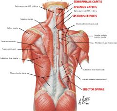
splenius capitus and splenius cervicis N: dorsal rami of SN: laterally bend and extend neck. rotate to same side |
|
|
trapezius
N: CN 11 (accessory n)
elevates, retracts and rotates depress scapula
Test nerve for damage by asking or to elevate shoulders against resistance |
|
G |
latissimus dorsi C6-8 (thoracodorsal n) extend, adduct, medial rotate at shoulder joint |
|
pin plus related |
rhomboid major-pinned rhomboid minor-above levetor scapulae-above minor C5-dorsal scapular nerve elevate rotate scapula |
|
|
serratus posterior superior Intercostal 2-5 accessory inspiration muscle |
|
|
serratus posterior inferior T9-12 accessory inspiration muscle |
|
|
infraspinatus
Suprascapular n C5-6
laterally rotates arm |
|
|
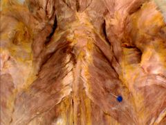
erector spinae: illiocostalis longisimis spinalis dorsal rami of SNs postoral muscle; extends back **most common complaint of back pain due to strain from overextension and rotation |
|

lines above H |
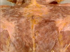
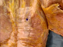
thoracolumbar fascia |
|
|
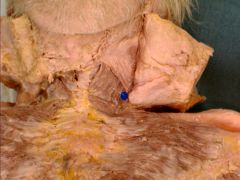
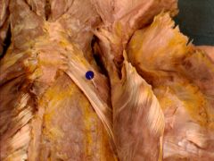
triangle of auscultation |
easiest to hear lung sounds. borders are medial border of scapula, dorsal border of lats, lateral border of traps |
|
|
Peripheral nerves numbers for each vertebral level and other level |
CN-12 SN-31 C8 T12 L5 S5 C1 |
|
|
where do SN exit and how does that affect herniations |
cervical nerves exit above their vertebrae all else exit below herniation between C or L vertebrae will damage lower nerve (C4/C5-damage c5 nerve) T herniation will damage higher nerve (T4/T5-T4 nerve damaged |
|
|
neuron types for somatic sensory somatic motor visceral sensory visceral motor |
sensory are pseudo unipolar motor are multipolar |
|
|
common dermatomes |
C6-thumb c7; middle and pointer c8 pinky and ring t4; nipples t10 umbilicus L1-4; anterior medial thigh S2-4 peritoneum |
|
|
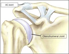
pectoral girdle attachments |
4 joints: sternoclavicular joint: only bony attachment for girdle. stabilized by sternoclavicular ligament acromioclavicular joint: stabilized by coracoacromial ligament scapulothoracic (17 muscles attach to scapula) glenohumoral: sit bump: posterior support (supraspinatous, infraspinatous, teres minor on greater tubercle). subscapularous anteriorly to lesser tubercle |
|
|
clavicle fracture |
most fractured long bone. Medial portion goes up, lateral goes down. worry about damage to brachial plexus |
|
|
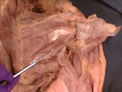
rotator cuff injuries |
overuse injuries.
Supraspinatus-must likely to get injured Infraspinatus Teres minor
Subscapularis
caused by repeated flexion and abduction. tear to ligament from rubbing on accordion and coracoacromial ligament. |
|
|
muscles of pectoral girdle. location and basic innervation |
most are dorsal but innervated by ventral rami because the muscles belong to pectoral girdle not the back. C5-T1 |
|

|
pectorals major
C5-6- 7lateral pectoral n C8-t1 medial pectoral
flexion, abduction, medial rotation at shoulder joint |
|
|
deltoid C5-6: axillary nerve anterior: flexion posterior: extension middle: abduction at shoulder |
|
|
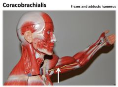
corocobrachialis musculocutaneous nerve (C5-8)- pierced by it! flexion (C5-7) adduction (C6-8) of shoulder **PRACTICAL HINT: musculocutaneous nerve pierces corocobrachiallis. also origin is on COROCOid process |
|
|
supraspinatus suprascapular n C5-6 abduction of shoulder also stabilizes glenohumoral joint in SIT bump. |
|
|

teres major lower subscapular nerve (C5-6) adduction and medial rotation of shoulder joint |
|
|
subscapularis
lower and upper sub scapular nerve (C5-6)
medial rotation at shoulder also stabalizes glenohumoral joint with SIT bump but on lesser tubercle |
|
|
teres minor axillary n C5-6 lateral rotation and stabilizes glenohumoral at sit bump |
|
|
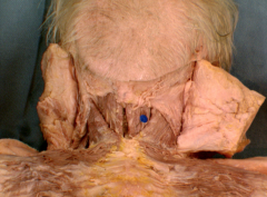
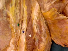
1. intervertebral disk 2 vertebral body 3. dura mater 4 epidural space 5 spinal cord with arachnoid mater 6. epidural space |
|
|
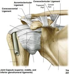
name acromioclaviclar ligament coracoclavicular ligament coracoid process acromion corocoacromion |
|
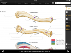
5 things plus bone |
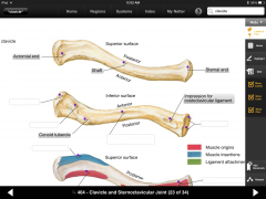
clavicle: acromial end conoid tubercle impression costoclavicular lig shaft body sternal end |
|
|
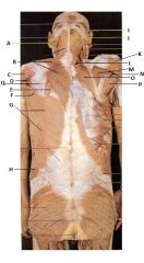
structure and function of mammary gland -nipple location -composition of breast -what does breast sit on/ whats between it |
nipple is over 4th intercostal space in undeveloped breast and men -composed of; -mammary gland has 15-20 lobes-lobules are modified sweat glands -suspensory ligaments (coopers) support breast -lactiferous ducts lead from lobules to nipple (15-20 openings on nipple) -fat tissue -breast sits on deep pectoral fascia; allowing some movement over pect. retromammary space between breast and facia |
|
|
Pregnancy changes to breast |
due to increase in placental estrogen and progesterone darkening of areola hyperplasia of lactiferous ducts and mammary gland lobules secretion of lubricant by areolar sebaceous glands |
|
|
premenstral breast swelling and tenderness menopause relation |
caused by estrogen cycles which increase hydration of connective tissue decreased estrogen cycling in post menopause causes regression of secretary apparatus which is replaced by fatty tissue. collogen decreases |
|
|
breast innervation |
cutaneous branches of intercostal n 4-6 |
|
|
artery supply and venous drainage to breast |
arterial supply: thoracoacromial lateral thoracic internal thoracic posterior intercostal artery; comes out at all vertebral levels of breast. venous drainage; axillary vein internal thoracic vein |
|
|
axillary and breast lymph system |
axillary group: drains into subclavian lymphatic trunk. nodes: humeral (lateral); primary upper limb central (near 3rd section of arterial artery) subscapular (posterior) pectoral (anterior): 75% of breast lymph apical: near first section of axillary artery breast: initial lymph drainage to subareolar lymphatic plexus -75% pectoral (anterior) -->central-->apical-->main lymphatic trunk-->venous system -rest to parasternal lymph nodes -sometimes to abdominal or opposite breast |
|
|
breast cancer |
typically adenocarcinomas from epithelial cells of lactiferous ducts of mammary gland. typically spread through lymph system; pectoral (anterior) node brings cancer cell in to be destroyed. more cancer cells, lymph swells -pectoral node swelling is early indication of metastatic breast cancer |
|
|
peau d orange |
orange coloration of breast tissue during inflammatory breast cancer. as tissue pushed out from tumor, suspensory (coopers) ligaments still attached so you get dimpling of skin. |
|
|
contents of axilla |
brachial plexus axillary artery axillary vein axillary lymph nodes |
|
|
clavipectoral fascia |
deep fascia allows muscles to slide over each other over pect minor |
|
|
axillary artery |

subscapular: forms anastomosis in scapula with axillary artery. -has circumflex scapular artery in triangular space -thoracodorsal; really long and travels with thoracodorsal nerve posterior circumplex humoral is in quadrangular space. -forms anastomosis with anterior circumflex humoral anastomosis allows for collateral flow |
|
|
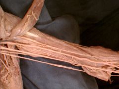
compartments of upper arm-whats in them. what separates them and functions |
separated by inter muscular septum; deep fascia between anterior and posterior portions anterior: elbow flexors: musculocutaneous n (c5-8). (pierces corocobrachialis, under biceps) -biceps -brachiallis -corocobrachialis posterior; elbox extensors; radial n -anconeous -triceps: medial, long and lateral head |
|
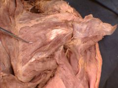
blue line |
anconeous elbow extensor innervated by radial n |
|
|
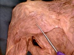
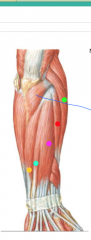
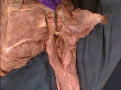
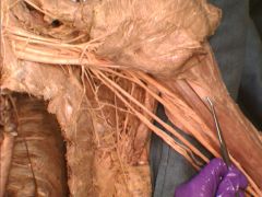
quadrangular space: long head and lateral head of triceps, teres major, teres minor axillary nerve comes through posterior circumflex humoral artery picture of nerve |
|
|
triangular space teres minor, teres major, long head of triceps circumflex scapular artery |
|

|
circumflex scapular artery. from anterior side; located in triangular space between teres minor teres major and long head of triceps |
|
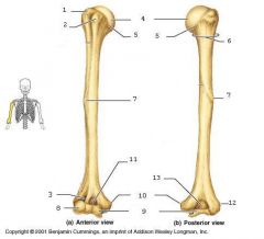
X3, 5,11, -space between1 and 2 -just behind 10 |
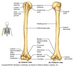
humerus -intertubercular sulcus -groove for ulnar nerve |
|
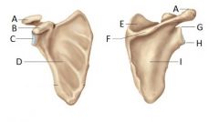
*not shown; dip next to B on inside |
A: acromion B: coracoid process C; glenoid cavity D; subscapular fossa E; F: spine G; supraglenoid tubercle H; infraglenoid tubercle I; infraspinous fossa ** suprascapular notch: where superior transverse scapular ligament crosses |
|
and related |
Biceps brachia-long head -short head is just medial innervated by musculocutaneous from deep side flexes elbow and strongest supinator |
|

|
bicipital aponeurosis |
|

|
brachialis innervated by musculocutaneous our strongest elbow flexor |
|

|
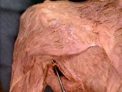
serratus anterior
innervated by long thoracic n. (runs with lateral thoracic artery)
holds scapula to thoracic wall. attaches to ribs 1-9 and scapula
***winged scapula if damage to long thoracic nerve. can be seen if dorsal pressure applied while flex at shoulder |
|
|
where is subclavius |
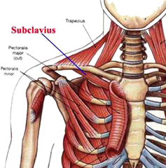
innervated by subclavian nerve (C5-6) depresses shoulder |
|

|
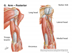
triceps and annconeous extensors of elbow. innervated by radial n |
|
|
|