Reading...
![]()
Play button
![]()
Play button
![]()
Use LEFT and RIGHT arrow keys to navigate between flashcards;
Use UP and DOWN arrow keys to flip the card;
H to show hint;
A reads text to speech;
50 Cards in this Set
- Front
- Back
|
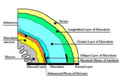
4 GI tract layers
|
Page 956
* Mucosa - Epithelium Lamina propria Muscularis mucosae * Submucosa * Muscularis externa Inter circular layer outer longitudinal layer * Serosa areolar mesothelium |
|
|
saliva composition
|
P961
99.5 Water Mucus amylase lipase lysozyme immunogolbulin A electrolytes PH 6.8-7.0 |
|
|
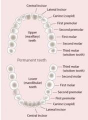
dental formula
|
Page 960
2132 = 32 Teeth R & L quad 2 Incisor 1 Canine 2 PreMolar 3 Molar |
|
|
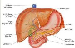
liver function
|
Page 975 &
Page 1021 - T26.6 Metabolism - Carbs, Protiens, lipids Synthesis - Cholesteroil - 1g per day Bile albumin - transports * breaks down -hormones ammonia toxic sub Drugs Storage - Fe, Cu,Gy lycogen,Vitamins |
|
|
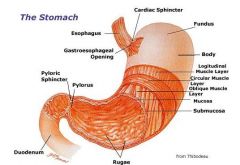
stomach function
|
Page 965
The stomach lies between the oesophagus (food and liquid throat tube) and the duodenum (the first part of the small intestine). It is on the left side of the abdominal cavity. The top of the stomach lies against the diaphragm. Lying beneath the stomach is the pancreas, and the greater omentum which hangs from the greater curvature. Two smooth muscle valves, or sphincters, keep the contents of the stomach contained. They are the esophageal sphincter (found in the cardiac region) dividing the tract above, and the Pyloric sphincter dividing the stomach from the small intestine. The stomach is surrounded by parasympathetic (stimulant) and orthosympathetic (inhibitor) plexuses (networks of blood vessels and nerves in the anterior gastric, posterior, superior and inferior, celiac and myenteric), which regulate both the secretions activity and the motor (motion) activity of its muscles. In humans, the stomach has a relaxed, empty volume of about 45 ml. It generally expands to hold about 1 litre of food,[5] but can hold as much as 1.5 liters. Used for Stroage and Mixing Controled by Vagus Nerve •Mucous cells: secrete an alkaline mucus that protects the epithelium against shear stress and acid •Parietal cells: secrete hydrochloric acid! •Chief cells: secrete pepsin, a proteolytic enzyme •G cells: secrete the hormone gastrin |
|
|
ANS stomach
|
sympathetic and parasympathetic coming from the vagus
parasympathetic -controls water secretions. Histamine and gastrin acting as neuro transmitters. Kime enters the duodenum and stops secretions. |
|
|
pancreas function
|
Page 978
The pancreas is an integral part of the digestive system. The flow of the digestive system is often altered during the surgical treatment of pancreatic cancer. Therefore it is helpful to review the normal flow of food before reading about surgical treatment. Food is carried from the mouth to the stomach by the esophagus. This tube descends from the mouth and through an opening in the diaphragm. (The diaphragm is a dome shaped muscle that separates the lungs and heart from the abdomen and assists in breathing.) Immediately after passing through the diaphragm's opening, the esophagus empties into the stomach where acids that break down the food are produced. From the stomach, the food flows directly into the first part of the small intestine, called the duodenum. It is here in the duodenum that bile and pancreatic fluids enter the digestive system. Includes enzymes that hydrolasize carbs,protiens,fats,acids secretion of insulin and glucagon |
|
|
urea cycle
|
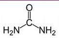
Page 1024
Urea is the major end product of nitrogen metabolism in humans and mammals. Ammonia, the product of oxidative deamination reactions, is toxic in even small amounts and must be removed from the body. The urea cycle or the ornithine cycle describes the conversion reactions of ammonia into urea. Since these reactions occur in the liver, the urea is then transported to the kidneys where it is excreted. |
|
|
bile salts
|
Page 978
Bile acids are steroid acids found predominantly in the bile of mammals. Bile salts are bile acids. An increase in bile flow is exhibited with an increased secretion of bile acids. The main function of bile acid is to facilitate the formation of micelles, which promotes processing of dietary fat. Bile acids are steroid acids found predominantly in the bile of mammals. Bile salts are bile acids conjugated to glycine or taurine. In humans, taurocholic acid and glycocholic acid (derivatives of cholic acid) represent approximately eighty percent of all bile salts. The two major bile acids are cholic acid, and chenodeoxycholic acid. Bile acids, glycine and taurine conjugates, and 7-alpha-dehydroxylated derivatives (deoxycholic acid and lithocholic acid) are all found in human intestinal bile. An increase in bile flow is exhibited with an increased secretion of bile acids. The main function of bile acid is to facilitate the formation of micelles, which promotes processing of dietary fat. Upon eating a meal, the contents of the gallbladder are secreted into the intestine, where bile acids serve the purpose of emulsifying dietary fats. Bile acids serve other functions, including eliminating cholesterol from the body, bile pigments - bilribin phospholipids ions bicarb |
|
|
digestive enzymes
|
Page 969
Stomach - Pepsin, amylase, lipase Small intestine - maltase, surcose, lactase. pancreatic - amylase Enzymes ae protien breakdown and reused. |
|
|
lipid metabolism
|
Page 986
fatty acids, cholesterol bile emulisfies lipids sugar fats amino acids urea cycle F,C,P are burned in Creb Cycle |
|
|
aerobic respiration
|
x
|
|
|
celiac trunk
|
The celiac artery supplies oxygenated blood to the liver, stomach, abdominal esophagus, spleen and the superior half of both the duodenum and the pancreas.
|
|
|
mesenteric arteries
|
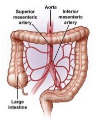
Page 787
Supplies blood to the bowls. Superior mesenteric artery (SMA) arises from the anterior surface of the abdominal aorta, just inferior to the origin of the celiac trunk, and supplies the intestine from the lower part of the duodenum through two-thirds of the transverse colon, as well as the pancreas. Inferior mesenteric artery, often abbreviated as IMA, supplies the large intestine from the left colic (or splenic) flexure to the upper part of the rectum, |
|
|
ANS small intestine
|
Page 955 & 980
Myenteric Plexus (Auerbach) - Parasympathetic & sympathetic Submucosal Plexus (Meissner)- parasympathetic |
|
|
blood function
|
Page 680 & 683 T18.2
O2 Hemiglobin CO2 - Bicarb H2CO3, CO2 Acidic Removes Waste Immunities - Blood cells Coagulation - platelets Messengers - Hormones Ph - 7.4 Temp - 100.4 Hi point of specificity |
|
|
blood structure
|
Page 683
Water - albumins, globulins, fibrinogen Carbs - glucose, amino acids, lipids, fatty acids, triglycerides High denisity lipoprotien - protein Low denisity lipoprotein- mostly fat WASTE - urea, uric acid, ammonia, Bilrubin |
|
|
hematopoiesis
|
page 684
Hematopoietic stem cells (HSCs) reside in the marrow and have the unique ability to give rise to all of the different mature blood cell types. HSCs are self renewing: when they proliferate, at least some of their daughter cells remain as HSCs, so the pool of stem cells does not become depleted. Formation of a blood cell in bone marrow, all come from one stem cell. All go thru a blast stage. reticulocyte is an inmature RBC |
|
|
differential
|
Page 690
|
|
|
antibodies
|
x
|
|
|
WBC function
|
x
|
|
|
clot formation
|
x
|
|
|
RBC function
|
x
|
|
|
H2CO3
|
x
|
|
|
ph
|
x
|
|
|
blood supply brain
|
x
|
|
|
blood distribution
|
x
|
|
|
levels of organization
|
x
|
|
|
macromolecules
|
x
|
|
|
hydrolysis/dehydration
|
x
|
|
|
nitrogenous wastes
|
x
|
|
|
capillary function
|
x
|
|
|
cardiac structures
|
x
|
|
|
cardiac valves
|
x
|
|
|
blood proteins
|
x
|
|
|
blood lipids
|
x
|
|
|
hemoglobin
|
x
|
|
|
blood electrolytes
|
x
|
|
|
AV/SA nodes
|
x
|
|
|
ABO blood groups
|
x
|
|
|
cardiac ANS
|
x
|
|
|
baro/chemo receptors
|
x
|
|
|
cardiomyopathy
|
x
|
|
|
anemia
|
x
|
|
|
vagus nerve
|
x
|
|
|
Kupffer cells
|
x
|
|
|
hepatic sinusoids
|
x
|
|
|
appendix
|
x
|
|
|
digestive processes
|
x
|
|
|
EKG
|
x
|