Reading...
![]()
Play button
![]()
Play button
![]()
Use LEFT and RIGHT arrow keys to navigate between flashcards;
Use UP and DOWN arrow keys to flip the card;
H to show hint;
A reads text to speech;
86 Cards in this Set
- Front
- Back
|
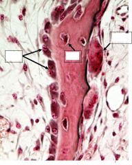
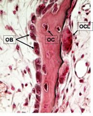
Osteoblasts (OB) are Cuboidal to columnar bone building cells
Deposit ___ (clear region between cells), which then becomes ___ . |
-osteoid
-calcified bone |
|
|
Osteocytes (OC)
|
Mature bone maintaining cell trapped in lacunae
|
|
|
Osteoclasts (OCL)
|
Large multinucleated cells responsible for bone resorption
|
|
|
Calcium phosphate homeostasis is regulated by 3 hormones:
|
parathyroid hormone (PTH)
calcitonin calcitriol (vitamin D) |
|
|
Parathyroid hormone (PTH) does what? How?
|
-increases bone resorption, increases serum Ca+ levels
-Binds to receptors on osteoblasts. Osteoblasts then secrete a protein (osteoclast-stimulating factor/RANKL), which binds to a receptor on osteoclasts/RANK, causing osteoclasts to resorb bone and thereby increase serum calcium concentration -PTH inhibits OPG production; (OPG inhibits maturation of osteoclasts) -PTH can, when given as a drug (Forteo, recombinant human PTH), act to build bond as a treatment for osteoporosis – this is counterintuitive and is not well understood by researchers. -Can also cause osteocytes to resorb bone (osteocytic osteolysis) |
|
|
Calcitonin does what? Where?
|
-inhibits osteoclasts --> decreased bone resorption --> inhibits Calcium release from bone --> decreases serum Ca+
-from the parafollicular cells of the thyroid |
|
|
Vitamin D :
|
-Increases Ca++ and phosphate absorption in the gut
-increases bone resorption, taking Ca++ and phosphate from old bone in order to mineralize new bone -reduced excretion of Ca++ |
|
|
-In osteoporosis, bones ___
-Who is most at risk? -Prevention & treatment should include: |
-lose mass (they lose organic matrix and the mineral component of bone)
-Postmenopausal women -Weight bearing exercise Vitamin D Calcium intake early in life and throughout life Vitamin K from green leafy vegetables |
|
|
-Estrogen maintains ___
-it inhibits ___ -Where is estrogen produced in men? |
-bone density in both sexes
-bone resorption -testes and adrenals |
|
|
There is a rapid loss of bone density under what two conditions:
|
-after menopause
-during periods of disuse during immobilizaton |
|
|
Bone, a specialized ___ tissue characterized by a ___ extracellular matrix
|
-connective
-mineralized |
|
|
The predominant structural protein of bone
|
Type I collagen
|
|
|
Both collagen and the ground substance becomes mineralized with calcium phosphate in the form of
|
hydroxyapatite crystals (Ca10 (PO4)OH 2 min
|
|
|
vitamin K dependent Ca binding protein that is 15-20% of the non collagen protein of bone
|
Osteocalcin (also called Gla protein and abrieviated MGP)
|
|
|
Bone morphogenic proteins (BMPs) induce the differentiation of ___ into ___.
Recombinant BMP can be used by surgeons after spinal fusions and repair of bone defects |
-mesenchymal cells
-osteoblasts |
|
|
|
|
|
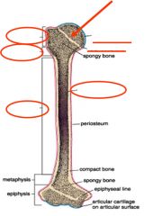
A bone (femur, radius, ulna, etc…) is made up of:
- - - - - - |
Bone tissue
Hemopoieitic tissue Fat tissue Blood vessels Nerves Hyaline cartilage (synovial joints) |
|
|
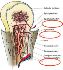
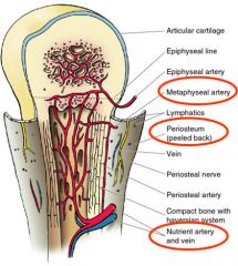
Source of pain from a bone fracture
|
Periosteal nerve
|
|
|
|
|
|
Classification of the following bones:
-vertebrae -skull -carpals -humerus |
-irregular
-flat -short -long |
|
|
What sites are used for marrow transplantation and biopsy?
|
-iliac crest & sternum
|
|
|
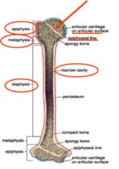
Site of developing blood cells and is found throughout the body during development and growth
|
red bone marrow
|
|
|
In the adult most of the red marrow is replace by ___ (except in the ___)
|
-yellow (fatty) marrow
-sternum and iliac crest |
|
|
if the need arises (after blood loss) what can convert to red blood cell producing marrow?
|
-yellow (fatty) marrow
|
|
|
Types of bone tissue:
-Compact (___) -Spongy (___, ___) |
-cortical
-cancellous, trabecular |
|
|
Under the microscope,
Both compact bone and the thicker trabeculae of spongy bone have ____ |
Haversian systems
|
|
|
Concentric lamellae and the Haversion canal constitute the
|
Osteon
|
|
|
Volkmann’s canals link
|
Haversion canals
|
|
|
interstitial lamellae are the result of ___ and lie between complete osteons
|
bone remodeling
|
|
|
|
|
|
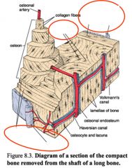
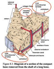
Periosteum: covers external surfaces of bones; has two parts:
(1) an outer fibrous layer of ___ (2) an inner cellular layer of ___. |
-dense C.T.
-osteoprogenitor (The cells of the inner layer can differentiate into osteoblasts.) |
|
|
Endosteum: lines internal surfaces; thin layer of ___
|
osteoprogenitor cells
|
|
|
Collagen fibers of the periosteum run ___ to the surface of a bone
|
parallel
|
|
|
Sharpey’s fibers are ___ fibers found at the site of a ___ into bone. These fibers, which run through the periosteum, are continuous with the ___ of bone tissue
|
-collagen
-tendon insertion -collagen fibers |
|

Gound section or EM?
|
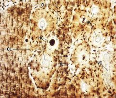
Ground section
O – osteon HC – Haversion canal VC – Volkman’s canal CL – circumferential lamellae IL – interstitial lamellae |
|
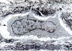
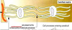
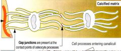
What is it? Where is it? What is it doing?
|
Osteocyte in lacunae
Osteocytes extend cytoplasmic processes into canaliculi can be visualized in the electron micrograph |
|
|
Lacunae -
|
a cavity, space, or depression, especially in a bone, containing cartilage or bone cells.
|
|
|
|
|
|
Haversian systems (osteons) consist of:
___ made of collagen fibers and bone matrix, parallel collagen fibers |
-Concentric lamellae
|
|
|
Haversian systems (osteons):
___ containing osteocytes located between or in the lamellae |
-Lacunae
|
|
|
Haversian systems (osteons):
___ are small canals allowing the extended cytoplasmic processes of an osteocytes trapped in lacunae to make contact with other osteocytes and communicate via ___ |
-Canaliculi
-gap junctions |
|
|
Haversian systems (osteons): The center of a Haversian system is an ___-lined canal containing blood vessels, nerves and loose connective tissue
|
endosteum
|
|
|
Haversian systems run ___ to the long axis of the bone. They are not restricted to compact bone and can also be observed in the thicker trabeculae of ___.
|
-parallel
-spongy bone |
|
|
What provides communication between haversian systems and marrow cavity? They run ___ to Haversian systems
|
-Volkmann's canals
-perpendicular |
|
|
Outer circumferential lamellae: located under
|
periosteum
|
|
|
Inner circumferential lamellae: located around
|
marrow cavity
|
|
|
Interstitial lamellae: remnants of remodeled
|
Haversian systems
|
|
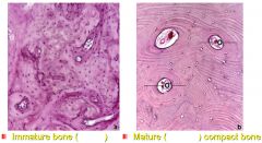
Decalcified H & E Bone Section
Compare and contrast: -# of cells/unit area -matrix layered or not |
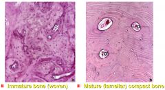
Immature bone (woven)
More cells Matrix is not layered (osteons) v.s. Mature (lamellar) compact bone Fewer osteocytes per unit area Osteons with concentric lamellae Interstitial lamellae |
|
|
In adults, ___ is found in the tooth sockets, near the sutures of the flat bones of the skull and at the insertions of some tendons.
|
primary bone
|
|
|
Two stages of bone formation
|
primary and secondary
|
|
|
Primary (___) bone:
Immature bone. characterized by an irregular array of ___ fibers, a low mineral content and a greater number of ___. |
-woven
-collagen -osteocytes |
|
|
Secondary (lamellar) bone:
More typical of ___. Collagen fibers are numerous and organized into parallel or concentric ___ around ___ |
-adults
-lamellae -blood vessels |
|
|
pink = bone
clear = osteoid |
|
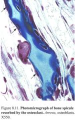
-Arrows
-Light blue -Dark blue Multinucleated osteoclast on the resorption side of this trabecula has a ___ border, depression in bone is called ___; clear area surrounding this is rich in actin filaments for attachment to bone matrix |

Special Stain: Mallery-Azan
Arrows are pointing to inactive bone lining cells/osteoprogenitor cells (flat/thin morphology) Light blue is calcified cartilage and dark blue is bone Multinucleated osteoclast on the resorption side of this trabecula has a ruffled border, depression in bone is called Howship’s lacunae clear area surrounding this is rich in actin filaments for attachment to bone matrix |
|
|
Osteoprogenitor cells:
have a ___ cytoplasm, have a ___-like appearance and give rise to ___ |
-basophilic
-fibroblast -osteoblasts |
|
|
Osteoblasts:
secrete ___ (organic matrix); they are located at ___; they are ___ or columnar shaped cells |
-osteoid
-bone surfaces -cuboidal |
|
|
Osteoclasts:
derived from the fusion of blood-derived ___ , thus creating large ___ cells |
-monocytes
-multinucleated |
|
|
Osteoclasts:
resorb bone matrix and in the process create depressions in the bone surface termed ___ . They are characterized by a ___ border at the bone resorbing end of the cell. |
-Howship's lacunae
-ruffled |
|
|
Osteoclasts:
Around the ruffled border is a region devoid of organelles, but rich in actin microfilaments. This ___ allows attachment of the osteoclast to the ___ and creates a microenvironment into which the cell secretes: |
-clear zone
-bone matrix -H+ (via an H+ ATPase) & Cl-, which dissolves inorganic matrix Lysosomal enzymes (collagenase, gelatinase, metalloproteinase) degrade organic matrix (collagen & other proteins). |
|
|
Bone Matrix
Organic (osteoid): collagen ___ fibers,___ and some ___ (sialoprotein and osteocalcin) These proteins play a role in ___ |
- type I
- proteoglycans -glycoproteins -binding Ca+ |
|
|
Bone Matrix
Inorganic: calcium and phosphorus in the form of ___ and amorphous ___ |
-hydroxyapatite crystals
-calcium phosphate |
|
|
Osteomalacia - ___ impaired; (soft bones) due to ___ deficiency, which causes deficient ___ absorption in the gut.
|
-mineralization of bone matrix
-vitamin D -calcium & phosphorus |
|
|
In children/growing bone vitamin D deficiency leads to
|
Rickets
|
|
|
Osteogenesis imperfecta, “Brittle Bone Disease”
Genetic disorder of ___ collagen characterized by repeated ___ after minor trauma, thin skin, weak tendons, and in certain subtypes blue sclerae |
-type I
-fractures |
|
|
Osteoporosis - decreased ___ - frequent problem of post-menopausal women. However, the___ in osteoporosis is normal. Can also occur in patients who are confined to bed for long periods. The bone is characterized by ___ than normal bone.
|
-bone mass
-ratio of minerals to organic matrix -spicules that are thinner |
|
|
Vitamin C deficiency
Poor bone growth and fracture repair due to impaired ___ synthesis |
collagen
|
|
|
There are two distinct ways in which bones can develop:
|
-Intramembranous ossification
-Endochondral ossification (within cartilage) |
|
|
Intramembranous Ossification
ECT – embyronic connective tissue ___ differentiate into osteoblasts. This site of osteoblast formation becomes a ___ center |
-mesenchymal cells
-primary ossification |
|
|
Intramembranous Ossification
Osteoblast lay down ___, which encapsulates osteoblasts and calcifies. Osteoblasts become ___ located in lacunae. Spicules of ___ form and these grow together forming trabeculae. Compact bone forms outer plates. Inner bone around the developing marrow cavity becomes spongy bone. |
-bone matrix
-osteocytes -woven bone |
|
|
Endochondral ossification (___):
Mode of development of ___ and most short bones |
-within cartilage
- long bones |
|
|
Endochondral ossification
Hyaline cartilage model of bone, forms in the embryo and is ___ by bone during development. |
replaced
(NOTE: Hyaline cartilage does not become bone.) |
|
|
Endochondral ossification consists of two phases:
|
-Hypertrophy and degeneration of chondrocytes
-Osteogenic bud of osteoprogenitor cells and blood vessels penetrates the space left by the dead chondrocytes |
|

|
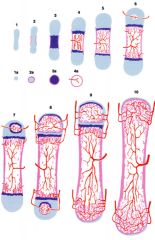
(1) Formation of hyaline cartilage model
Primary ossification center (2) Bone collar formation is the first sign of ossification (3) cartilage matrix in the shaft begins to calcify (4) Blood vessel invasion and connective tissue cell erode the calcified cartilage, creating a primary marrow cavity Osteoblasts secrete matrix on cartilage scaffold Secondary ossification center |
|
|
Bone collar formation, is actually a form of ___, i.e. bone formation within the perichondrium.
|
intramembranous bone formation
|
|
|
Formation of the primary ossification center (pre- or postnatal)?
|
prenatal; Formation of secondary ossification centers is postnatal
|
|
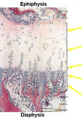
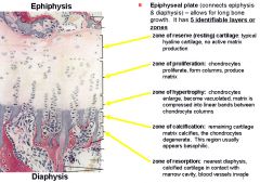
Epiphyseal plate (connects epiphysis & diaphysis) – allows for ___
|
|
|
|
Bone formation is taking place at which surface of the epiphyseal plate?
|
Bone formation is taking place at both the epiphyseal and diaphyseal surface of the epiphyseal plate. Growth rate is much greater on the diaphyseal side
|
|
|
What advances and resorbs bone in a bone remodeling unit?
|
Cutting cone (osteoclasts)
|
|
|
What deposits new bone in a bone remodeling unit?
|
Closing cone (osteoblast)
|
|
|
In bone growth and remodeling does the epiphyseal plate enlarge or decrease in size?
|
epiphyseal plate maintains a constant thickness during growth phase
|
|
|
Growth in width (___)
due to bone formation by osteoprogenitor cells within the inner ___ around the diaphysis. This is actually a form of ___. the marrow cavity increases in diameter due to the resorption of bone from internal surfaces by osteoclasts |
-appositional growth
-periosteum -intramembranous ossification. |
|
|
Pituitary Dwarfism
Deficiency of ___ during the developing years leads to failure of ___. |
-growth hormone (somatotropin)
-bone growth |
|
|
Gigantism
Excess ___ during the developing years (prior to the closure epiphyseal plate) leads to an abnormal increase in the length of bones. |
growth hormone
|
|
|
Acromegaly
Excess ___ in ___ leads to thickening of the bones since the growth (epiphyseal) plates have already closed. |
-growth hormone
-adulthood |
|
|
Fracture Repair (___ weeks):
1. A ___ forms at site of fracture 2. ___ (granulation tissue) of periosteum and endosteum proliferate 3. ___ forms in this connective tissue 4.___ forms at site by both endochondral and intramembranous bone formation 5. ___ (dense CT and cartilage) temporarily unites fracture 6. ___ from the periosteum divide and become ___, they invade the callus and deposit new bone 7. ___ occurs with healing (spongy bone is replaced with compact bone) |
-6-12
1. blood clot 2. connective tissue 3. hyaline cartilage 4. primary bone 5. bone callus 6. Osteoprogenitor cells --> osteoblasts 7. remodeling |
|
|
The interaction of the RANK receptor with RANK ligand (RANKL) is necessary for
|
osteoclast differentiation
|