![]()
![]()
![]()
Use LEFT and RIGHT arrow keys to navigate between flashcards;
Use UP and DOWN arrow keys to flip the card;
H to show hint;
A reads text to speech;
35 Cards in this Set
- Front
- Back
|
describe the greater pelvis |
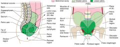
area from the iliac crest to the pelvic floor |
|
|
describe the lesser pelvis |
region of greater pelvis from promentory/pubic symphysis to pelvic floor |
|
|
what are the major boney components of the pelvis
|
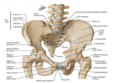
iliac crest iliac fossa/ala pubis ischium greater/lesser sciatic notch anterior superior/inferior iliac spine coccyx obturator foramen promentory-- sacrum/L5 pubic symphysis pectineal line-- arcuate line |
|
|
what are the differences between male and female pelvises |
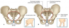
male: heavier build, deeper interior and conical in shape, heart shaped inlet, subpubic angle of 50-60 degrees, sharper coccyx and ischial spines female: interior is cylindrical, circular inlet, subpubic angle of 80-85 degrees |
|
|
describe the linea terminalis |
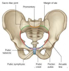
inlet of lesser pelvis pubic crest, pecten pubis (pectineal line), arcuate line, margin of ala, promentory |
|
|
what are the important measurements/distances to know for the pelvis |
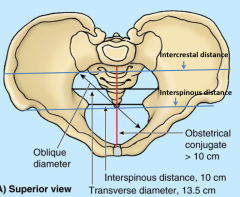
intercrestal distance interspinous distance (iliac spines) interspinous distance (ischius spines) oblique diameter transverse diameter obstetrical conjugate (>10cm) |
|
|
what are the 3 different conjugates and which is the most important |
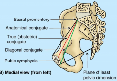
anatomical (superior pubic symphysis to promentory) **true (obstetric) (median pubic symphysis to promentory) baby passes here diagonal (inferior to promentory) can measure here |
|
|
what are the 2 proper orientations for the baby to emerge safely |
face to anus or face up largest/safest diameter for head to move through |
|
|
what are the largest diameters through the birth canal |
inlet: transverse (12cm) cavity: oblique (12cm) outlet: sagittal (9.5cm) like an elbow pipe coccyx can shift backwards to allow head through-- 12cm |
|
|
what are the major ligaments of the pelvis |
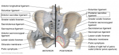
connecting sacrum to pelvis iliolumbar ligament anterior/posterior sacro iliac ligaments other ligaments (sacrotuberous/sacrospinous etc) create greater/lesser sciatic foramen like a suspension bridge |
|
|
describe the obturator foramen |
almost completely filled by obturator membrane which gives attachment to muscles obturator nerve passes through |
|
|
describe the greater sciatic foramen |
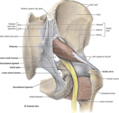
penetrated by piriformis muscle creating supra and infra piriform hiatus sciatic nerve runs through |
|
|
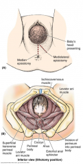
describe the floor of the pelvis |
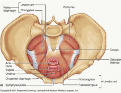
pelvic diaphragm; levator ani muscle and its fascia funnel with a beak (anus), hole in anterior side (vagina/urethra) function: supports pelvic viscera and resists downward thrust from increased abdominal pressure (coughing, defacation, labor, heavy lifting) |
|
|
what are the components of the levator ani muscle |
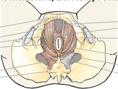
puborectalis- median, pubis to coccyx around anus (external anal sphincter) forms urogenital hiatus anteriorly (left and right crus) central tendon anterior to anus pubococcygeus- medial, pubis around anus to pubis iliococcygeus- lateral, attaches to obturator internus muscle with tendinous arch of levator ani muscle |
|
|
what are the contents of the female pelvis |
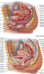
uterus and ovaries (fallopian tubes, round ligament off fundus) rectum bladder vagina 2 pouches: rectouterine and vesicouterine pelvic diaphragm (levator ani and superior/inferior fascia) |
|
|
describe ischiorectal fossa |
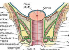
mostly fatty tissue and skin below the pelvic diaphragm triangle in shape, on both sides of vagina/prostate between ischium and rectum |
|
|
describe paracolpium |

above pelvic diaphragm surrounding vagina tissue |
|
|
what are the contents of the male pelvis |
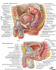
rectum bladder prostate gland (below bladder, surrounds urethra) seminal vesicles ampulla of vas deferens 1 pouch: rectovesicle pelvic diaphragm (levator ani and superior/inferior fascia) |
|
|
describe the urogenital membrane |
provides support for structures against abdominal pressure in the uruogenital hiatus forms external sphincter of urethra |
|
|
describe the uterus |
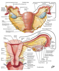
intraperitoneum broad ligament of the uterus-- membrane holds uterus in place, attaches to pelvis (parametrium- close to uterus, caudinal ligament- at base, hold vessels, mesosalpinx- broad ligament above ovary, mesovarium- attaches to ovary) fallopian tubes (isthmus, ampulla, infundibulum, fimbriae) ligament of ovary suspensory ligament of ovary with gonadal artery and vein triangle shaped cavity, isthmus is narrowing, cervix is muscular entrance, protrudes into vaginal canal (creates anterior/posterior pouch) ovary is naked- no peritoneum (capsule) |
|
|
describe the position of the uterus |
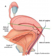
lays almost horizontal on bladder angle between body and cervix angle between cervix and vagina anteflexed and anteverted position |
|
|
describe uterine prolapse |
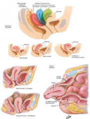
uterus moves down through/out of vagina because of a lack of support from the pelvic floor |
|
|
describe the blood supply to the uterus |
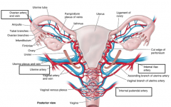
uterine artery to uterus (from internal iliac/hypogastric artery) in floor of broad ligament vaginal artery (from internal pudendal artery [from internal iliac]) ovarian artery is special, from abdominal aorta within suspensory ligament, developed higher and descended |
|
|
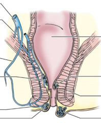
describe the rectum |
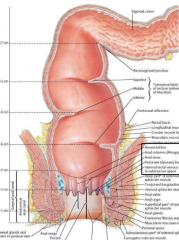
most posterior component of pelvis 5 inches long, begins at middle sacrum and follows sacral curve ends 1 inch beyond coccyx bending backwards external sphincter= levator ani air-tight closure achieved by columns/sinuses |
|
|
what is the difference between the surgical and the anatomical anal canal |
surgical canal includes the anal columns and sinuses anatomical is just below the pectinate line |
|
|
define the ampulla of the anus |
lower dilated part |
|
|
describe the innervation of the pelvis |
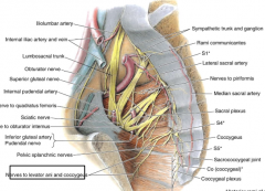
somatic sacral plexus (superior gluteal, inferior gluteal, sciatic, internal pudendal, posterior femoral cutaneous, pudendal nerves) inferior hypogastric plexus- mixed nerves some small sympathetic (off trunk-- sacral splanchnic) and parasympathetic (pelvic splanchnic) |
|
|
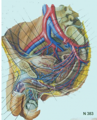
describe the blood supply to the pelvis |
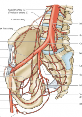
internal iliac artery visceral and parietal branches anterior: superior/inferior vesicle, uterine/prostatic, superior rectal posterior: median sacral, lateral sacral, iliolumbal, superior/inferior gluteal |
|
|
describe the venous drainage of the pelvis |
accompany arteries but with greater variation networks and plexi-- named by region rectal venous plexus etc. |
|
|
describe the blood supply to the rectum |
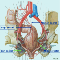
superior rectal artery (from inferior mesenteric) middle rectal artery (from internal iliac) inferior rectal artery (from internal pudendal) median sacral artery (from aorta) |
|
|
describe the venous drainage of the rectum |
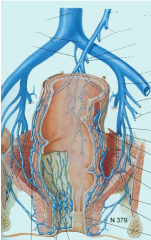
mirrors arteries drains partly to portal system via inferior mesenteric veins partly to caval system via internal iliac, internal pudendal and median sacral inferior rectum drains to caval porto-caval anastomoses (rectal/anal, esophageal, umbilical, retroperitoneal) |
|
|
describe hemorrhoids |
portal hypertension is one cause internal: covered by mucous membrane, painless external: covered by skin, very painful, may prolapse |
|
|
what are classic signs of portal hypertension |
hemorrhoids esophageal bleeding caput medusa (umbilical veins filling, distending) |
|
|
describe the lymphatic drainage of the pelvis |
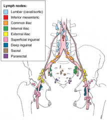
follow veins many lymph nodes named based on location and association skin and superficial fascia to superficial nodes viscera to nodes and lymphatics along deep blood vessels vagina, urethra, fundus of uterus, and anal canal to superficial inguinal nodes |
|
|
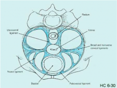
describe endopelvic fascia |
pelvic viscera lateraly enclosed by fibrous, fatty fascia with is packing and anchoring visceral nerves and vessels travel through it thickened sections are named ligaments female: pubovesical, transverse cervical (cardinal), uterosacral |