Reading...
![]()
Play button
![]()
Play button
![]()
Use LEFT and RIGHT arrow keys to navigate between flashcards;
Use UP and DOWN arrow keys to flip the card;
H to show hint;
A reads text to speech;
54 Cards in this Set
- Front
- Back
|
what are the three areas of spinal curves:
1. 2. 3. |
1. cervical lordosis
2. thoracic kyphosis 3. lumbar lordosis |
|
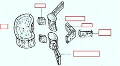
identify the labeled structures:
|
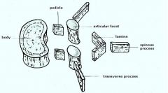
(see figure)
|
|
|
what distinguishes the lumber vertebrae:
1. 2. 3. |
1. larger vertebral body
2. wider and deeper than rest of spine 3. spinous process is quadrangular shaped |
|
|
the lumbar vertebrae have enlarged ... because there are lots of peripheral nerves to ...
|
spinal canal
lower extremities |
|
|
lumbar spineThoracic Facet Orientation
|
slide 7 - NEED QUESTION
|
|
|
... orientation of articular facets contributes to large ROM in ...
|
parasagittal
flexion/extension |
|
|
Superior facet faces ... and is ... to the inferior facet of above vertebra
|
posteromedially
lateral |
|
|
Lumbar spine follows ... Laws of spinal mechanics
Type I curves involve ... restrictors and ... vertebral segments Type II curves involve ... restrictors and ... vertebral segments |
Fryette’s
large several short single |
|
|
what muscles are in the superficial layer of the back:
1. 2. 3. 4. |
1. Trapezius
2. Latissimus dorsi 3. Rhomboid minor 4. Rhomboid major |
|
|
what muscles are in the intermediate layer in the back:
1. 2. 3. 4. 5. |
Erector spinae:
1. Iliocostalis lumborum 2. Longissimus 3. Spinalis 4. Serratus posterior inferior 5. Lumbodorsal fascia |
|
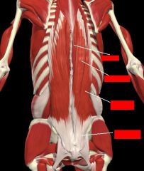
identify the labeled structures:
|
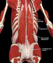
(see figure)
|
|
|
what are the deep layer back muscles:
1. 2. 3. they are involved with what type mechanics: |
1. Semispinalis
2. Multifidus 3. Rotatores a. longus b. brevis type II |
|
|
what are the lumbar muscle connections to the lower extremities:
1. 2. |
1. psoas
2. quadratus lumborum |
|
|
the Lumbosacral aponeurosis connects ... to ...
|
lumbar spine
sacrum |
|
|
Abdominal diaphragm crura anchors to ...
Increased lordosis keeps diaphragm in ... position |
L1-3
inhalation |
|
|
11th and 12th rib lesions can produce spasm of this muscle
|
Lumborum
|
|
|
Quadratus lumborum tension will transfer to ipsilateral
Strain transfers down to ..., ..., and ... |
tensor fascia latae (TFL)
knee lateral leg foot |
|
|
Lesions of 11th and 12th ribs must be considered with ... strain
|
TFL/knee
|
|
|
Erector spinae muscles originate in ... and traverse to the ...
Through ... and ... muscles the lumbars are connected to the anterior abdominal wall |
lumbar spine
head and neck transverse abdominus oblique |
|
|
... connects lumbar spine to upper extremity
... blends with lumbar portion of thoracolumbar fascia |
Latissimus dorsi
Serratus posterior inferior |
|
|
somatic dysfunction at lumbar plexus will effect ..., patient may complain primarily of problems to ... without lumbar pain
|
lower extremity
legs |
|
|
Lumbar spinal canal contents are ... and ...
Frequently lumbar disk herniation will compress ... within spinal canal |
conus medullaris
cauda equina nerve roots |
|
|
Upper lumbar spine involved in ... innervation to ...
Pre ganglionic fibers go to ... |
sympathetic
lower abdominal organs inferior mesenteric ganglion |
|
|
at what spinal cord level does the preganglionic neuron originate for the below listed visceral organs:
1. colon and rectum 2. kidney and ureters 3. urinary bladder 4. testicle and epididymus 5. uterus 6. prostate |
1. T8-L2
2. T10-L1 3. T10-L1 4. T9-T10, L1-L2 5. T10-L1 6. L1-L2 |
|
|
you can can have visceral problems that manifest as ...
and areas feeding into the lower thorax are also going to be involved in ... |
chronic back pain
lumbar mechanics |
|
|
somatic dysfunctions may result from things like:
1. 2. 3. 4. |
1. trauma
2. visceral disease 3. mechanical asymmetries 4. chronic asymmetric motions |
|
|
when doing a structural exam, you are going to look at a sagittal view of:
1. 2. 3. 4. 5. |
1. thoracic kyphosis
2. lumbar lordosis 3. cervical curves 4. sacral curves 5. pelvic tilt |
|
|
how do you find the L4-L5 interspace:
|
place hands on top of iliac crests and thumbs will be level with the interspace
|
|
|
what does the following describe:
1. Long restrictor muscles which cross more than one segment 2. Multiple segments involved in curve 3. Occurs in NEUTRAL position 4. Sidebending/Rotation occur in OPPOSITE directions |
type 1 mechanics
|
|
|
what does the following describe:
1. Short restrictor muscles which cross one joint 2. One segment involved in lesion 3. Occurs in FLEXED or EXTENDED position 4. Sidebending/Rotation occur to SAME side |
Type 2 Mechanics
|
|
|
The ... plays a major role in lumbar curvature
We can use the lower extremity as a lever to help increase or decrease ... through ... |
sacrum
lordosis extension/flexion |
|
|
(extension/flexion) of L-spine increases lumbar lordosis
|
extension
|
|
|
As top of sacrum tilts back,
lumbar lordosis decreases this is relative (extension/flexion) of L-spine |
flexion
|
|
|
Muscle Energy is (direct/indirect) treatment
|
direct
|
|
|
to correct a type 1 lesion using muscle energy what would you do:
L3-5 SRRL |
we sidebend L (and rotate R) while maintaining neutral position
|
|
|
when Correcting a Type I Lesion by laying patient on side, what side do we lay the patient on:
|
the side they sidebend to
|
|
|
how would you correct a type II lesion in the seated position using muscle energy technique:
lesion: L1-3 ESR RR |
for extended lesion we FLEX patient SB to L, Rotate to L
|
|
|
Keeping lower leg in extension will help ... lumbar spine
Bending lower leg will help to ... spine |
extend
flex |
|
|
... degeneration and deformity of the joints of two or more adjacent vertebral bodies
|
Spondylosis
|
|
|
osteophytes is also called ...
|
spinal arthritis
|
|
|
... inflammation of a vertebra
|
Spondylitis
|
|
|
... is primary type of spondylitis. is is ... arthritis primarily effecting spine and SI joints
Young adults (<35 yo) Severe chronic pain |
Ankylosing spondylitis
an autoimmune |
|
|
spondylopathy
|
pathologic condition of the spine
|
|
|
... crack in vertebra due to excessive lordotic force
|
Spondylolysis
|
|
|
... anterior displacement of one vertebrae relative to the one immediately below
|
Spondylolisthesis
|
|
|
... is the “closing in” of spinal canal which compresses nerves
|
Spinal stenosis
|
|
|
a Herniated/Bulging nucleus pulposis (disc)is most concerning when nerve root ...
|
impinged
|
|
|
... = pain originating in peripheral nerve proximal to the area of pain perception. Pain follows along path of nerve
|
Radiculopathy
|
|
|
Sciatica symptoms:
1. 2. 3. |
1. pain radiating down back of leg
2. weakness in ankle dorsiflexors 3. numbness over dorsal foot |
|
|
when do you get an MRI:
1. Radiculopathy- rule out a herniated disc impinging on a nerve root within the neural foramen 2. MRI can help diagnose diseases of the soft tissues 3. diagnosing somatic dysfunction of the low back |
1. yes
2. yes 3. no |
|
|
what is the most common cause of low back pain:
|
somatic dysfunciton
|
|
|
Conditions such as herniated/ bulging discs and arthritis are often:
|
mistakenly blamed for low back pain which is actually from somatic dysfunction
|
|
|
If lower back pain is from somatic dysfunction they will get better with OMM quickly, if they do not reevaluate for ...
|
pathology
|
|
|
What do you do with the patient after you have “fixed” them?
|
Get them moving!
Walking Stretches |