Reading...
![]()
Play button
![]()
Play button
![]()
Use LEFT and RIGHT arrow keys to navigate between flashcards;
Use UP and DOWN arrow keys to flip the card;
H to show hint;
A reads text to speech;
67 Cards in this Set
- Front
- Back
|
The intricate structure of the heart develops during the fist ___ of life
|
eight weeks
|
|
|
From what type of tissue does the heart develop:
|
mesoderm
|
|
|
When does the heart begin to beat:
|
on the 22nd day
|
|
|
Where does the heart start forming:
|
rostral to the oral plate, adjacent to the septum transversum
|
|
|
what happens on day 18 in the development of the heart:
|
small islands of blood and endothelial tissue begin to differentiate out of the mesoderm
|
|
|
When does blood and endothelial tissue begin to differentiate out of the mesoderm:
|
day 18
|
|
|
blood islands unite to form ___ shaped tube when during embryonic development: (what day)
|
horseshoe
days 18-19 |
|
|
what happens in the embryo heart in days 21-22: (x2)
|
Venous end receives blood from yolk sac
(body’s first functional veins) Arteries begin to pump to the head, via early aortic arches |
|
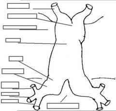
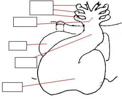
identify the structures:
|
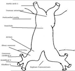
(see figure)
|
|
|
As the heart grows why does it bend into an ‘S’ shape:
|
the heart grows at a faster rate than the pericardial cavity
|
|
|
The heart grows at a faster rate than the pericardial cavity, what is the result:
|
it bends into an ‘S’ shape
|
|
|
what is the name of the bulge in the 'S' shaped heart:
|
bulboventricular loop
|
|
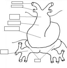
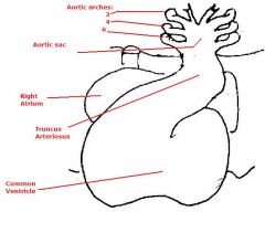
identify the labeled structures:
embryonic heart age: |
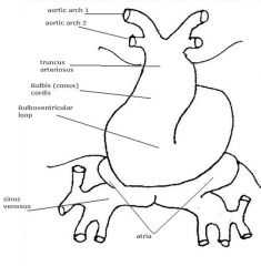
(see figure)
6th week |
|
identify the labeled structures:
age of the embryonic heart: |
(see figure)
8-9 weeks |
|
|
As the heart develops, systemic veins move to drain into the ___ side of heart, systemic arteries move to arise from the ___ side of heart
|
right
left |
|
|
how do the developing embryonic atria fuse and expand:
|
by incorporating veins into atrial walls
|
|
|
The rough-walled part of the atrium comes from ___ while the smooth-walled (sinus venarum) part comes from ___
|
embryonic atrium
venous origin |
|
|
the __ develops in the center of the single atrial and ventricular cavity from ant. to post.
|
endocardial cushion
|
|
|
the ___ initially separates the 2 atria but there is open communication via the ___
|
septum primum
foramen primum |
|
|
during embryonic development, the ___ separates the 2 ventricles and the ___ is the opening between them
|
interventricular septum
interventricular foramen |
|
|
As foramen primum shrinks, a second foramen connecting the two atria forms called ___ and a second septum froms called the ___
|
foramen secondum
septum secondum |
|
|
what happens in the final separation of the atria and ventricles: (x3)
(what's closed/open) |
1. atria leave an opening called the foramen ovale
2. interventricular septum closes 3. truncus arteriosus closes |
|
|
what is the function of the foramen ovale:
|
for blood to go from the IVC to the L. atrium
|
|
|
when the interventricular septum closes the membranous part arise from what:
|
endocardial cushion
|
|
|
the truncus arteriosus closes with a ___, separating the ___ from the ___
|
spiral septum
aorta pulmonary trunk |
|
|
in the fetus, SVC carries (high/low) oxygen blood from where:
IVC carries (high/low) oxygen blood from where: |
low
head and upper limb high umbilical vein |
|
|
how is blood routed through the right atrium from SCV:
IVC: |
SVC blood goes directly through right atrium to right ventricle
IVC blood goes through foramen ovale to left atrium, and hence to left ventricle |
|
|
the two Aorta and Pulmonary trunk separate out from a common truncus by means of a ___
|
spiral septum
|
|
|
the right ventricle is anterior, while the pulmonary trunk has to arise from the ___ of the aortic sac
|
posterior part
|
|
|
the left ventricle is posterior, while its Aorta has to arise from the ___ of the aortic sac
|
anterior part
|
|
|
The final result of the spiral septum is what:
|
aorta and pulmonary trunk that twist around each other
|
|
|
what are the great vessels:
from where do they arise: |
aorta and pulmonary trunk
aortic arches |
|
|
what are the aortic arches that develop in the human embryo:
arches ___ involute early, and leave no major remnant |
1,2,3,4,and 6
1 and 2 |
|
|
what is the function of the umbilical artery:
what is the function of the vitelline artery: |
supply deoxygenated blood from the fetus to the placenta
an artery carrying blood to the yolk sac from the embryo |
|
|
___ that is high in O2 brings blood back from the maternal circulation and continues up to the liver as the ___ where it meets up with the ___ that is low in O2
|
umbilical vein
ductus venosus portal vein |
|
|
the ___ is brining some low O2 content blood from the GI tract up to the liver where it mixes with the high O2 blood from the ___ to form the ___
|
portal vein
ductus venosus inferior vena cava |
|
|
the ___ is brining fairly high O2 content blood to the right atrium, where the majority of the blood with squirt through the ___ into the ___
|
inferior vena cava
foramen ovale left atrium |
|
|
from the left atrium, the blood will go to the ___ and out the ___ and to the rest of the body from there
|
left ventricle
aorta |
|
|
the ___ brings low O2 blood from the body and brings it in to the ___ where it then goes to the ___ and then out the ___
|
superior vena cava
right atrium right ventricle pulmonary trunk |
|
|
the lungs aren't working yet so the blood from the ___ goes through the ___ and mixes with the aortic blood where it feeds all the tissues of the body and then leaves the body through the ___
|
pulmonary trunk
ductus arteriosus umbilical arteries |
|
|
what causes the foramen ovale to close: (4 steps)
|
1. stopping of umbilical circulation
2. (↓)IVC blood flow causing (↓)right atrial pressure 3. blood flows to expanding lungs through pulmonary circulation 4. (↑)blood flow to left ventricle |
|
|
___ and other factors cause smooth muscle in ductus arteriosus and ductus venosus to contract, sealing off these ducts
|
oxygen tension
|
|
|
atrial septal defects occur in the ___ and allow what:
|
interatrial wall
mixing of blood between left and right atria |
|
|
what is a probe-patent foramen ovale:
|
minor atrial septal defect
|
|
|
what is a high atrial septal defect:
|
hole up near the SVC, connecting the two atria
|
|
|
a typical atrial septum defect is caused by either 1 of 2 things:
|
1. too large of a foramen secundum
2. too large a fossa (foramen) ovalis |
|
|
what is the cause of a septum primum defect:
what is the result of this defect: |
failure of septum primum to completely fuse with endocardial cushion
a variably sized hole connecting the two atria |
|
|
This septum defect is frequently found in patients with Down’s syndrome:
|
septum primum defect
|
|
|
what is the cause of the membranous ventricular septal defect:
|
failure of fusion of the interventricular septum with the endocardial cushion
|
|
|
(atrial/ventricular) septum defects are the most common:
|
ventricular
|
|
|
(membranous/muscular) septal defects are less common:
|
muscular
|
|
|
what are truncus defects caused by:
|
failure of the spiral septum of the truncus arteriosus
|
|
|
what are the affected structures in truncus defects: (x3)
|
ascending aorta
pulmonary trunk semilunar valves |
|
|
what is the cause of persistent truncus arteriousus:
what is the result in the newborn: |
failure for truncus to separate at all
no separation of blood flows no aortic or pulmonary valves |
|
|
what is occurring in the defect transposition of great vessels: (x3)
why does this occur: |
right ventricle pumps to aorta
left ventricle pumps to pulmonary trunk no transfer of oxygen to systemic circulation spiral septum is straight and not spiral |
|
|
what is occurring in unequal division:
what does this usually involve so that a baby could survive: |
one vessel has a large opening, the other is small (aorta/pulmonary trunk)
a VSD so rest of right ventricular blood can get into aorta |
|
|
what is occurring in tetralogy of fallot:
|
1. pulmonary stenosis
2. VSD (ventricular septal defect) 3. Over-riding aorta (aorta sits right over the interventricular septum, collecting blood from both ventricles) 4. hypertrophy of right ventricle |
|
|
what is ectopia cordis:
|
failure of sternum to fuse, producing a heart outside of the thoracic cavity
|
|
|
what is dextrocardia:
when is this not a big problem: |
reverse rotation
situs inversus |
|
|
what is persistent ductus arteriosus:
|
pulmonary by-pass to continues functioning
|
|
|
what is occurring in ductus arteriosus: (think pressure)
|
Flow reverses, so blood going from high pressure to low → goes from aorta to pulmonary trunk (with resulting pulmonary hypertension).
|
|
|
what is coarctation of the aorta:
what are the 2 forms: |
inexplicable narrowing of the aorta near the ductus arteriosus
preductal forms and postductal forms |
|
|
what is occurring in coarctation of the aorta: (blood pressure)
|
blood pressure to upper extremities is increased
blood pressure to lower extremities is minimal. |
|
|
how do you detect coarctation of the aorta:
|
compare brachial pulse to femoral pulse in newborn
|
|
|
in coarctation of the aorta:
in which form does ductus arteriosus act to supply low-oxygen blood to lower extremities |
preductal form
|
|
|
in coarctation of the aorta:
in which form does blood from aorta use ductus arteriosus as pressure relief valve: |
postductal forms
|
|
|
what happens if blood from the aorta uses ductus arteriosus as a pressure valve:
|
pulmonary hypertension
|