![]()
![]()
![]()
Use LEFT and RIGHT arrow keys to navigate between flashcards;
Use UP and DOWN arrow keys to flip the card;
H to show hint;
A reads text to speech;
37 Cards in this Set
- Front
- Back
|
Compare & Contrast lower limb rotation with upper limb rotation during development |
Rotation causes the embryological relationship to be opposite that of the upper limb
Dorsal embryological origin = anterior leg = extensors (& lateral rotators and abductors)
Ventral embryological origin = posterior leg = flexors (& medial rotators and adductors) |
|
|
Describe the superficial fascia |
loose connective tissue, fat, superficial veins & nerves. |
|
|
Describe the Deep fascia |
Fascia lata - Deep fascia of the thigh Iliotibial tract - Thickened laterally & reinforced with longitudinal fibers, continuous inferior to the knee Crural fascia - deep fascia of the leg |
|
|
Describe the Great Saphenous vein |
begins in medial aspect (dorsal venous arch), courses anterior to medial malleolus, terminates in the femoral vein. - Most common site for transfusion of blood & injection of fluids in infants. - has 1-way valves to limit retrograde blood flow - saphenous nerve runs with the greater saphenous vein in the lower leg. |
|
|
Describe the femoral triangle |
neurovascular structures enter the anterior thigh deep to the inguinal ligament
The triangle is formed by the inguinal ligament, sartorius, and adductor longus.
It contains NAVEL. Concern: lacunar ligament herniation |
|
|
Describe the popliteal fossa |
Medial boundary: semimembranosus & medial head of gastrocnemius Lateral boundary: biceps femoris & lateral head of gastrocnemius All neurovascular structures traveling from thigh to leg pass through this space. Contents: end of the small saphenous vein, popliteal artery & vein, tibial & common fibular nerve, lymph nodes. |
|
|
Describe the posterior compartment of the Pelvic girdle |
Gluteal region, ABductors & extensors, Deep gluteal are medial rotators.
Innervation: posterior division of the lumbosacral plexus, superior & inferior nerves. |
|
|
Describe the anterior compartment of the pelvic girdle |
Iliopsoas - flexor of the thigh Innervation - posterior division nerves from lumbosacral plexus Femoral triangle
|
|
|
Describe the posterior thigh compartment |
Muscles: hamstrings Function: flexor of the knee Nerve: tibial division of the sciatic nerve Artery: Deep femoral artery |
|
|
Describe the Anterior thigh compartment |
Muscles: Rectis Femoris, Vastus lateralis, Vastus medialis, Vastus intermedius, Sartorius Function: extensor of the knee except for Sartorisu (flexes knee) Nerve: femoral nerve Artery: femoral artery |
|
|
Describe the Medial thigh compartment |
Muscles: Adductor Brevis|Longus|Magnus, Pectineus, Gracilis Function: adduction & medial rotation Nerve: Obturator n Artery: Obturator a. & Deep Femoral a. |
|
|
Describe the Deep Posterior leg compartment |
Muscles: Flexor hallucis longus, Flexor digitorum longus, Tibialis posterior, Popliteus, Plantaris Function:plantar flexion of the foot Nerve: Tibial n Artery: Posterior Tibial a. |
|
|
Describe the Superficial Posterior leg compartment |
Muscles: Gastrocnemius & Soleus Function: Plantar flexion of the foot Nerve: Tibial n Artery: Posterior Tibial a. |
|
|
Describe the Anterior leg compartment |
Muscles: Tibialis anterior, Extensor digitorum longus, Extensor hallucis longus Function: Extensors, Dorsiflexion of toes Nerve: Deep fibular n. Artery: Fibular a. |
|
|
Describe the Lateral leg compartment |
Muscles: Peroneus, Fibularis Longus|Brevis|Tertius Function: eversion, weak plantar flexion Nerve: Superficial fibular n. Artery: Fibular a. |
|
|
List the compartments of the foot |
Lateral Central Interosseous Dorsal Medial |
|
|
Describe the muscles in the medial compartment of the foot |
Nerve: Medial plantar nerve
Artery: Posterior Tibial a.
Muscles: 1 LAFF |
|
|
Define 1 LAFF |
First Lumbrical m. Abductor hallucis m. Flexor hallucis brevis m. Flexor digitorum brevis m. |
|
|
Describe the muscles in the Lateral compartment of the foot |
Nerves: Lateral Plantar n. Muscles: Abductor digiti minimi m., Flexor digiti minimi m., |
|
|
Describe the muscles in the Central compartment of the foot |
Nerves: Lateral plantar n. & a.
Muscles: Flexor digitor brevis, Quadratus plantae, Lumbricals, Adductor hallucis, digital tendons |
|
|
Describe the muscles in the Interosseous compartment of the foot |
Nerves: Deep plantar
Muscles: Dorsal & Plantar Interossei |
|
|
Describe the muscles in the Dorsal compartment of the foot |
Nerves: Lateral & Medial plantar a. & branches
Muscles: Extensor digitorum brevis, Extensor hallucis brevis |
|
|
Describe the 3 arches of the foot |
Medial Longitudinal arch
Lateral longitudinal arch
Transverse arch |
|
|
What tarsals create the transverse arch and which tendons support it |
Cuboid & Cuneiform
Tibialis posterior & Fibularis longus |
|
|
Describe the location of the L1 dermatome |
Inguinal line |
|
|
Describe the location of the L2 dermatome |
Anterior-medial thigh |
|
|
Describe the location of the L4 dermatome |
Anterior Knee |
|
|
Describe the location of the L5 dermatome |
Antero-lateral leg
Plantar & Dorsal center of surface of the foot |
|
|
Describe the location of the S1 dermatome |
Postero-lateral leg
Lateral Foot (anterior & posterior) |
|
|
Describe the location of the S2 dermatome |
Posterior Axial line |
|
|
List the sequence of nerves of the lumbosacral plexus. |
Subcostal n., Iliohypogastric n., Ilioinguinal n., Genitofemoral n., Lateral Cutaneous n. of the thigh., Femoral n., Obturator n., Sciatic n [Sup. & Inf. Gluteal n.] Posterior cutaneous n. of the thigh., Pudendal n. |
|
|
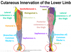
Describe the areas of innervation provided by the lumbosacral plexus |
|
|
|
Muscles arising from the ilium are innervated by what? |
Femoral nerve. |
|
|
Muscles arising from the ischium are innervated by what? |
Tibial n. |
|
|
Muscles arising from the pubis are innervated by what? |
Obturator n. |
|
|
Muscles arising from the posterior femur are innervated by what? |
Common fibular n. |
|
|
Define Tom, Dick, ANd Harry |
Tibial posterior m. Flexor Digitorum longus m. Posterior Tibial A. Tibial Nerve Flexor Hallucis longus m. |