Reading...
![]()
Play button
![]()
Play button
![]()
Use LEFT and RIGHT arrow keys to navigate between flashcards;
Use UP and DOWN arrow keys to flip the card;
H to show hint;
A reads text to speech;
83 Cards in this Set
- Front
- Back
|
E. MHC
|
Major Histocompatibility Complex (MHC) molecules: MHC molecules function in the presentation of antigens to cells of the immune system.
|
|
|
F. class I MHC
|
Proteins expressed in the cytoplasm (endogenous proteins) are broken up into peptides, picked up by MHC class I molecules, carried to the cell surface and displayed.
|
|
|
class II MHC
|
Extracellular proteins (exogenous proteins) are endocytosed, broken up into peptides, picked up by MHC class II molecules, carried to the cell surface and displayed.
|
|
|
H. allele
|
genes
|
|
|
I. polymorphism
|
This phenomenon of having multiple stable forms of a gene (referred to as alleles) in the population is known as genetic polymorphism.
|
|
|
J. HLA
|
Unfortunately, these different MHC molecules act as foreign antigens and cause graft rejection between genetically unmatched individuals. This is why the human MHC is often referred to as human leukocyte antigen (HLA). HLA is an older term that comes from transplantation studies in the 1930 and 1940s. Before their function in the immune system was understood, MHC molecules were recognized as antigens expressed on leukocytes that were associated with tissue graft rejection
|
|
|
K. co-dominantly expressed
|
each individual expresses all of the class I and class II MHC molecules that they inherit (i.e., the haplotype or set of genes they inherit from their father and the haplotype or set of genes they inherit from their mother).
|
|
|
L. haplotype
|
set of genes they inherit from their father and the haplotype or set of genes they inherit from their mother
|
|
|
Μ. β2m
|
The class I MHC molecule is expressed on the cell surface in noncovalent association with a small molecule called β2-microglobulin (β2m). β2m does not bind peptide, does not vary in sequence between individuals (it is nonpolymorphic) and is encoded on a different chromosome from the MHC genes.
|
|
|
N. somatic recombination
|
V(D)J recombination. This site-specific recombination reaction generates a diverse repertoire of T cell receptor (TCR) andimmunoglobulin (Ig)
|
|
|
O. RAG-1 and RAG-2
|
RAG-1 and RAG-2 are proteins (enzymes) at the ends of VDJ genes that separate, shuffle, and rejoin the VDJ genes
|
|
|
P. P-nucleotides
|
These additions create palindromes in the DNA; therefore, the nucleotides that are added are called palindromic (P)-nucleotides.
|
|
|
Q. TdT
|
Before joining occurs these DNA ends are also vulnerable to nucleotide removal by cellular exonucleases, and to further nucleotide addition by an enzyme called terminal deoxynucleotidyl transferase (TdT). Since TdT does not require a DNA template, the nucleotides that it adds are called nontemplated (N)-nucleotides.
|
|
|
R. N-nucleotides
|
|
|
|
S. combinatorial diversity, junctional diversity
|
The combinatorial diversity that comes from the V, D and J gene segments, and the junctional diversity imprecise cutting and nucleotide additions/subtractions, all contribute to the variability of the antigen-binding region of antibody molecules.
|
|
|
U. CDR1and CDR2
|
While the V region gene sequence is the major diversification factor for CDR 1 and CDR2
|
|
|
V. CDR3
|
CDR3 is subject to a high degree of both combinatorial and junctional diversity.. CDR3 shows the greatest variability as it is encoded by a recombination of the VJ in the case of a light chain region and (VDJ in the case of heavy chain) regions.
|
|
|
W. nonproductive rearrangement
|
One consequence of somatic recombination is that frame shifts may occur creating stop codons so that the RNA transcribed from the recombined DNA cannot be translated into a functional protein. In this case the recombination is called a nonproductive rearrangement.
|
|
|
X. allelic exclusion
|
Since there are two copies of each chromosome, each B cell first attempts to rearrange one of copy (one allele). If that rearrangement is successful (productive), it stops. This process is called allelic exclusion and is true for both the heavy and light chain loci
|
|
|
Y. idiotype
|
These regulatory processes ensure that each B cell expresses antibody with only one antigenic specificity (only one idiotype).
|
|
|
Z. class switch recombination
|
One of the puzzles in the molecular genetics of antibody production was how different antibody classes (heavy chain isotypes, i.e. IgG1, IgA, IgM, etc.) of identical antigenic specificity (identical idiotype) could be produced. The exact mechanism of this class switch (CS) recombination is still not fully understood. In CS recombination the previously recombined IGH variable region is moved to a new location that is directly upstream of a different IGH constant region gene segment.
|
|
|
|
|
|
|
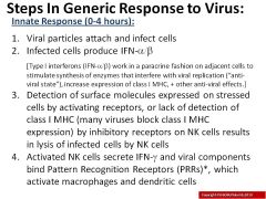
Where are antigens concentrated?
|
peripheral lymphoid organs (lymph nodes and spleen)
|
|
|
How do antigens arrive in the lymph nodes?
|
via lymph or DCs
|
|
|
How do antigens arrive in the spleen?
|
blood
|
|
|
Where do immature DCs reside?
|
epithelia and most organs
|
|
|
Are immature DCs phagocytic?
|
yes
|
|
|
Do immature DCs express PRRs(TLRs)?
|
yes
|
|
|
What do DCs phagocytose?
|
microbes
|
|
|
How are DCs activated?
|
signaling through their PRRs
|
|
|
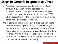
Where are chemokines recognized by CCR7 produced/
|
in the paracortex
|
|
|
What does CCR7 do?
|
causes DCs to travel through the lymph to the lymph node paracortex and to secrete IL-12
|
|
|
What do DCs do as they travel to the paracortex? What do they arrive secreting
|
mature (they arrive secreting cytokines like IL12)
|
|
|
What are required to activate naïve T cells?
|
mature DCs
|
|
|
Are Mature DCs potent antigen presenting cells?
|
yes
|
|
|
What activates T cells?
|
DCs
|
|
|
Are mature DCS required to initiate T-dependent adaptive immune responses?
|
yes
|
|
|
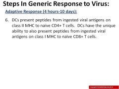
When does an adaptive immune response begin?
|
when the antigen receptors of lymphocytes bind antigens
|
|
|
Where do T cell antigen (TCRs) receptors bind peptides?
|
in Major HistoCompatibility (MHC) molecules on antigen presenting cells
|
|
|
Do TCRs make contact with both the peptide and MHC?
|
Yes
|
|
|
What are MHC molecules also called?
|
human leukocyte antigens (HLAs)
|
|
|
Where are MHC molecules ecpressed?
|
on cell surface
|
|
|
What is the function of an MHC molecule?
|
to present peptides from antigens to T cells
|
|
|
What is the structure of MHC 1?
|
Peptide-binding cleft formed by single chain (alpha). Invariant molecule, b2M, completes structure. Binds small peptides (8-11 amino acids long)
|
|
|
What is the structure of MHC 2?
|
Peptide-binding cleft formed by two chains (alpha and beta). Binds larger peptides (10-30 amino acids long)
|
|
|
What are CDs
|
Cluster of Differentiation (really means nothing)
|
|
|
What is the MHC 1/peptide complex bound by?
|
CD8+ cytotoxic T cells
|
|
|
Where does CD8+ bind to MHC 1?
|
on the a3 domain
|
|
|
What does MHC2/peptide complex bind?
|
CD4+ helper T cells
|
|
|
Where does CD4 bind?
|
B2 domain
|
|
|
Each MHC molecule binds :(3)
|
only peptides from antigens (not whole antigens), many different peptides, only one peptide at a time
|
|
|
Peptides bound to MHC: (3)
|
are acquired during assembly, stabilize MHC moleculre for transport to cell surface, have slow off rate at the surface
|
|
|
MHC 1 =
|
HLA-A, B and C
|
|
|
MHC 2 =
|
HLA-D (DR, DP, and DQ)
|
|
|
Where are MHC 1 and MHC 2 molecules encoded?
|
close together on human chromosome 6 (inherited as a unit)
|
|
|
How are MHC molecules expressed?
|
codominantly (both haplotypes are expressed)
|
|
|
Are MHC genes polymorphic?
|
Very
|
|
|
Macrophages and MHCs
|
Macrophages in tissues phagocytose microbes and display peptides from these antigens on MHC II to CD4+ effector or memory (not naïve) helper T (TH) cells
|
|
|
Are viruses intracellular or extracellular antigens?
|
intracellular
|
|
|
B cells and MHCs
|
B cells take up antigens by receptor-mediated endocytosis and display peptides from these antigens on MHC II to CD4+ effector or memory helper T (TH) cells in peripheral lymphoid tissues (i.e. lymph nodes)
|
|
|
Any nucleated cell and MHCs
|
Any nucleated cell can present peptides derived from intracellular antigens (i.e. viruses) on MHC I to CD8+ effector or memory cytotoxic T cells (CTLs)
|
|
|
What cells express MHC2?
|
DCs, macrophages, and B cells
|
|
|
Can other cells be induced to express MHC 2?
|
yes
|
|
|
Where is MHC 1 expressed?
|
on all nucleated cells and platelets
|
|
|
Where are peptides from extracellular microbes presented?
|
on MHC II to CD4+ helper T (TH) cells
|
|
|
Where are peptides from intracellular microbes presented?
|
on MHC I to CD8+ cytotoxic T cells (CTLs)
|
|
|
Describe the MHC 2 pathway
|
During MHC II molecule synthesis, CLass II Invariant chain Peptide (CLIP) binds/blocks peptide binding cleft until MHCIIs arrive in vesicles containing peptides from extracellular microbes. Lysosomal enzymes (called cathepsins) break down incoming microbes into peptides. HLA-DM* removes CLIP so these exogenously derived peptides can load onto MHCIIs
|
|
|
Describe the MHC 1 pathway
|
Proteins from intracellular microbes (i.e. virus), produced in cytosol, are degraded by proteosome into peptides. Transporter associated with Antigen Processing (TAP) injects peptides into endoplasmic reticulum (ER). Aminopeptidases trim peptides to correct size. Peptides are loaded onto MHCIs as they are synthesized
|
|
|
Describe pathway differences
|
|
|
|
Presentation of MHC2 results in
|
antigen-specific helper CD4+ T (TH) cells which produce cytokines that activate macrophages and stimulate B cells to produce antibodies. (extracellular microbes like Group A Strep)
|
|
|
Presentation by MHC1 results in
|
antigen-specific CD8+ cytotoxic T cells (CTLs) which can directly kill infected cells. (intracellular microbes like Rhinovirus)
|
|
|
Cross presentation by DCs
|
|
|
|
How are DCs special in relation to antigen presentation?
|
do not have to be virally infected to present viral peptides on MHC I
|
|
|
Are both CD4 and CD8 T cells activated or "primed" by DCs
|
Yes
|
|
|
What kind of response to viruses are produced by DCs?
|
Both antibody and CTL(cytotoxic lymphocyte)
|
|
|
Pro-avtivation of NK cells
|
molecules on stressed cells bind NK cell activating receptors
|
|
|
Anti-activation of NK cells
|
MHC1 molecules on target cells bind to Killer Inhibotory Receptors (KIRs) (to protect host cells)
|
|
|
How is NK cell activation balanced?
|
?????????? Stress signals are expressed on virally infected cells. The virus decreases MHC 1 expression. The NK cell can detect the decrease in MHC1 and the increase in stress molecules on the cell surface.
|
|
|
What does the inhibitory receptor on NK cells recognize?
|
MHC1
|
|
|
|
|
|
|
|
|
|
|
|
.
|