![]()
![]()
![]()
Use LEFT and RIGHT arrow keys to navigate between flashcards;
Use UP and DOWN arrow keys to flip the card;
H to show hint;
A reads text to speech;
174 Cards in this Set
- Front
- Back
|
PAS + vitreous layer surroundig entire follicle
thick and wrinkled during which stage? |
glassy membrane
becomes thick and wrinkled during CATAGEN |
|
|
4 regions of hair follicle
|
1. infundibulum
2. ithmus 3. supra-bulbular 4. bubular |
|
|
Infundibulum keratins
|
basal: CK 5/6, 14
suprabasal: 1,10,4,14 keratohyaline granules |
|
|
Ithmus keratins
|
CK 5/6, 14, 17, 19
NO keratohyaline granules |
|
|
buldge keratins
|
CK 15
CK 19 B1 Integrin |
|
|
buldge stem cells
|
ORS
|
|
|
CK15 stain periph of ?
|
trichoepitheliomas
sebaceous adenomas normal basaloid cells periph of sebaceous glands more diffuse BCC and trichoblastoma |
|
|
greatest density of merkel cells hair follicle?
|
isthmus
|
|
|
tumors w increase merkel cells?
|
ithmus tumors
trichoblastomas trichoepitheliomas fibroepithelioma of Pinkus |
|
|
melanocyte ratio buldge compared to nl epi
|
buldge 1:4
epi 1:10 larger with longer dendritic processes |
|
|
bulb keratin?
|
kHa5
|
|
|
widest region of hair bulb?
|
line of Auber
separates growing bulb from cornification |
|
|
3 main layers surround fibrous sheath
|
IRS
ORS Companion Layer |
|
|
ORS keratins
|
2: K5, K6a, K6b
1: K14, K15, K16, K17, K19 |
|
|
IRS keratins
|
2: K 1 - 4
1: IRSa 1- 3 |
|
|
Companion Layer keratins
|
K6hf, K6a, K16
|
|
|
Hair Shaft keratins
|
Hb 1-6, Ha 1- 9
|
|
|
ORS cells
|
clear cells glycogen
keratinize at ISTHMUS (CK 5/6, 8, 15) DOES NOT CORNIFY BELOW ISTHMUS |
|
|
3 layers
|
1. cuticle 2 layers: Huxely, Henle
|
|
|
first area of hair follicle to cornify (before hair shaft cornifies)?
|
Henle's layer of cuticle of IRS
|
|
|
IRS granules?
|
trichohyalin granules
|
|
|
IRS desquam at?
|
midportion of isthmus
(between arrector pili attachemtn and sebaceous duct) immature if below arrector pili = cicatricial alopecia, scarring alopecias |
|
|
Hair shaft 3 layers
|
Outer cuticle (integrity)
Cortex (bulk) Medulla (hard keratins) |
|
|
EMA stains?
|
Epidermal membrane antigen
mature sebocytes > seboblasts |
|
|
Androgen receptor stains?
|
seboblasts > sebocytes
|
|
|
Sebaceous glands lipid stains with?
section? |
Sudan black
oil Red O frozen sections |
|
|
Sebaceous glands stain with?
|
EMA (sebocytes)
CK 7 (seb and appocrine) CK 15 periph CK 10 CK 17 Androgen receptor (seboblasts) |
|
|
Ectopic sebaceous glands
|
LIP and BUCCAL MUCOSA: Fordyce glands
AREOLAE: Montgomery's tubercles PENIS: Tyson's glands EYELID: glands of Zeis and Meibomian lgands LABIA MINORA AND VAGINA Harmatomas nevus sebaceous follicular induction: DF, Becker's melanosis |
|
|
Apocrine duct opens?
|
superior to sebaceous gland
|
|
|
apocrine gland location in skin? epi? dermis?
|
deep dermis and subcutis
deeper than eccrine glands |
|
|
Apocrine glands located?
|
axillae, genitals, periumbilical, periareolar
rarely face and scalp |
|
|
Eyelid apocrine glands?
|
Moll's glands
|
|
|
Ear canal aprocrine glands
|
ceruminous glands
|
|
|
modified apocrine gland?
|
breast
|
|
|
special apocrine glands
|
eyelids: Moll's glands
ear canals: ceruminous glands breast |
|
|
Apocrine glands structure?
|
single layer of tall columnar cells
PAS-DR + esoinophilic granular cytoplasm w basal vesicular nuclei surrounded by discontinuous lyaer of myoepithelial S100+ cells |
|
|
Apocrine granules cytoplasmic granules
cytoplasm brown to yellow pigment? |
lysosomes
SIALOMUCIN, LIPIDS, IRON brown to yellow pigment LIOFUCIN AND MELANIN GRANULES |
|
|
Apocrine secrete?
|
cholesterol esters, tg, squalene
|
|
|
Apocrine glands stain for?
|
Acid Phosphatase
B-glucouronidase Indoxyl acetate esterase |
|
|
Apocrine express?
|
estrogen/progesterone receptors
GCDFP-15+ human milk fat globulin 1 |
|
|
Aporcine Lumen Cells?
|
CK 6, 7, 8, 18, 19
CEA+ |
|
|
Aprocrine vs Eccrine lumen
|
apocrine 7 - 10 x larger
|
|
|
Eccrine spirled duct in epi
|
acrosyringium
|
|
|
No eccrine glands?
|
nail bed
glans penis labia minora |
|
|
Eccrine 2 types of cells
|
1. pale to clear basaloid: glycogen
2. darker: apical surrounded by myoepithelial cells |
|
|
Eccrine dark cells secrete
|
mucoid sialomucin
|
|
|
Eccrine basal cells secrete
|
hypotonic saline
|
|
|
Eccrine secretions + for?
|
Phosphyorlase
Succinic dehydogenase |
|
|
Eccrine inervation?
|
sympathetic
respond to achetylcholine |
|
|
Eccrine glands periph cells +?
|
EMA epidermal membrane antigen
|
|
|
Eccrine Intermdiate cells
|
CK10
|
|
|
Luminal cells
|
CK 6,8,18,19, CEA +
|
|
|
Eccrine glands + for?
|
S-100
IKH-4 |
|
|
Apocrine and Eccrine ducts?
|
same
2 layers of cells inner flat cuboidal outer basophilic plumper cuboidal cell Thin PAS+ bm no myoepithelial cells intraepi keratohyaline granules |
|
|
Myoepithelial cells stain?
|
H caldesmon
calponin vimentin CK 5, 6 HMWCK p63 alpha smooth muscle actin and myosin S100+ benign keep myoepithelial layer ca lose layer |
|
|
H Caldesmon
|
myoepithelial cells
|
|
|
Calponin
|
myoepithelial cells
|
|
|
hair follicle nevus histo?
|
normal vellus hairs w OWN STROMA clefting
|
|
|
Trichofolliculoma
|
trichohyaline granules
fibrotic stroma vellus hairs opening into mother hair |
|
|
folliculosebaceous cystic hamartoma
|
sebaceous trichofolliculoma
|
|
|
trichoadenoma of ?
|
nickolowski
trichoepithelioma variant horn cysts w FIBROUS STROMA nodule w multiple milia |
|
|
trichepithelioma
|
LOOSE STROMA w fibroblasts
horn cysts papillary mesenchymal bodies K7 - |
|
|
trichoepithelioma K7?
|
negative
|
|
|
brooke-spiegler
inheritance? tumors? chromo? |
AD
cylindromas, spiradenomas, trichoepitheliomas (TE) chromo 16 |
|
|
Rombo
|
multiple trichoepitheliomas (TE)
atrophodermal vermiculatum milia BCCs hyotrichosis |
|
|
syndromes with trichoepitheliomas
|
brooke-spiegler
rombo |
|
|
desmoplastic trichoepithelioma
|
keratin cysts
papillary menenchymal bodies |
|
|
desmoplastic trichoepitheliomas CK20?
|
CK 20 + bc of MERKEL CELLS
bcl-2 neg stromomelysin 3 neg CEA neg involucrin + |
|
|
desmoplastic trichoepitheliomas involucrin?
|
involucrin +
|
|
|
morehaform BCC
bcl2? stromelysin 3? |
bcl-2 +
stromelysin 3 + fibroblasts |
|
|
desmoplastic trichoepithelioma ddx?
|
morpheaform BCC (bcl2 +, stromelysin 3 +)
syringoma (CEA+, involucrin neg) MAC |
|
|
syringoma
|
CEA+
involucrin neg |
|
|
desmoplastic trichoepithelioma clinical
|
plaque w annular border and central on face young woman
|
|
|
Trichoblastoma
|
like trichoepithelioma but DEEPER IN DERMIS or SUBCUT
NO EPIDERMAL ATTACHMENT broccoli heads papillary menenchymal bodies well-circumscribed |
|
|
Trichoblastoma clinical
|
SCALP
mc benign tumor nevus sebaceous |
|
|
Cutaneous lymphadenoma
|
trichoblastoma variant w basolid islands of clear cells
lo fibrous stroma |
|
|
panfolliculoma
|
trichoblastoma and matricoma
diff toward all follicle elements |
|
|
dilated pore of winer
|
granular layer w hyperkeratosis
buds of epi no hair follicles INFUNDIBULAR |
|
|
infundibular origin
|
dilated pore of winer
inverted follicular keratosis |
|
|
inverted follicular keratosis
|
squamous eddies
looks like seb k but no string sin INFUNDIBULAR |
|
|
tumor of the follicular infundibulum
|
variant of trichilemmonma or seb k
anastomosign strands similar to fibroepitehlioma of Pinkus but NOT BASALOID grasshopper bridgin fo follicles parallel to epidermsi ISTHMIC |
|
|
pilar sheath acanthoma
|
similar to dilated pore of winer but bigger epidermal buds
thick epidermal buds no hair shafts OUTER ROOT SHEATH ISTHMIC |
|
|
pilar sheath acanthoma clinical?
|
upper lip elderly
central pore w keratin plug |
|
|
Trichilemmoma
|
clear cells
glycogen PAS+ w periph basaloid palisading |
|
|
Trichilemmoma assoc w syndrome?
|
cowden
|
|
|
cowdens
|
AD
PTEN tumor supressor multiple hamartomas trichilemmomas sclerosing fibromas palmoplantar punctate keratoses oral papillomatosis Lhermitte-Duclos breast and thyroid ca |
|
|
Lhermitte-Duclos
|
cowdens
|
|
|
lhermitte-Duclos
|
cerebellar tumor
AD cowden's |
|
|
trichilemmoma
|
hyperkeratosis
papillamtosis lobule of clear cells thin rim of basaloid cells |
|
|
PILOMATRICOMA
|
calcifying epitehrlioma of Malherbe
2 cells 1. dark basaloid w calcification 2. ghost cells giant cells and calicification |
|
|
multiple pilomatricoma
|
myotonic dystrophy (Steinert syndrome)
|
|
|
Steinert syndrome
|
multiple pilomatricoma
|
|
|
multiple pilomatricoma
|
steinert syndrome
Turner's Rubinstein-Taybi sydnrome Gardner's (multiple EICs w focal PMX like changes) |
|
|
sporadict pilomatricoma
|
activating B-catenin mutations
|
|
|
fibrolliculoma
|
no hair follicles
cords of 2 to 4 cells radiating from follicular structure |
|
|
Hornstein-Knickenberg syndrome
|
Birt-Hogg syndrome
AD FOLLICULIN mult fibrofolliculomas trichodiscomas SKIN TAGS spontaneous PTX renal cell ca |
|
|
sebaceous hyperplasia
|
seb lobules around follicle
grapes androgen receptor |
|
|
cyclosporin
|
10 - 15 % pts seb hyperplasia
|
|
|
nevus sebaceus
|
organoid nevus
papillomatosis seb glands enlarged NOT ASSOC W HAIR FOLLICLE apocrine glands trichoblastoma 5 syringocstadenoma pailliferum 5% BCC < 1 % |
|
|
Nevus Sebaceous Syndrome
|
large linear nevus on scalp
CNS SKELETAL |
|
|
sebaceous adenoma
|
lobular tumor
> 50% sebocytes immature basoalid cells MUIR-TORRE SYNDROME |
|
|
MUIR-TORRE SYNDROME
|
AD
MSH2/MLH1 mismatch repair genes microsattelite instability assoc w seb neoplasms KAs colon CAs (allelic to HNPCC) |
|
|
Sebaceoma
|
sebaceous epithelioma
|
|
|
sebaceous epithelimoa
|
sebaceoma
|
|
|
sebaceoma
|
few mature sebocytes
>50% immature basaoloid cells looks like BCC assoc w MUIR-TORRE Rapini BCC with seb diff |
|
|
Sebaceous Carcinoma
|
Pagetoid spread ocular tumors
EMA+ eyelid from Meibominan or Zeis glands mets 30% MUIR-TORRE |
|
|
Sebaceous Carcinoma EMA?
|
EMA+
|
|
|
Syringocystadenoma papilliferum
|
apocrine
stroma w bvs plasma cells papillomatous red plaque on SCALP with vescile-like inclusions 1/3 assoc w nevus sebaceous 10% w BCC or trichoblastoma eleted PTCH gene or p16 |
|
|
syringocystadenoma papilliferum
|
open to epidermis
cystic dilation plasma cells papillary projections |
|
|
Hidrandoma papilliferum
|
deeper dermal nodule
MAZE-LIKE apocrine solitary lesion on women's vulva |
|
|
eccrine acrospiroma
|
apocrine hidradenoma
|
|
|
apocrine hidradenoma
|
eccrine acrospiroma
|
|
|
eccrine acrospiroma
|
solitary nodule w solid and cystic componets
single type of basaloid cells eccrine ducts w celar cells and dark cells squamous diff keratin cysts firm nodule on head/neck men |
|
|
mixed tumor of skin
|
chondroid syringoma
|
|
|
chondroid syringoma
|
epi islands w small to medicum sweat ductal structuers within mucinous stroma w blue chondroid appearance
bone formation |
|
|
mucinous carcinoma
|
islands floating in snot
dermal tumor light and dark cells (ductal structures) SIALOMUCIN fibrous septae dx of exclusion must r/o metastatic breast cancer and GI islands EMA and CEA + EYELID |
|
|
mucinous carcinoma islands +
|
EMA +
CMA + |
|
|
most common location mucinous carcinoma?
|
eyelid
|
|
|
erosive adenomatosis of nipple
|
thick epidermis
smooth muscle laciferous glands papilliferum like tubular ducts connect to suface w apocrine decap lymphoplasmacytic infiltrate BENIGN crusted papule or plaque on nipple resembles Page's clincally |
|
|
Papillary Eccrine Adenoma
|
dermal tumor
glandular spaces w eccrine and occ apocrine diff papillary projections into lumina focal or no connection w epi CEA + S100+ dermal nodule on extremities of AA women |
|
|
Tubular apocrine adenoma
|
apocrine diff
dermal tumor with glandular spaces and projections within lumen No specific location (unlike papillary eccrine adenoma) |
|
|
syringoma
|
superficial ddermal tumor
multipl small eccrine ductal structures 2 cell layers taadpole horn cysts fibrous sclerotic stroma multiple DOWN's clear cell variant w DM II |
|
|
Syringoma clear cell assoc?
|
DM II
|
|
|
multiple syringomas
|
Down's
|
|
|
cylindroma
|
dermis
thick eosinophilic cuticles "cylinders" TYPE IV COLLAGEN smooth red nodules scalp |
|
|
multiple cylindromas syndromes?
|
brooke-spiegler
familial cylindromatosis |
|
|
brooke-spiegler
|
AD
chromo 16 multiple cylindromas spiradenomas trichoepitheliomas |
|
|
Familiarl cyldindromas
|
CYLD1 mutations
chromo 16 |
|
|
Eccrine spriadenoma
|
blue balls in dermis
rosettes PAINFUL |
|
|
BLEND AN EGG
|
blue rubber bleb
Leiomyoma Eccrine spiradenoma Neuroma DF Angiolipoma Neurillemmoma Endometriosis Granular Cell tumors Glomus tumor |
|
|
Eccrine Poroma
|
glycogen containing PAS+
basaolid or cuboidal smaller than ko moat aroun it looks like seb k on low power clinical PG SOLE SCALP |
|
|
mulitple promomas
|
ecrine poromatosis
palms soles assoc with hidrotic ED |
|
|
Hidraoacanthoma Simplex
|
intraepidermal poroma
looks like clonal seb k (Borst-Jadassohn) |
|
|
Dermal Duct Tumor
|
dermal prooma or eccrine acrospiroma
NO CONNECTION TO EPIDERMIS |
|
|
Syringofibroadenoma
|
poroma variant
lattice thin epithelial cords connected to undersurface of epidermis similar to fibroepitheklioma of Pinkus but NO BASALOID BUDS |
|
|
multiple syringofibroadenoma
|
Schopf syndrome
hidrotic ED |
|
|
Schopf Syndrome
|
multiple syringofibroadenomas
|
|
|
hidrotic ectodermal dysplasia
|
Clouston's
|
|
|
Clouston's
|
hidrotic ectodermal dysplasia
AD CONNEXIN 30 French Canadians PPK w transgrediens nail dystrophy sparse hair tuffed phalanges HPV-10 |
|
|
Schopf-Schultz-Passarge
|
adult onset focal dermal hypoplasia w multiple hidrocystomas
multiple syringofibroadenoma |
|
|
Clouston's HPV?
|
HPV-10
|
|
|
Microcytic Adenxal Carcinoma
|
superficial cysts
ductal diff deeper smaller islands and cords dense halinized stroma muscle and perinerual invasion rare mitsoses littly atypia solar elastosis sweat glands low in dermis UPPER LIP recurs 50% rarely mets |
|
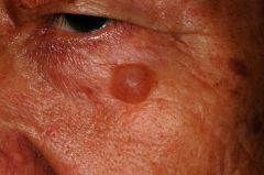
?
|
apocrine hydrocystoma
|
|
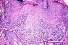
?
|
trichilemmoma
|
|
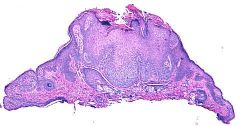
?
|
trichilemmoma
|
|
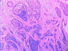
?
|
cutaneous lymphadenoma
|
|
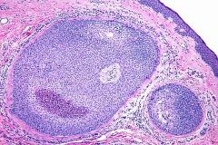
?
|
dermal ductal tumor
|
|

?
|
digital papillary carcinoma
|
|
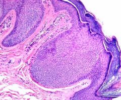
|
hidroacanthoma simplex
|
|
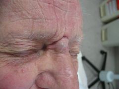
|
chondroid syringoma
|
|
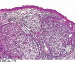
|
eccrine acrospiroma
|
|

|
fibroepitheliioma of Pinkus
|
|
|
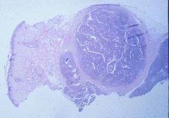
eccriine spiroadenoma
|
|
|
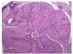
fibrofolliculoma
|
|

|
fibrofolliculoma of pinkus
|
|
|
angiofibroma
|
activated stellate fibroblasts (factor 13a +)
increase collagen increase bvs concentric fibrosis around bv and adenxa |
|
|
koenan tumor
|
TS
periungual fibroma angiofibroma |
|
|
penile peraly papules
|
angiofibromas
|
|
|
acral fibrokeratoma
|
angiofibroma
|
|
|
TS
|
chromo 9q34
HAMARTIN and 16p13 TUBERIN angiofibromas |
|
|
MEN-1
|
Wermer Syndrome
11q13 MENIN parathyroid pancreatic pituitary assoc w angifibromas |
|
|
Wermer's
|
MEN-1
11q13 MENIN parathyroid pancratic pituitary assoc w angiofibromas |
|
|
Birt-Hogg Dube
|
17p11
FOLLICULIN skin tags trichodiscomas fibrofolliculomas renal tumors |
|
|
fibrofolliculomas
|
birt-hogg dube
|
|
|
trichodiscomas
|
birt-hogg dube
|
|
|
skin tag
|
bv fat
|
|
|
pleomorphic fibroma
|
skin tag w scattered dark bizarre appearing multinuclated fibroblasts looksl ike mal sarcoma
|
|
|
sclerotic fibroma
|
storiform collagenoma
plywood hypocellular Cowden's |
|
|
storiform collengoma
|
sclerotic fibroma
plywood hypocellular Cowden's |
|
|
Fibromatoses
|
dense collegen
bland fibroblasts Gardner's Dupuytren's Ledderhose Peyronie's Knuckle Pads Desmoid tumor (abdominal wall) |
|
|
Gardner's
|
5q12
APC binds B-catenin fibromas colorectal polyps |
|
|
Nodular fascitis
|
tissue culture pattern
increasedd myofibroblasts w wispy mucinous increased bv extravasated RBCs no atypia basophillic similar to gangion cells young person w enlarging painful mass upper extremity/dorsal fingers |
|
|
digital fibromatosis of childhood
|
inclusion body fibromatosis
dome shaped acral dermal tumor w whorled appearance eccrine gland like structures centers of swrils perinuclear red dot inclusions on high power actin fibers actin + PTAH + red with Masson tirhrom stain fingers and toes of infants no thubm or great toe spon regress |
|
|
inclusion body fibromatosis
|
digital fibromatosis of childhood
|

