Reading...
![]()
Play button
![]()
Play button
![]()
Use LEFT and RIGHT arrow keys to navigate between flashcards;
Use UP and DOWN arrow keys to flip the card;
H to show hint;
A reads text to speech;
42 Cards in this Set
- Front
- Back
|
Bone is __% mineral with a _:_ Ca:P ratio
|
90; 5:3
|
|
|
Bone matrix is composed of:
|
Collagen (mostly type I) - required for tensile strength
Alk Phos - Isoenzyme produced by osteoblasts |
|
|
Cementum is
|
mineralized glue between new and old bone
|
|
|
Chondrodysplasia is due to abnormal __.
|
Fibroblast growth factor 4
|
|
|
Abnormal FGF-4 predisposes to:
|
IVDD - premature degeneration of the Nucleus pulpous
DJD - d/t abnormal limb angulation |
|
|
Chondrodysplasia in Scottish folds is weird because:
|
Autosomal incomplete dominance
Heterozygotes - folded ears, short limbs, stiff gait Homozygotes - thick, stiff tails, lameness, distal limb bone proliferation |
|
|
About osteogenesis imperfecta...
|
Rare inherited defect of collagen --> fragile bones
|
|
|
Pathogenesis of Osteogenesis imperfecta
|
Pathogenesis: Collagen defect > brittle bones > fractures > decreased weight bearing > decreased bone mineralization/density (often difficult to separate primary from secondary lesion) difficult to diagnose, even microscopically
|
|
|
Physeal dysplasia most common in __ and leads to __.
|
Young large male cats
Atraumatic unilateral or bilateral slipped capital femoral epiphyses (although all physes affected) Due to persistent disorganized growth plates |
|
|
Amelia is ___
|
- absence of limb
|
|
|
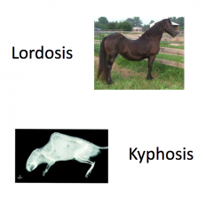
Spinal anomalies include:
|
Spinal anomalies: lordosis = lean straighter , kyphosis = humpback , scoliosis = S
|
|
|
About Vitamin A Toxicity
|
a. Due to over supplementation or abnormal diet (liver)
b. Deforming cervical spondylosis in cats (rare today) c. Vitamin A is toxic to osteoclasts |
|
|
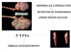
Primary Hyperparathyroidism
|
Primary - uncommon; idiopathic parathyroid hyperplasia or tumor secretes excess PTH osteoclastic bone resorption replacement with fibrous tissue and woven bone
|
|
|
All about the 2ndary types of HyperPTH
|
i. Renal secondary hyperparathyroidism = renal rickets, rubber jaw
Renal disease decreased phosphorus excretion and decreased activation of vitamin D “hypocalcemia” parathyroid gland hyperplasia excess PTH osteoclastic bone resorption replacement with fibrous tissue and woven bone (fibrous osteodystrophy) Young dogs - enlarged very firm maxilla Older dogs - “rubber jaw” ii. Nutritional secondary hyperparathyroidism - rare in dogs and cats Big head or bran disease of horses; metabolic bone disease of reptiles Low calcium/high phosphorus diet excess PTH fibrous osteodystrophy |
|
|
2 types of vitamin D deficiency
|
a. Osteomalacia (bone softening) - adult animals; rare
b. Rickets - growing animals; especially primates |
|
|
Osteoporosis leads to__.
|
thin trabeculae and cortices
|
|
|
About FQ arthropathy
|
Articular cartilage necrosis with vesicle formation
Quinolone antibiotics (such as Baytril) contraindicated in growing animals |
|
|
About LCP DZs
|
Necrosis and collapse of femoral capital epiphysis; due to ischemia
Young small breed dogs, especially terriers and poodles Necrotic bone has empty lacunae; the osteocytes are dead |
|
|
Stages of Fracture repair (6):
|
1. Hematoma
2. Inflammation 3. Granulation tissue 4. Soft callus: fibroblasts collagen osteoblasts osteoid chondroblasts cartilage 5. Hard callus >2 weeks primary callus - mineralized woven bone secondary callus - mature (lamellar) bone 6. Remodeling - months to years, return to original shape |
|
|
What is a callus, really?
|
connective tissue that bridges fracture fragments
1. Forms both inside (internal; endosteal callus) and outside (external; periosteal callus) 2. Movement and low oxygen favors chondrocyte differentiation (cartilage within callus) Cartilage within a fracture site can look histologically very scary; history is important |
|
|
Pathologic fractures are typically due to one of the following:
|
Osteogenesis imperfecta
Osteoporosis (disuse, starvation, advanced age) Metabolic bone diseases Osteomyelitis Neoplasia (lytic osteosarcoma or other tumor) |
|
|
Complications of fracture healing:
|
1. Osteomyelitis - most common in open fractures
Inflammation does not indicate infection 2. Sequestrum - necrotic bone fragment too big to be resorbed Necrotic bone has empty lacunae; the osteocytes are dead 3. Malunion - bony union in an abnormal position 4. Nonunion - repair has ceased, but only fibrous tissue bridges fragments 5. Premature physeal closure - due to trauma that injures germinal layer of physeal cartilage Distal ulnar physis in large breed dogs continued growth of radius causes bowing |
|
|
Cartilage fibrillation
|
fraying of the articular cartilage due to excessive friction and wear
|
|
|
Cartilage erosion
|
loss of cartilage due to excessive friction and wear
|
|
|
Osteophytes
|
periarticular bony proliferations
|
|
|
Eburnation
|
polishing and osteosclerosis of subchondral bone after the cartilage has been completely lost; literally “to become ivory like”
|
|
|
Thickened joint capsule
|
increased fibrous connective tissue and synovial proliferation
|
|
|
Cranial cruciate rupture
|
Very common; Large breed middle aged dogs
Osteophytes common, with and without repair May predispose to histiocytic sarcoma in some breeds |
|
|
Luxating patella. All the fun stuff.
|
Medial luxation most common
Shallow trochlear groove (result, not cause) plus osteophytes |
|
|
DJD in Cats
|
90% of cats over 11 years old have DJD
DJD of shoulders and/or stifles in Siamese should raise suspicion of mucopolysaccharidosis |
|
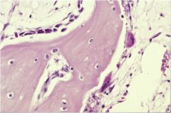
Name the cells.
|
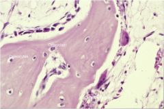
|
|
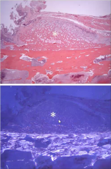
Describe this bone.
|
Woven and cancellous
|
|
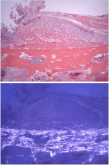
Describe this bone.
|
Lamellar and compact.
|
|
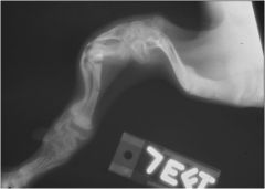
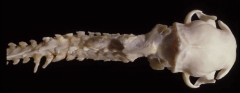
What's wrong with this picture? What does it lead to? What breeds are most common?
|
Chondrodysplasia.
Leads to IVDD (degen of nucleus pulposus) and DJD (d/t abnormal angulation of limb) Common in Basset, Corgi, Dachshund |
|
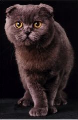
What is the significance of this cat?
|
It is a heterozygotic Scottish fold. The trait is autosomal incomplete dominant and leads to chondrodysplasia
Hetero - short limb, stiff gait, fold ears Homo - thick tail, lame, periosteal bone proliferation on distal limb |
|
What is this and what is its pathogenesis?
|
Osteogenesis Imperfecta.
Genetic Collagen defect causing fragile bones. Collagen defect>brittle bones>fractures>decr. weight bearing>decreased bone density |
|
|
Differentiate between lordosis and kyphosis.
|
|
|
This is an example of what? How is it caused?
|
Cervical spondylosis from Vitamin A toxicosis.
Caused by all liver diet or over-supplementation. The vitamin A kills the osteoclasts (which normally clean up the old bone) |
|
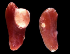
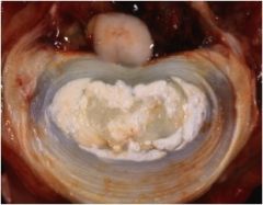
What is this? What would it lead to and how?
|
Parathyroid Adenoma.
Would cause extreme increase in PTH > Fibrous Osteodystrophy |
|
|
Mechanism for Renal 2ndary HyperPTH
|
|
|
|
What's the difference in FOD in an old vs. young animal?
|
Young - swollen muzzle
Older - rubber jaw |
|
|
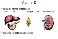
What is the pathway for Calcium absorption?›
|
|