Reading...
![]()
Play button
![]()
Play button
![]()
Use LEFT and RIGHT arrow keys to navigate between flashcards;
Use UP and DOWN arrow keys to flip the card;
H to show hint;
A reads text to speech;
25 Cards in this Set
- Front
- Back
|
Medium, concentration, and dose for upper GI series
|
Liquid Barium Sulfate
37% wt/vol 6mL/lb (1oz/5lb) |
|
|
Basic, general cause of restrictive-type pyloric outflow obstruction
|
Thickening of the pyloric wall. Doesn't allow pyloric sphincter to open adequately. Causes pyloric outflow obstruction.
|
|
|
Dog with pancreatitis would have what type of obstruction? Describe this in terms of distribution/severity of distention/peristalsis in affected segment.
|
Inflammatory/Traumatic
Focal/regional 3-4x absent |
|
|
String of pearls vs. Linear foreign body
|
String of pearls - even throughout - contractions give the hole thing a very symmetrical appearance. Only in cats. Alternating contraction/dilation.
Linear foreign body - random, haphazard folding of intestines. Dogs or cats. |
|
|
3 potential Ddx for a focal, caudal thoracic soft tissue opacity associated with the esophagus
|
Neoplasia
Hiatal hernia Foreign body |
|
|
What's the advantage of using barium/food to using barium paste in an esophagram?
|
Better for visualization of esophageal stricture. Fluid will flow right through.
|
|
|
Differentiate between restrictive and obstructive pyloric outflow obstruction.
|
Restrictive is an infiltrative disease that involves the wall of the pylorus preventing outflow through the pylorus
Obstructive is intraluminal. |
|
|
Define Tracheal stripe sign.
|
Thickening of the dorsal tracheal wall caused by the superimposition of the ventral aspect of the gas distended esophagus.
|
|
|
5 yo canine w/vomiting and abdominal pain. Abdominal rads show a moderate fluid dilation of a focal segment of SI. What are your 2 main Ddx, and how can you tell them apart?
|
Mechanical
Traumatic/inflammatory Functional would be generalized, so it's not that. To differentiate, compare the dilation to rib width. Inflammatory is 3-4x dilation vs. Mechanical 5-6x Contrast study may be done, but mechanical is a surgical emergency. Mechanical should have stacked bowel loops and hairpin turns. |
|
|
Two causes of paralytic/functional/adynamic ileus
|
Parvo
Dysautonomia |
|
|
2 Roentgen signs of Infiltrative bowel disease
|
bowel before area will be distended
Wall thickened |
|
|
Roentgen signs for Tracheal stripe
|
air in the esophagus so the wall of esophagus can be visualized.
"Stripe" is the thick stripe between the esophagus and trachea cranial to the heart because the two walls are close together and there is a gas interface on either side. |
|
|
preferred contrast medium for perforation of GI/esophagus? What are the contraindications for its use?
|
Organic iodine
Bronchoesophageal fistula or aspiration |
|
|
Radiographic finding in an esophagram of an animal with hiatal hernia that permits definitive diagnosis?
|
Gastroesophageal junction will be seen cranial to the diaphragm.
|
|
|
What are the 3 Types of ileus/degrees of distention/distribution/character of bowel loops?
|
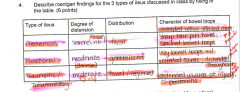
|
|
|
How can soft tissue opacity gastric foreign body be differentiated from a focal mucousal mass?
|
Contrast media. It will completely surround a FB to show a filling defect. Will only be seen on 3 sides of a mucosal mass, and not seen between the mass and mucousa.
|
|
|
Intussusception would be what type of ileus?
|
Obstructive/mechanical
|
|
|
What's the best way to administer an upper GI series/positive contrast gastrogram?
|
Orogastric tube.
Causes more rapid full distention of the stomach making visualization of marginations easier. Since stomach is filled all at once, you can more accurately determine gastric emptying time. |
|
|
Fluid will localize in the pylorus when animals are in __ lat recumbency and __ recumbency.
|
Right
Ventral |
|
|
3 distinct Roentgen signs of pancreatitis
|
Visualization of pancreas
Lateral displacement of duodenum Left displacement of gastric axis |
|
|
Decreased opacity in the liver or gall bladder is commonly associated with ?
|
Infection
|
|
|
Presence of an irregularly shaped periosteal reaction on the ventral margins of the cd lumbar vertebrae is indicative of what?
|
neoplastic mets of pelvic canal.
|
|
|
Enlarged mineralized adrenal gland is most likely associated with what process?
|
Cushing's
|
|
|
With generalized hepatomegaly, the stomach is deviated __, __, and to the __.
|
Caudally, dorsally, and to the left.
|
|
|
Granular peritoneal surfaces are seen with __ & ___.
|
ruptured viscus
Peritonitis |