Reading...
![]()
Play button
![]()
Play button
![]()
Use LEFT and RIGHT arrow keys to navigate between flashcards;
Use UP and DOWN arrow keys to flip the card;
H to show hint;
A reads text to speech;
59 Cards in this Set
- Front
- Back
|
Sclera
|
Outer white layer of the eye, composed of fibrous tissue, covered by conjunctival epithelium
|
|
|
Conjunctiva
|
Stratified epithelium lining the inner eyelids and covering the sclera.
|
|
|
Cornea
|
Anterior, transparent structure, contributes to most of the eyes refractive power
|
|
|
Choroid
|
Vascular layer b/t sclera and retina, provides oxygen and nutrients to the outer retina
|
|
|
Ciliary body
|
Contains ciliary muscle involved in the lens accommodation response, epithelial coating produces aqueous humor
|
|
|
Lens
|
Transparent biconvex structure that alters curvature to change focal distance (accommodation)
|
|
|
Iris
|
Pigmented smooth muscle, controls pupil diameter.
|
|
|
Aqueous humor
|
Gelatinous, transparent fluid similar to interstitial fluid, located in anterior and posterior chambers, provides ocular pressure to inflate eyes and provides nutrients to avascularized structures.
|
|
|
Retina
|
Inner, neural layer in the posterior eye; responsible for phototransduction and transmission of visual signals to the brain.
|
|
|
Fovea
|
Depression in the inner retinal surface where light focused; cone photoreceptor-rich; responsible for sharp, central vision.
|
|
|
Optic disc
|
Where ganglion cell axons coalesce before exiting the eye; responsible for the ‘blind-spot’.
|
|
|
Vitreous humor
|
Transparent, gelatinous mass that fills the space between the lens and the retina; presses the retina in place.
|
|
|
Autonomic innervation of the eye
|
Innervates iris and ciliary body
Both parasympathetic and sympathetic |
|
|
Parasympathetic innervation of eye
|
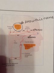
Parasympathetic preganglionic fibers arise in the Edinger-Westphal nucleus and project to the ciliary ganglion
|
|
|
Sympathetic innervation of eye
|
Sympathetic preganglionic fibers arise in T1 spine and project to superior cervical ganglion
|
|
|
Edinger-Westphal nucleus
|
Located in rostal midbrain (upper midbrain).
|
|
|
Pupillary light reflex
|
Parasympathetic response. Some light intensity signals from the optic nerve terminate in the pretectal area, which in turn projects to the EW nucleus, which then stimulates parasympathetic preganglionic fibers
|
|
|
Pretectum
|
Midbrain structure involved in visual function
|
|
|
Parasympathetic activity of the eye
|

Causes contraction of the circular muscle, constricting the pupil (miosis). Mediated by release of Ach onto a muscarinic receptor.
|
|
|
Sympathetic activity of the eye
|
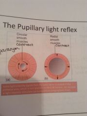
Not part of pupillary reflex, causes contraction of the radial/dilator muscles (mydriasis)
|
|
|
What stimulates accommodation?
|
Stimulated by chromatic aberration, convergence of eyes, differences in focus in fovea vs edges of retina. Near vision directed by stimulation from parasym. fibers in EW nucleus.
|
|
|
Accommodation
|
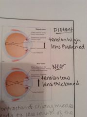
Contraction of ciliary body reduces suspensory ligaments tension and lens rounds up.
Object nearby - tension is low, lens thickened, rounds up Distant object - tension is high, lens flattened |
|
|
Chromatic aberration
|
A type of distortion in which there is a failure of a lens to focus all colors to the same convergence point, occurs b/c lenses have a diff refractive index for diff wavelengths of light.
|
|
|
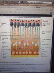
Organization of retina
|

Outer layer to inner layer:
Pigments cells, rods and cones, amacrine cells, horizontal cells, bipolar cells, ganglion cells. |
|
|
Pigment cells
|

Epithelial cells which prevent light scatter, phagocytose discs from rods, and recycle all-trans-retinal to 11-cis-retinal.
|
|
|
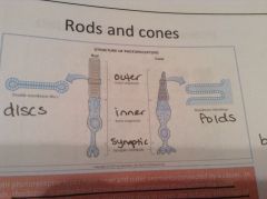
Rods and cones
|

Photoreceptors which transduce light to MEMBRANE potentials.
|
|
|
Amacrine cells
|

Regulate bipolar cells; provide most synaptic input to ganglion cells.
|
|
|
Horizontal cells
|

Mediate lateral inhibition of visual fields (see discussion in later part of this presentation).
|
|
|
Bipolar cells
|

Transmit ELECTRICAL potentials from photoreceptors to ganglion cells.
|
|
|
Ganglion cells
|
Send visual signals, in the form of ACTION potentials, to the brain. Note that all other visual signals up to this point are graded changes in membrane potential. This is possible b/c within the retina itself, electrical signals do not have great distances to travel.
|
|
|
Rods
|
Periodically shed renew and shed their discs; much higher rhodopsin content, and, thus, sensitivity to light.
|
|
|
Cones
|

Relatively low rhodopsin content; each cell requires several hundred photons to be activated; responsible for color vision by variable expression of one of three different photopigments.
|
|
|
Rhodopsin
|
Made up of opsin protein (GPCR) and retinal ligand
In rods - embedded across double-membrane discs w/n the outer segment cytoplasm. In cones - are actually infoldings of the outer segment cell membrane. |
|
|
Different types of rhodopsin
|
3 types of photopigments (red, green and blue) can be expressed in cones.
People w/colorblindness have a mutation where they only express 2 photopigments |
|
|
Distribution of photoreceptors
|
Cones are highly concentrated in the fovea (visual sharpness & acuity), rods are distributed throughout the peripheral retina (sensitive to low light conditions)
|
|
|
What is the best way to see a dim object?
|
Peripheral vision, where you use more rods than cones b/c they are more sensitive in low light and better for night vision.
|
|
|
Rods convergence
|
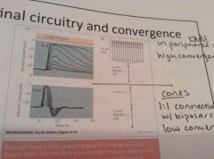
Rod cells have a much greater degree of convergence onto bipolar cells. 15-30 rods per 1 bipolar cell=high convergence. This results in a much larger receptive field for contacting bipolar cells (more sensitive) and lower visual acuity (not as sharp).
|
|
|
Cones convergence
|

1:1 ratio = low convergence, low sensitivity, small receptive field but high acuity
|
|
|
Retinal
|
Part of rhodopsin (along with opsin protein-GPCR) that responds to light by changing from cis to trans conformation.
|
|
|
Photoisomerization
|
The process of light converting retinal from cis to trans form. Once in trans form, the retinal causes the opsin protein to undergo a conformational change, which activates the coupled G protein and phototransduction occurs.
|
|
|
Returning retinal to cis form
|
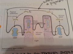
Once in trans form, retinal has to go through many steps to get back to cis form, is taken up by pigment cells and transformed back to cis form. This process requires vitamin A. A vitamin A deficiency will lead to night blindness.
|
|
|
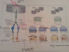
Phototransduction steps
|
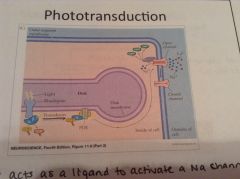
1. Rhodopsin is isomerized by light and opsin GPCR undergoes conformation change.
2. Transducin G protein senses this shift and is activated. 3. Transducin alpha subunit stimulates phosphodiesterase (PDE) embedded in disc membrane. 4. PDE hydrolyzes cyclic GMP to GMP. 5. Less cAMP is available to open cell membrane Na+ channels. 6. Cell is hyperpolarized and releases less neurotransmitter onto bipolar cells. |
|
|
Phototransduction in dark conditions
|

cGMP levels higher, Na+ channels open, cell depolarized and releasing neurotransmitter
|
|
|
Bipolar cell postsynaptic potentials
|
Photoreceptors only release glutamate onto bipolar cells, hyperpolarization or depolarization can both occur, it depends on the type of glutamate receptor expressed on the bipolar cell postsynaptic membrane.
|
|
|
On-center bipolar and ganglion cells
|

Fire when light is shone in center of receptive field
|
|
|
Off-center bipolar and ganglion cells
|

Fire when center of receptive field is dark.
|
|
|
Contrast
|
On-center and off-center cells make it possible to perceive contrasting light levels in the visual field. Contrast information makes up a huge proportion of what we see. Color requires encoding of much less information.
|
|
|
Lateral inhibition
|
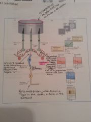
Horizontal cells antagonize the effects of the direct pathway (central cone to ganglion), release the inhibitory neurotransmitter, GABA, which causes center cone to release even more glutamate, which hyperpolarizes the bipolar cell even more, leading to less glutamate release onto ganglion cell=hyperpolarization of ganglion cell
|
|
|
Central visual pathway
|
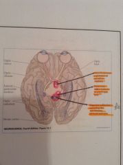
Ganglion cell axons form optic nerve, some cross at optic chasm.
Collateral branches end in hypothalamus and midbrain (pretectum and superior colliculus). Optic nerve fibers synapse in LGN of thalamus and neurons of the LGN project to visual cortex. |
|
|
Hypothalamus
|

Receives visual info, needed for photo-entrainment of light dark cycles (circadian rhythms)
|
|
|
Pretectum
|

Receives visual info for pupillary reflex and accommodation.
|
|
|
Superior colliculus
|

Receives visual info for orienting movements of head and eyes.
|
|
|
Lateral geniculate nucleus
|
Neurons project into the visual cortex
|
|
|
Retinal fields
|
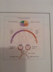
Left peripheral visual field sensed by left nasal retina; right peripheral visual field sensed by right nasal retina. Binocular (central )visual field sensed by both temporal retinas.
|
|
|
Optic chiasm
|

Decussation of axons from nasal retinas at the optic chiasm; right visual field represented in the left visual cortex and vice versa. Understand what this means for lesions of the pathway.
|
|
|
Hemianopia
|

Loss of vision in half of the visual field of one or both eyes.
|
|
|
Macular sparing
|
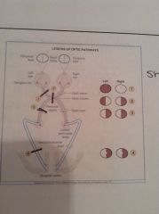
Caused by a lesion in the geniculocalcarine tract, missing part of vision, but area of macula is spared and can still see.
|
|
|
Primary visual cortex (V1)
|
Tissue has striped appearance, often V1 called the ‘striate cortex’; find neurons tuned to very basic components of a visual scene, such as line orientation (see figure).
|
|
|
Areas outside of V1
|
Note that visual areas just outside the visual cortex (extrastriate) are involved in sensing movement (MT area), attending to objects, and processing color (V4). Neural cells in extrastriate areas are typically responsive to visual information from both eyes.
|