![]()
![]()
![]()
Use LEFT and RIGHT arrow keys to navigate between flashcards;
Use UP and DOWN arrow keys to flip the card;
H to show hint;
A reads text to speech;
67 Cards in this Set
- Front
- Back
- 3rd side (hint)
|
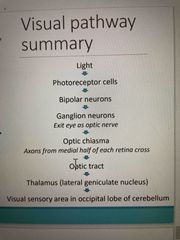
Visual Pathway Summary |
Receptor cells located in complex organs in the head region (ears, eyes, tastebuds, olfactory)what is our dominant
|
|
|
|
What is our dominant sense? |
Vision! 70% of body’s sensory receptors are in the eye and 30% cerebral neurons process visual information |
|
|
|
What does the eye consist of? |
Accessory Structures and the eyeball |
|
|
|
What are the Accessory Structures? |
Eyebrows, lids, conjunctiva, lacrimal apparatus, extrinsic eye muscles |
|
|
|
What is/does the Conjunctiva do? |
A transparent membrane that is surrounded by the inner surface of the eyelids and anterior surface of sclera *Produces lubricating mucus that prevents the eye from drying out |
|
|
|
Lacrimal Apparatus |
Contains the lacrimal gland (secretes dilute saline solution - tears) small ducts (drains excess fluid into nasolacrimal duct) *Produces lacrimal fluid (tears) (that contains antibodies, mucus, lysozyme and cleanses, moistens & protects eyes) |
|
|
|
What is the Pathway of Tears? |
Lacrimal gland -> lacrimal ducts -> lacrimal fluid flows over eye > lacrimal punctum - lacrimal canaliculus -> lacrimal sac -> nasolacrimal duct -> nasal cavity |
|
|
|
What controls movement of the eyeball? |
6 extrinsic eye muscles that are innervated by oculomotor, abducens, and trochlear nerves Functions: • Maintain eyeball shape, • Hold it in orbit • Precise eye movement |
|
|
|
What are the 3 layers of the eyeball? |
Fibrous Layer (outermost layer) Vascular Layer (middle layer) Retina Layer (inner layer) These layers enclose an internal cavity filled with fluids (humours) |
|
|
|
What are Humours? |
Internal cavities filled with fluids Function - Help maintain shape |
|
|
|
Fibrous Layer |
*outmost layer, is avascular Contains: -Sclera (opaque white) - maintains shape of eye and protects inner surface -Cornea (clear) - allows light to enter the eye and refracts light to focus on light rays on the retina |
|
|
|
Vascular Layer |
*middle layer, highly vascularized, contains melanin in the choroid Contains: -Choroid - darkly pigmented region that absorbs excess light and blood vessels that nourish posterior surface of retina -Ciliary Body - includes ciliary muscle (regulates lens shape), Ciliary Process (have blood capillaries that secrete aqueous humour) and suspensory ligaments that extend from ciliary processes to hold lens in shape *allows accommodations of lens for near or far vision -Iris - the pigmented part of the eye (contains melanocytes) and contains circular & radial smooth muscle that reflexively controls pupil size & amount of light that enters the eye |
|
|
|
Pupil constricts and dilated when… |
Constricts - as sphincter pupilae muscles contract (parasympathetic) due to bright light Dilates - as dilator muscles contract (sympathetic) due to dim light |
|
|
|
What is the retina? |
At the back of the eye where you have projections of light |
|
|
|
What is the retina? |
At the back of the eye where you have projections of light |
|
|
|
What are the 2 layers of the retina? |
Pigmented Layer - absorbs excess light to reduce scattering (is closest to choroid)
Neural Layer - the visual layer with 3 layers of retinal Neurons (photoreceptor layer, bipolar cell layer, ganglion cell layer). |
|
|
|
What does the photoreceptor layer of the neural layer do? |
The Photoreceptor Layer contains 2 diff photoreceptors - Rods & Cones |
|
|
|
What are the aspects & functions of Rods? |
Aspects - rods are highly sensitive, more numerous than cones Functions - rods are used in peripheral vision, are suited for night vision Can’t - resolve colour or sharp images |
|
|
|
What are the aspects & functions of Rods? |
Aspects - rods are highly sensitive, more numerous than cones Functions - rods are used in peripheral vision, are suited for night vision Can’t - resolve colour or sharp images |
|
|
|
What are the special aspects & functions of Cones? |
Aspects - cones are less sensitive, and 3 types (blue, red, green) Function - provide high resolution colour vision and are best adapted to bright light Poor in dim light |
|
|
|
Special parts of the retina’s Neural Layer includes… |
1) Optic Disc - blind spot at the back of the retina, where there are NO photoreceptors and the optic nerve exits the eye 2) Macula Lutea - is lateral to the blind spot and at its centre contains the Fovea Centralis, which has the highest density of cones for detailed colour vision, and can move our eyes to focus image on fovea* |
|
|
|
The Bipolar Layer of the retina’s Neural Layer does what? |
Bipolar cells in the layer relay info from photoreceptors (rods & cones) to ganglion cells |
|
|
|
What does the Ganglion Layer of the retina’s Neural Layer do? |
Ganglion cells in the layer take info from bipolar cells then group together (converge) at the optic disc and exit the eye through the optic nerve to send info to brain |
|
|
|
The eyeball’s Lens - characteristics, function |
Characteristics - the lens is avascular & biconvex structure, is held in place by suspensory ligaments, is flexible Function - changes shape to focus image *cells contain transparent crystalline protein |
|
|
|
What is a cataract? |
The clouding of the lens due to changes in the crystalline proteins (clumping) |
|
|
|
What determines the shape of the lens? |
Whether the suspensory ligaments are contracted or relaxed |
|
|
|
What 2 main parts are there in the interior of the eyeball? |
1) Anterior Cavity - 2) Posterior Cavity - |
|
|
|
What are the characteristics/function of the Anterior Cavity? |
Characteristics - anterior cavity is in front of the lens and contains a continuous supply of Aqueous Humor that is continuously removed (provides oxygen & nutrients to lens, cornea; removes wastes) Function - maintains intraocular pressure to support eyeball internally |
|
|
|
What causes Glacoma? |
When the drainage of aqueous humor is blocked, it leads to increased pressure that compresses the retina & optic nerve, leading to blindness |
|
|
|
What are the characteristics/function of the eyes Posterior Cavity? |
Characteristics - posterior cavity is behind the lens, contains Vitreous Humor (gel-like substance formed during embryonic life and isn’t replaced. Also holds the retina in place) Function - it maintains intraocular pressure and holds the retina in place |
|
|
|
What contributes to Image Formation? |
1) Refraction of Light Rays - light refracts as it passes through objects of differing optical densities *diff in these optical densities b/w air & cornea is what helps to bend the light 2) Focusing light rays on the retina - involves the cornea & lens -Cornea - has greatest role in focusing image (75%). A large diff in optical density bw air & corneal tissue. Cornea has a fixed curvature -Lens - fine tunes the focus (25%). The transparent, flexible structure is able to change shape to allow precise focusing of light onto retina |
|
|
|
What does laser eye surgery do? |
Corrects how your cornea is bending the light |
|
|
|
What happens when looking at a distant object- Distant Vision? |
-Ciliary Muscles are relaxed (which decreases their width & increases their length), the ciliary muscles increase tension on suspensory ligaments, which causes the lens to flatten Far point of vision - the distance beyond which no change in lens shape (accomodation) is required. 6m in a normal eye |
|
|
|
When does the direction of light waves change? |
When there’s a change in the distance of the object (whether the object is far or close to you) As the object gets closer. The light has to bend further |
|
|
|
Close Vision - What happens when looking at a close object? |
There are 3 adjustments: 1) Accommodation of lens - thickens & increases light refraction. Involves the contraction of ciliary muscle (increases width), loosening tension on suspensory ligaments, and lens becomes more rounded. 2) Constriction of Pupils - better directs light to lens 3) Convergence of Eyeballs - allows object to remain focused on foveae |
|
|
|
When does Near Point of Vision occur? |
It’s when your eyes can’t reflect light any further. Occurs at the point of maximal thickening of lens (about 10cm/4” |
|
|
|
What are 3 main types of vision problems |
Myopia (near sighted) Hyperopia (far sighted) Astigmatism |
|
|
|
Myopia (near-sighted) |
Objects focus in front of retina & the Eyeball is too long or lens too curved/thick • Close objects are seen clearly; distant objects blurred • Corrected by decreasing refraction (concave lens) |
Bends light too much |
|
|
Hyperopia (far-sighted) |
• Objects focus behind retina • Eyeball too short or lens too flat/thin • Distant objects seen clearly; close objects blurred • Correct by increasing refraction (convex lens) |
Doesn’t bend light enough |
|
|
Astigmatism |
• Irregular curvature of lens or cornea; produces blurred (out of focus) images |
|
|
|
VISION II |
January 6th |
|
|
|
What form does Electromagnetic Radiation exist in? |
In waves - long radio waves to short X-rays |
|
|
|
What is wavelength? |
The distance b/w waves. Particularly important for vision. We can see 400-700 nm wavelengths b/c the pigment in our eyes is designed to detect those wavelengths. |
|
|
|
We perceive diff wavelengths as… |
Different colours!! Visible light is - 400-700nm Violet is 400nm and reds are 700nm |
|
|
|
Colours are reflections of wavelengths b/c… |
Objects absorb some wavelengths and reflect the colours we see.
Ex - grass is green, bc all other colours were absorbed by the object, and green is reflected and absorbed by our green cones |
|
|
|
What is Phototransduction? |
A process in which light energy produces graded receptor potentials - yielding nerve impulses in rod & cone cells in the retina |
|
|
|
What are Photoreceptors? (Rods & Cones) |
Modified neurons w/ photoreceptive ends inserted into the pigmented layer of retina. They detect light. * contain photopigments that change shape as they absorb light (found in folds (cones) or discs (rods)) *are destroyed by intense light *outer-segment is renewed every 24 hours |
|
|
|
Rods during Phototransduction |
Rods have a single pigment (perception of one colour) -many rods converge into one ganglion - creating a fuzzy, in distinct image As light comes in it activates photopigments in rods, which activates bipolar cells, then ganglion cells. Ganglion Cells’ axons form the optic nerve that sends msg to the brain. Involves integration - taking info from diff areas, sending it out, producing a response. Convergence - bipolar cells pool info into ganglion cells. Going from a large number of cells to a smaller number of cells * Convergence of info creates a less clear image |
|
|
|
Cones during Phototransduction |
Cones involve: 3 pigments (vivid colour detection) Less convergence (sometimes having own ganglion cells) - results in high resolution *less sensitive Connects w a single bipolar cell and a single ganglion cell. NO convergence |
|
|
|
Visual Pigments in Phototransduction include |
Photopigments (retinal & opsin) Rods - contain a single opsin mixed w retinal, rhodopsin Cones - opsins named after wavelengths they absorb, also combined w retinal (red, green, blue) |
|
|
|
What is Retinal? |
Retinal is a light absorbing pigment (chromosphere; vitamin A derivative) |
|
|
|
What is Opsin? |
Opsin is a G Protein-coupled receptor. Called this because it activates something else It changes shape in molecule which activates other things *the diff in amino acid sequences of diff opsins creates the sensitivity to diff wavelengths |
|
|
|
Phototransduction - when cone wavelengths overlap…. |
One wavelength can activate more than one cone and perceive a variety of hues! Each cone detects a specific range, which can overlap with other cones wavelengths. Therefore we activate multiple cones |
|
|
|
Red & Green Colorblindnesscyc |
You lack red or green cones due to a mutation in either gene. If you’re missing one cone, you won’t be able to distinguish bc red & green cones overlap so much Genes for red & green opsins are located very close to one another on the X chromosome • High overlap between perceived wavelengths |
|
|
|
Process of Visual Transduction/Phototransduction includes 4 steps |
1) Isomerization - cis-retinal changes to trans-retinal when it absorbs light (physically changes shape, which leads to… 2) Bleaching - trans-retinal (colour portion) separates from opsin - goes from being coloured to white, bc the pigment leaves 3) Conversion - Retinal isomerase (an enzyme that allows trans-retinal to convert back to cis-retinal - allows it to eventually rejoin opsin) 4) Regeneration - Once converted back to cis-retinal it rebinds to opsin |
|
|
|
Key parts of Visual Transduction |
There is a molecular change in the retinal that leads to bleaching & being able to see again. You need the molecule to go back to the original formation & the enzyme, Retinal Isomerase to help with that. And 2 molecules that make up photopigments (opsin and cis-retinal) rejoin to absorb light again |
|
|
|
The Visual Field |
Visual field detected by different portions of retina • Left visual field strikes right part of retina • Right visual field strikes left part of retina Retina is divided into lateral & medial sections • Neurons from medial retina cross over at optic chiasm • Left lateral geniculate nucleus -> right visual field • Right lateral geniculate nucleus -> left visual field * Involves pooling info from your visual field to parts of brain What is on left of visual field projects to right side, and right to left. Parfocal. Can divide retina in half, and half of neurons cross over, but other half stay on same side. You're reorienting the neurons to diff pathways so your grouping together diff portions of your visual field. |
|
|
|
Binocular Zone |
• Overlap between right & left visual fields • Area processed on both sides of brain -> allows for comparison of image properties from each eye • One of the processes that underlies depth perception *Both of your eyes are able to visualize at once. B/c each of your eyes is seeing parts of the same image - the amount each eye is seeing of that image, helps your brain to understand depth. |
|
|
|
What happens if you’re in continual bright light? |
Molecules won’t go back to original spot until we leave bright light |
|
|
|
Review of Membrane Potentials |
Neuron communication signals: Graded potentials - usually incoming signals that travel short distances. Vary in magnitude (de/hyper-polarizing) Action potentials - all-or-none signal that travel long distances along axon. Same size & duration Changes from resting membrane potential: • Depolarization - less negative (excitatory) • Hyperpolarization - more negative (inhibitory) |
|
|
|
Membrane Potentials & Special Senses |
This model of graded potentials leading to action potential is found throughout the sensor system Rods & cones are the exception • Dark - depolarized at rest (more positive) about -30 • Light - hyperpolarized when activated (more negative) |
|
|
|
Photoreceptors in the Dark |
Photoreceptors are slightly depolarized • Ion (Na*) channels are open - cell is more positive • Channels are opened in response to high cGMP levels (produced by enzyme) Rods & cones release inhibitary neurotransmitter (when cell is depolarized) (glutamate) which activates voltage gated calcium channels (calcium is the trigger for the release of the NT from the synaptic vesicles). • Inhibits Bipolar cell cannot stimulate the ganglion cell (inhibits visual response in the dark) |
|
|
|
Photoreceptors in the Light |
When light is present it activates a chain of events leading to NT being released. Light inhibits the response, creating a Hyperpolarization in the cell. It does this by activating the gprotein, transducin, which activates enzyme, phosphodiesterase (breaks down cyclic GMP). Closes sodium channels, leading to less sodium entering and the cell hyperpolarizing (becoming more negative). With less NT released, the bipolar cell gets less of an inhibitory stimulation & is excited. Bipolar cell can now stimulate the ganglion cell, sending a msg to the brain. Bleaching of pigment hyperpolarizes the photoreceptors • Ion (Na*) channels are closed -> cell is more negative • Phosphodiesterase is activated - enzyme that breaks down cGMP • Channels are closed in response to low cGMP levels Decreased release of inhibitory neurotransmitter to bipolar cell • Bipolar cell stimulates the ganglion cell (results in visual response) |
|
|
|
**Concept b/w Dark and Bright Light |
*There's an inhibitory neurotransmitter released by rods and cones acting on bipolar cells, then ganglion - amount released depends on whether it's dark or light, and how bright that light is. In dim light a hyperpolarization occurs (less inhibitory signal). In bright light it's more hyperpolarized - a higher degree of action potentials that flow to the brain. |
|
|
|
Photoreceptors in Light |
Extent of light determines the magnitude of the response (receptor potential) Dim light - partially turn off inhibitory NT release Bright light - turn off inhibitory NT more completely |
|
|
|
Light & Dark Adaptation |
Has to do with us, switching from using cones and rods and vice versa When we walk from dark to bright light - rods are very sensitive and absorb all the light, becoming completely saturated, quickly becoming bleached. (Completely saturated by bright light). Light adaptation (darkness -> bright light) • Initially, both rods & cones strongly stimulated • Large amounts of photopigment broken down - glare • Pupils constrict to decrease light reaching retina • ~5-10 mins - increase visual acuity & decrease retinal sensitivity as rods turn off Going from bright light to darkness - rods need time to reactivate. Transretinal needs to go back to cisretinal and bind to opsin. Dark adaptation (bright light -> darkness) • Cones cannot function in low-intensity light • Previously bleached rods require time to reactivate • Pupils dilate to increase light reaching retina • increase rhodopsin in dark, max increase visual acuity at 20-30 min |
|
|
|
Visual Perception involves… |
Optic nerve - retinal ganglion cells merge in back of eyeball Optic chiasma - crossing of optic nerves Optic tracts - from optic chiasma to thalamus • Lateral geniculate nucleus of thalamus Optic radiations - project to primary visual cortex in occipital lobes for visual processing |
|