![]()
![]()
![]()
Use LEFT and RIGHT arrow keys to navigate between flashcards;
Use UP and DOWN arrow keys to flip the card;
H to show hint;
A reads text to speech;
38 Cards in this Set
- Front
- Back
|
What is the overall purpose of the visceral nervous system? |
To maintain homeostasis within our body. |
|
|
General visceral afferent (GVA) fibers carry information from the body core, from our internal organs, but not from the _____. |
Periphery |
|
|
What are the two components of the general visceral efferent system?
Which of the two innervates core and periphery? Which only core?
Which system is responsible for gut motility? |
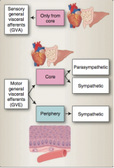
Parasympathetic (core only) and sympathetic (core and periphery) ==> autonomic nervous system
Enteric nervous system |
|
|
What are the three visceral receptors? What does each detect? |
Nociceptors (pain) Mechanoreceptors (fullness) Specialized receptors (internal physical or chemical environment) |
|
|
Where are the unipolar cell bodies of visceral afferent neurons located? |
Ganglia of spinal nerves (T1-L2; S2-S4) Ganglia of cranial nerves IX and X |
|
|
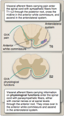
Afferents carrying pain are carried mainly along ______ efferents, whereas information from both the mechanoreceptors and specialized receptors related to physiological functions are carried mainly along ______ efferents. |
Afferents carrying pain are carried mainly along sym- pathetic efferents, whereas information from both the mechanoreceptors and specialized receptors related to physiological functions are carried mainly along parasympathetic efferents. |
|
|
The cell bodies of the visceral afferents related to pain are localized pri- marily in the spinal ganglia from T1 to L2 and travel along sympathetic efferent fibers. Afferent fibers enter the posterior horn, and the majority cross the midline and travel with the ______ on the con- tralateral side (Figure 4.2). These fibers terminate in the ventral postero- lateral nucleus of the thalamus, from which they project to the _______, where the sensation of ______ is interpreted. Like other vis- ceral sensations, visceral pain is poorly localized and often experienced as referred pain to superficial somatic structures (dermatomes). |
The cell bodies of the visceral afferents related to pain are localized pri- marily in the spinal ganglia from T1 to L2 and travel along sympathetic efferent fibers. Afferent fibers enter the posterior horn, and the majority cross the midline and travel with the anterolateral system on the con- tralateral side (Figure 4.2). These fibers terminate in the ventral postero- lateral nucleus of the thalamus, from which they project to the insular cortex, where the sensation of visceral pain is interpreted. Like other vis- ceral sensations, visceral pain is poorly localized and often experienced as referred pain to superficial somatic structures (dermatomes). |
|
|
Where can visceral afferents concerned with physiological functions arise?
What is unusual about these? |
Pelvic nerves (S2-S4) or CNs
Enter spinal cord along parasympathetic efferents through ANTERIOR ROOT |
|
|
Fibers carrying visceral afferent information from sacral areas ascend _____ through the anterolateral system and _____ through the spinoreticular system, as described for the fibers enter- ing the spinal cord at T1–L2. |
Fibers carrying visceral afferent information from sacral areas ascend contralaterally through the anterolateral system and bilaterally through the spinoreticular system, as described for the fibers enter- ing the spinal cord at T1–L2. |
|
|
Visceral afferents from the thoracic and most abdominal viscera (down to the left colic flexure) are carried by the ______, which also provides the visceral efferent (motor, parasympathetic) innervation of these organs. The associated mechanoreceptors are located in the smooth muscles of the thoracic and abdominal organs and detect _____ and _____. CN IX, the _____ nerve, carries visceral afferent information from chemoreceptors and baroreceptors in the carotid sinus, which detect changes in blood gases and blood pressure. CN IX also carries visceral afferent information from the pharynx and is the afferent limb of the _____ reflex. |
Visceral afferents from the thoracic and most abdominal viscera (down to the left colic flexure) are carried by the vagus nerve (CN X), which also provides the visceral efferent (motor, parasympathetic) innervation of these organs. The associated mechanoreceptors are located in the smooth muscles of the thoracic and abdominal organs and detect fullness and cramps. CN IX, the glossopharyngeal nerve, carries visceral afferent information from chemoreceptors and baroreceptors in the carotid sinus, which detect changes in blood gases and blood pressure. CN IX also carries visceral afferent information from the pharynx and is the afferent limb of the gag reflex. |
|
|
Where do signals of the visceral motor system originate? What can influence autonomic neurons? |
Hypothalamus
Relay nuclei, amygdala |
|
|
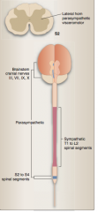
Preganglionic cell bodies of the sympathetic system can be found in the lateral horn of the spinal cord from levels __ to __. The preganglionic parasympathetic cell bodies are found both ___ and ____ to these sympathetic cell bodies (“para” to the sympathetics) in the brainstem and in the sacral spinal cord at S_–S_. |
Preganglionic cell bodies of the sympathetic system can be found in the lateral horn of the spinal cord from levels T1 to L2. The preganglionic parasympathetic cell bodies are found both rostral and caudal to these sympathetic cell bodies (“para” to the sympathetics) in the brainstem and in the sacral spinal cord at S2–S4 |
|
|
Moreover, as the vis- ceral afferents (sensory fibers) travel through the visceral ganglia, a cer- tain degree of ______ integration takes place in these ganglia. This allows visceral motor fibers to respond directly to visceral sensory input without influence from the ______, which gives the visceral nervous system additional autonomy and the ability to respond more quickly. |
Moreover, as the vis- ceral afferents (sensory fibers) travel through the visceral ganglia, a cer- tain degree of sensory-motor integration takes place in these ganglia. This allows visceral motor fibers to respond directly to visceral sensory input without influence from the CNS, which gives the visceral nervous system additional autonomy and the ability to respond more quickly. |
|

|
|
|
|
True or false:
Sympathetic ganglia are located close to the CNS, whereas parasympathetic ganglia are typically located close to or within the organs they will innervate. |
True |
|
|
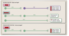
What do the sympathetic and parasympathetic systems use to activate their postganglionic neurons? |
Acetylcholine |
|
|
Sympathetic neurons that project to the periphery to innervate sweat glands, blood vessels, and the erector pili muscles also use _____ as their neurotransmitter. By contrast, postganglionic sympathetic fibers to the core (internal organs) use the neurotransmitter _____, which decreases gut motility and increases cardiac muscle contraction |
Sympathetic neurons that project to the periphery to innervate sweat glands, blood vessels, and the erector pili muscles also use ACh as their neurotransmitter. By contrast, postganglionic sympathetic fibers to the core (internal organs) use the neurotransmitter noradrenaline, which decreases gut motility and increases cardiac muscle contraction |
|
|
One subset of sympathetic neurons projects directly to the adrenal medulla, without synapsing in a peripheral ganglion. These neu- rons use the neurotransmitter _____ and cause the secretion of hormones from the ______ cells in the adrenal medulla. Chromaffin cells are of the same embryological origin as the peripheral ganglia (from the neural crest) and secrete norepinephrine, _____, and endorphins into the blood stream, causing a widespread sympathetic (“fight-or-flight”) response. |
One subset of sympathetic neurons projects directly to the adrenal medulla, without synapsing in a peripheral ganglion. These neu- rons use the neurotransmitter ACh and cause the secretion of hor- mones from the chromaffin cells in the adrenal medulla. Chromaffin cells are of the same embryological origin as the peripheral ganglia (from the neural crest) and secrete norepinephrine, epinephrine, and endorphins into the blood stream, causing a widespread sympathetic (“fight-or-flight”) response. |
|
|
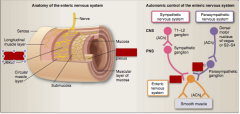
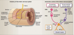
The ______ system comprises a complex plexus of neurons within the gastrointestinal system that is autonomous but under the influence of the parasympathetic and sympathetic nervous systems. |
The enteric nervous system comprises a complex plexus of neurons within the gastrointestinal system that is autonomous but under the influence of the parasympathetic and sympathetic nervous systems. |
|
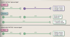
Identify which is core and which is periphery. |
|
|
|
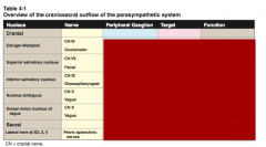
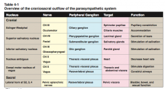
The first-order neurons in the parasympathetic system are located in the ____ and _____ regions of the CNS (craniosacral outflow) (Figure 4.5). Parasympathetic innervation to the eye is from the Edinger- Westphal nucleus (CN ___, oculomotor). The preganglionic parasympa- thetic neurons innervating the salivary glands in the head are found in the _____: the superior salivatory nucleus (CN ___, facial) and the inferior salivatory nucleus (CN ___, glossopharyngeal). |
The first-order neurons in the parasympathetic system are located in the cranial and sacral regions of the CNS (craniosacral outflow) (Figure 4.5). Parasympathetic innervation to the eye is from the Edinger- Westphal nucleus (CN III, oculomotor). The preganglionic parasympa- thetic neurons innervating the salivary glands in the head are found in the brainstem: the superior salivatory nucleus (CN VII, facial) and the inferior salivatory nucleus (CN IX, glossopharyngeal). |
|
|
The dorsal motor nucleus of the vagus contains the cell bodies of the parasympathetic neurons that will travel with CN __ (vagus) to all the thoracic and abdominal viscera (up to the left colic flexure). The ______ contains parasympathetic cell bodies for the innervation of cardiac muscle. These fibers also travel with CN __. This latter is an interesting exception, as the nucleus ambiguus primarily sends output to _____ muscles of the phar- ynx and larynx. |
The dorsal motor nucleus of the vagus contains the cell bodies of the parasympathetic neurons that will travel with CN X (vagus) to all the thoracic and abdomi- nal viscera (up to the left colic flexure). The nucleus ambiguus contains parasympathetic cell bodies for the innervation of cardiac muscle. These fibers also travel with CN X. This latter is an interesting exception, as the nucleus ambiguus primarily sends output to skeletal muscles of the phar- ynx and larynx (see Chapter 10, “Sensory and Motor Innervation of the Head”). |
|
|
The cell bodies of the preganglionic parasympathetic fibers that innervate the ____ colon and the pelvic viscera are located in the _____ horn of the spinal cord (S_–S_). Their second-order neurons are located in specific ganglia located close to or within the walls of the target organ. |
The cell bodies of the preganglionic parasympathetic fibers that innervate the descending colon and the pelvic viscera are located in the lateral horn of the spinal cord (S2–S4). Their second-order neurons are located in specific ganglia located close to or within the walls of the target organ. An overview of the peripheral ganglia can be seen in Figure 4.5. |
|
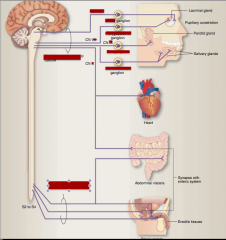
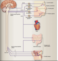
Identify the cranial nerve and the ganglion: CN III, VII, IX, X, submandibular, otic, pterygopalatine, ciliary, sacral parasympathetic outflow, cranial parasympathetic outflow.
|
|
|
|
Where does sympathetic outflow originate? |
Spinal cord from lateral horn (T1-L2) Preganglionic neurons leave through the anterior root. |
|
|
What connects preganglionic fibers to sympathetic trunk?
What contains cel bodies of postganglionic fibers? |
White communicating ramus Chain of ganglia |
|
|
Sympathetics destined for the head region will ascend in the sympathetic trunk and SYNAPSE in the ______ ganglion. From here, the _____ fibers will travel into the head region along the surface of the _____ artery. The axons to abdominal and pelvic viscera will travel through the sym- pathetic chain without _____ and proceed via _____ nerves to specific preaortic ganglia to synapse on their postganglionic target neurons. Sympathetics destined for the trunk and limbs synapse in the ganglia of the sympathetic chain and send postganglionic fibers via _____communicantes (gray because they are unmyelinated) to be distributed with the spinal nerves to their effector organs. |
Sympathetics destined for the head region will ascend in the sympathetic trunk and synapse in the supe- rior cervical ganglion. From here, the postganglionic fibers will travel into the head region along the surface of the internal carotid artery. The axons to abdominal and pelvic viscera will travel through the sym- pathetic chain without synapsing and proceed via splanchnic nerves to specific preaortic ganglia to synapse on their postganglionic target neurons. Sympathetics destined for the trunk and limbs synapse in the ganglia of the sympathetic chain and send postganglionic fibers via gray rami communicantes (gray because they are unmyelinated) to be distributed with the spinal nerves to their effector organs. |
|
|
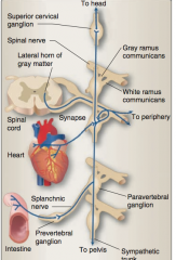
Identify superior cervical ganglion, spinal nerve, gray ramus communicans, white ramus communicans, splanchnic nerve, prevertebral ganglion, paravertebral ganglion, sympathetic trunk. |
|
|
Identify myenteric and submucosal plexus. Identify whether inhibitory or stimulatory and neurotransmitter. |
|
|
|
Which is the main controller of gut motility? Which is the main controller of absorption of fluids?
Myenteric, Submucosal |
Myenteric Submucosal |
|
|
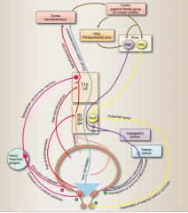
Descending pathways from the ________ regulate the visceral motor system. The hypothalamus sends projections to the visceral motor (parasympathetic) nuclei in the brainstem (of CNs VII, IX, and X) as well as to neurons in the lateral horn in spinal cord segments T1–L2 (sympathetic) and S2–S4 (parasympathetic). |
Descending pathways from the hypothalamus regulate the visceral motor system. The hypothalamus sends projections to the visceral motor (parasympathetic) nuclei in the brainstem (of CNs VII, IX, and X) as well as to neurons in the lateral horn in spinal cord segments T1–L2 (sympathetic) and S2–S4 (parasympathetic). |
|
|
The main tracts that influence the visceral motor neurons are the _____ and _______ . These fibers originate mainly from the _____ nucleus. They descend through the ______ (PAG) and nearby reticular formation of the midbrain and rostral pons and then move anterolaterally in the medulla. The hypothalamomedullary fibers terminate on all brainstem nuclei containing the cell bodies of GVE fibers (Table 4.1 and Figure 4.8). The _____ fibers continue through the _____ medulla into the lateral column of the spinal cord, ending in the _____ horn. These two tracts are an essential link between the hypothalamus and the autonomic nuclei of the brainstem and spinal cord. |
The main tracts that influence the visceral motor neurons are the hypothalamospinal and hypothalamomedullary tracts. These fibers originate mainly from the paraventricular nucleus. They descend through the periaqueductal gray (PAG) and nearby reticular formation of the midbrain and rostral pons and then move anterolater- ally in the medulla. The hypothalamomedullary fibers terminate on all brainstem nuclei containing the cell bodies of GVE fibers (Table 4.1 and Figure 4.8). The hypothalamospinal fibers continue through the anterolateral medulla into the lateral column of the spinal cord, end- ing in the lateral horn. These two tracts are an essential link between the hypothalamus and the autonomic nuclei of the brainstem and spinal cord. |
|
Complete |
|
|
|
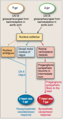
Where is blood pressure monitored?
What are the two cranial nerves involved with each location?
Where does the information travel? |
Baroreceptors in the carotid sinus and aortic arch
CN IX (carotid sinus) CN X (aortic arch)
To the nucleus solitaries |
|
|
Draw the pathway that follows increased or decreased blood pressure. Use the cranial nerves, nucleus solitarius, nucleus ambiguous, dorsal motor nucleus of vagus, rostral ventroloateral medulla, preganglionic sympathetic neurons in intermediate horn, etc. |
|
|
Draw this out. |

|
|

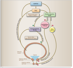
Draw the control of micturition |
|
|
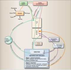
Draw this. |

|