Reading...
![]()
Play button
![]()
Play button
![]()
Use LEFT and RIGHT arrow keys to navigate between flashcards;
Use UP and DOWN arrow keys to flip the card;
H to show hint;
A reads text to speech;
131 Cards in this Set
- Front
- Back
|
What are the 3 layers of the eye?
|
FIBEROUS TUNIC is the outermost layer consisting of schelera and cornea
-VASCULAR TUNIC-middle layer- middle vascularized layer which includes the iris, ciliary body, and choroid. The choroid contains blood vessels that supply the retinal cells with necessary oxygen and remove the waste products NERVOUS TUNIC-the inner sensory which includes the retina |
|
|
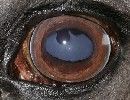
What is the sclera?
|
The sclera is the WHITE outer wall of the eye. It is a tough, fibrous tissue that extends from the cornea (the clear front section of the eye) to the optic nerve at the back of the eye. The sclera gives the eye its white color.
|
|
|
What is the cornea?
|
The clear front window of the eye that transmits and focuses light into the eye
*Although the cornea is clear and seems to lack substance, it is a highly organized group of cells and proteins. Unlike most tissues in the body, the cornea contains no blood vessels to nourish or protect it against infection. Instead, the cornea receives its nourishment from the tears and aqueous humor that fills the chamber behind it. The cornea must remain transparent to refract light properly, and the presence of even the tiniest blood vessels can interfere with this process. To see well, all layers of the cornea must be free of any cloudy or opaque areas. |
|
|
What is the uvea?
|
Part of the eye, consisting collectively of the iris, the choroid of the eye, and the ciliary body. Supplies nutrition to the eye, smooth muscles control--shape of lens and size of pupil.
The iris: The circular, colored curtain of the eye that surrounds the pupil. The choroid of the eye: The thin vascular middle layer of the eye that is situated between the sclera (the white of the eye) and the retina (the nerve layer that lines the back of the eye, senses light, and creates impulses that travel through the optic nerve to the brain). The ciliary body: A body of tissue that connects the iris with the choroid and includes a group of muscles which act on the lens of the eye to change its shape. |
|
|
What is the tapetum lucidum?
|
is a layer of tissue in the eye of many vertebrate animals, that lies immediately behind or sometimes within the retina. It reflects visible light back through the retina, increasing the light available to the photoreceptors. This improves vision in low-light conditions, but can cause the perceived image to be blurry from the interference of the reflected light.[citation needed] The tapetum lucidum contributes to the superior night vision of some animals
|
|
|
What is pecten?
|
a pigmented vascular membrane with parallel folds suggesting the teeth of a comb , projecting into the vitreous humor of the eye in birds and reptiles.
Distributes nutrition to the eye |
|
|
What is the ciliary body?
|
Thickest layer of vascular tunic-located between choroid and iris-
is a thin vascular (blood vessel-filled) middle layer of the eye that is situated between the sclera (the white of the eye) and the retina (the nerve layer that lines the back of the eye, senses light, and creates impulses that travel through the optic nerve to the brain). |
|
|
What are the ciliary muscles?
|
One of the muscles that relax the zonules to enable the lens to change shape for focusing.
The zonules are fibers that hold the lens suspended in position and enable it to change shape during accommodation. |
|
|
What is the ciliary process?
|
The ciliary processes produce aqueous humor.
This is an important junction where the iris and the sclera meet. Close by is the circular canal of Schlemm, which runs around the eye just below the limbus. Aqueous humour is exuded from secretory cells just below the pigment epithelium in the cauliflower-like ciliary processes. The aqueous humour drains through the Zonnules of Zinn to the posterior chamber and through the pupil to the anterior chamber. The fibrous Zonnules of Zinn, which support the lens, are attached to the valleys between the ciliary processes. |
|
|
What does the suspensory ligament of the eye do?
|
-Attach lens to ciliary body
-hold lens in place -Accomodation-controls shape of lens The suspensory ligament forms a hammock stretching below the eyeball between the medial and lateral check ligaments and enclosing the inferior rectus and inferior oblique muscles of the eye. The ligament supports the eye,[1] and prevents downward displacement of it. It can be considered a part of the bulbar sheath. |
|
|
What is the function of the Iris?
|
A colored circular muscle, the iris, which is beautifully pigmented giving us our eye's color (the central aperture of the iris is the pupil). This circular muscle controls the size of the pupil so that more or less light, depending on conditions, is allowed to enter the eye.
|
|
|
What is the function of the pupil?
|
The iris acts like the shutter of a camera. In the middle of a normal iris is the pupil, typically a circular hole, comparable to the aperture of a camera. The pupil regulates the amount of light passing through to the retina, which is at the back of the eye.
As the amount of light entering the eye diminishes (such as in a dark room or at night), the iris dilator muscle (which runs radially through the iris like spokes on a wheel) pulls away from the center, causing the pupil to “dilate.” This allows more light to reach the retina. When too much light is entering the eye, the iris sphincter muscle (which encircles the pupil) pulls toward the center, causing the pupil to “constrict” and allowing less light to reach the retina. |
|
|
What is the function of the retina?
|
is the nerve layer that lines the back of the eye, senses light, and creates impulses that travel through the optic nerve to the brain. There is a small area, called the macula, in the retina that contains special light-sensitive cells. The macula allows us to see fine details clearly.
|
|
|
What is the optic disc?
|
Area of retina where axons leave the eye to optic nerve. Is a blind spot that has no rods or cones.
The circular area in the back of the inside of the eye where the optic nerve connects to the retina. Also called the optic nerve head. |
|
What is the corpora nigra?
|
these are masses of modified iridal tissue attached at the pupillary border and composed of melanotic cells, blood vessels, and fluid-filled spaces. They usually have a lobulated appearance and are present only in herbivores. The dorsal ones usually are larger; the ventral ones may be poorly developed or absent.
|
|
|
What is the lens?
|
By changing its shape, the lens focuses light onto the retina. Through the action of small muscles (called the ciliary muscles), the lens becomes thicker to focus on nearby objects and thinner to focus on distant objects.
|
|
|
What is accomodation?
|
Increase in optical power by the eye in order to maintain a clear image (focus) as objects are moved closer. Occurs through a process of ciliary muscle contraction and zonular relaxation that causes the elastic-like lens to "round up" and increase its optical power.
|
|
|
What are rods sensitive to?
|
dim light and shapes
|
|
|
What are cones sensitive to?
|
color and sharpness
|
|
|
What is the aqueous humor and what is its function?
|
Clear, watery fluid that fills the space between the back surface of the cornea and the front surface of the vitreous, bathing the lens. Produced by the ciliary processes. Nourishes the cornea, iris, and lens and maintains intraocular pressure.
|
|
|
What is the vitreous humor?
|
Transparent, colorless gelatinous mass that fills the rear two-thirds of the eyeball, between the lens and the retina.
|
|
|
Accessory structures of the eye are called?
|
Adnexa
|
|
|
Photoreceptors are
|
the light sensitive first layer of the eye and is made up of rods and cones
|
|
|
What is the Macula lutea?
|
An area that is densely packed with rods and cones in caudal part of the retina
is located in central part of the fundus where light hits directly |
|

Eye and muscles
|
fyi
|
|

Eye structures
|
fyi
|
|
eye & eyelids
|
fyi
|
|
|
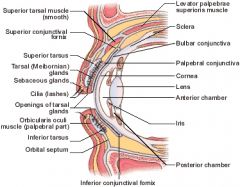
What is the difference between the Anterior and Posterior cavities?
|
ANTERIOR cavity is everything anterior/in front of the lens and is filled with AQUEOUS Humor.
POSTERIOR Cavity is everything between lens and retina and is filled with VITREOUS humor. |
|
|
What 2 structures make up the Anterior cavity of the eye?
|
ANTERIOR CHAMBER-comprised of anything between cornea and iris
POSTERIOR CHAMBER: Located between the iris, lens and suspensory ligaments |
|
|
The lens is its own area within the eye located caudal to the iris. The lens is __________ in shape and is a part of the ________________
|
biconvex
refractive mechanism of the eye |
|
|
What is refraction?
|
bending of light towards a central point, in the eye, this point is called the central fovea
|
|
|
What are some facts about aqueous humor?
|
-Fills anterior chamber
-has a watery consistency -secreted by ciliary process -flows through chamber into anterior chamber -Drains out of anterior chamber into blood |
|
|
What is the function of vitreous humor?
|
Located within the posterior cavity and it is a viscous, jelly like substance that holds the retina in place and maintains the shape of the globe.
|
|
|
What is the palpebrae?
|
eyelid
|
|
|
What is the canthus?
|
The angle formed by the meeting of the upper and lower eyelids at either side of the eye.
Medial canthi- canthus located at the midline side of the eye Lateral canthi- towards the lateral side |
|
|
What is the lacrimal caruncle?
|
a small reddish body at the medial angle of the eye, containing modified sebaceous and sweat glands.
|
|
|
What are cilia?
|
The eyelashes
|
|
|
What is the conjunctiva?
|
Transparent mucous membrane covering the outer surface of the eyeball except the cornea, and lining the inner surfaces of the eyelids.
|
|

eye parts
|
fyi
|
|
|
What are the two types of conjunctiva?
|
Palpebral lines the inner surface conjunctiva of the eyelid
BULBAR conjunctiva- holds globe in place |
|
|
What is the third eyelid/nictatating membrane?
|
a transparent or translucent third eyelid present in some animals that can be drawn across the eye for protection and to moisten the eye while also keeping visibility. Various reptiles, birds, and sharks have a full nictitating membrane, whereas, in many mammals, there is a small vestigial remnant of the membrane present in the corner of the eye.
-A fold of conjunctiva reinforced by cartilage reinforced by cartilage. -Located at medial canthus of the eye |
|
|
What are the 5 structures the make up the lacrimal apparatus?
|
-Lacrimal gland
-Lacrimal puncta -Lacrimal canaliculi -Lacrimal sac -Nasolacrimal Duct |
|
|
What is the function of the lacrimal gland?
|
Releases tears onto eye and is located on the dorsal lateral aspect of the eye.
|
|
|
What is the lacrimal puncta?
|
2 openings in medial canthus on upper and lower lid
The minute circular opening of the lacrimal duct on the margin of each eyelid near the medial commissure. |
|
|
What is the function of the lacrimal puncta?
|
drains tears, leads into lacrimal canaliculi
|
|
|
What is the lacrimal canaliculi?
|
Small canals that lead from puncta to lacrimal sac
|
|
|
What is the lacrimal sac?
|
the beginning of the nasolacrimal duct
|
|
|
What is the function of the nasolacrimal duct?
|
It is the duct from the lacrimal sac to nose and opens into nasal cavity.
|
|
|
What is exophthalmos?
|
Protrusion of one or both eyeballs; can be congenital and familial, or due to pathology, such as a retroorbital tumor (usually unilateral) or thyroid disease (usually bilateral).
|
|
|
What muscle of the eye affect accommodation?
|
the pupillary sphincter muscle affects accomodation and constriction of the pupil in bright light.
|
|
|
Iris is made up of what 2 muscles?
|
Pupillary sphincter
pupillary dialator |
|
|
What of the eye muscle closes the eyelids?
|
Orbulis oculi
|
|
|
What is the difference between serous, mucoid and purulent discharge?
|
Lacrimation is active serous discharge
- Serous-clear & watery, common in allergies -Mucoid-mucous based discharge, can be eye bugers Purulent discharge- countains WBC's, may need to scope to check |
|
|
What is microophthalmia?
|
smaller than normal eye
aka: Collie eye--smaller beady eye that may be normal for that breed |
|
|
What is macroophthalmia?
|
larger than normal eye
such as seen as seen in glaucoma |
|
|
What is enophthalmos?
|
Globe is sunken back into orbit
--maybe due to -microphthalmic eye -contraction of retractor bulbi muscle typically due to foreign body -absorption of post orbital fat (typical in aging) -dehydration Presents with: Acute corneal pain - |
|
|
What is exophthalmos?
|
Abnormal protrusion of the eye
Can be due to: Tumor behind the eye deformity of eye orbit bracheocephalic breeds is norm abscessed molar can cause tumor like reactions due to accumulation of pus |
|
|
What is esotropia?
|
A type of strabismus/ eye deviation:
Eye misalignment in which one eye deviates inward (toward nose) while the other fixates normally. . |
|
|
What is exotropia?
|
A type of strabismus/ eye deviation:
Eye misalignment in which one eye deviates outward (away from nose) while the other fixates normally. |
|
|
What is a reflex?
|
automatic involuntary response to stimulus
|
|
|
What is a stimulus?
|
a change in environment
|
|
|
The menace response test is used to:
|
observe the animals reaction to something moving rapidly towards their eye
Normal: blink, move away |
|
|
The tracking test checks the animals
|
ability of animal to follow object with theireyes. Quiet object w/o smell should be used.
|
|
|
A maze test maybe used
|
to test animals ability to navigate through a series of barriers --used to check night vision
|
|
|
Pupillary Light reflex test (PLR) checks
|
neurologic function of the eye
-determines if there may be lesions in neurologic portion of the eye -May be used to check for head trauma _should be done in a dim room w/ a pen light |
|
|
Trauma in Left side of the brain will cause issues with which eye?
|
right
|
|
|
What is a normal PLR test result?
|
Both pupils constrict when light is shown into one eye
Positive direct-eye that has light shown into it constricts Positive indirect-eye that is not having light shown into it constricts along with the eye that is having light shown in it. |
|
|
What is Direct ophthalmoscopy?
|
used to examine fundus of the eye
It is crucial in determining the health of the retina and the vitreous humor. -Method Pupil is dilated w/ midratic agent (Atropine) Look through pupil into back of eye w/ophthalmoscope |
|
|
What is indirect ophthalmoscopy?
|
-has a wider field of view
-Used to examine fundus -Eye is dilated and a hand lens and light source is used to examine eye -Image seen will be inverted and magnified 3x to 4x |
|
|
What does the Schirmer tear test measure? What is the procedure and what disease does it diagnose?
|
Measures tear formation
Procedure: Test strip is placed in eye between lower eyelid and cornea for one minute unless a normal reading of 17-22 is obtained before that time. This test for dry eye. AKA Keratoconjunctivitis Sicca” or “KCS" |
|
|
What is Keratoconjunctivitis Sicca” or “KCS"?
|
the technical term for a condition also known as 'dry eye.' Inadequate tear production is the cause. This may be due to injuries to the tear glands, such as infections or trauma. The nerves of these glands may also become damaged. Eye infections and reactions to drugs such as sulfonamides can impair the nerves and/or the glands.
The eyes typically develop a thick, yellowish discharge. Infections are common as the lack of the bactericidal tears allows bacterial organisms to overgrow on the eye. Additionally, inadequate lubrication allows dust, pollen, etc., to accumulate. As a result the eyes lose their ability to flush away foreign particles and protect themselves from bacteria. |
|
|
What is the Florscein stain test?
|
It evaluates the outer surface of the cornea for integrity.
-Checks for ulcers, abrasions, which will stain darker -checks for patency of nasolacrimal duct---will stained tears coming out of nose |
|
|
What is tonometry?
|
Measurement of intraocular pressure.
|
|
|
What is trichiasis?
|
medical term for abnormally positioned eyelashes that grow back toward the eye FROM A NORMAL FOLLICLE, touching the cornea or conjunctiva. Can result in a corneal abrasion. This can be caused by infection, inflammation, autoimmune conditions, congenital defects, eyelid agenesis and trauma such as burns or eyelid injury.
|
|
|
What is Distichiasis?
|
2 rows of eyelashes growing rather than one with one row growing toward cornea and has 2 rows of follicles. and may cause corneal abrasions
|
|
|
What is DistRichiasis?
|
2 lashes growing out of ONE follice
|
|
|
What is entropion?
|
the turning in of the edges of the eyelid (usually the lower eyelid) so that the lashes rub against the eye surface.
-can be acquired or congenital -3rd eyelid may come across to protect eye It occurs in a wide variety of purebred dogs, including the chow chow, English bulldog, Irish setter, Labrador retriever, St. Bernard, Chinese shar-pei, golden retriever, Great Dane, and Chesapeake Bay retriever. Tx-surgery |
|
|
What is acquired entropion?
|
Main cause is excessive blepharospasm due to corneal injury from eyelash, foreign body
Conjuctivitis may be a cause |
|
|
What is ectropion?
|
Lower eyelid is EVERTED
-common in some breeds such as bassets and bloodhounds who have loose skin -rare in cats & large animals -can be caused by fatigue of facial muscles "Hunting Dog Paralysis" -Trauma to facial nerve may cause -can be caused by scar tissue or over correction of entropion Tx- Temporary-corrects itself Permanent- enuculation Congenital-Sx - |
|
|
what is miosis?
|
CONSTRICTION of pupil in response to bright light
|
|
|
What is mydriasis?
|
DILATION of pupil in response to dim light
|
|
|
What is the function of the retina?
|
lines the fundus and it is a part of image formation of the eye
|
|
|
Where is the fundus located?
|
caudal portion of inside of the eye
|
|
|
What is the ora serrata?
|
where retina and ciliary body come together, has a scalloped boarder.
|
|
|
What are some clinical signs of conjunctivitis?
|
-Hyphemia-Red eyes
-discharge-thick or watery -Bilateral symptoms/discharge indicates systemic disease -Unilateral indicates a localized eye issue -Chemosis-swelling of conjunctiva may indicate a more serious issue |
|
|
What is conjunctivitis?
|
Inflammation of conjunctiva
|
|
|
Serous discharge tends to indicate what type of conjuctivitis?
|
Acute--viral, allergic, foreign body
|
|
|
Mucoid discharge indicates
|
a chronic conjunctivitis attributed to KCS
|
|
|
Purulent discharge is linked to
|
bacterial conjunctivitis and or foreign body in the eye
|
|
|
What are some causes of conjunctivitis?
|
-Bacterial-requires lab id
-Viral- serous discharge but may become purulent in a chronic viral discharge. Canine distemper & URI in cats will cause serous conjuntivitis -Fungal - Mycotic--often associated with a corneal lesion -Rickettsia organisms -External parasites puritis leads to self mutilation flies in large animals demodex in small animals -Allergies- -Antibiotic resistant conjunctivitis |
|
|
What is KCS aka Keratoconjuctivitis Sicca?
|
Also known as "Dry Eye"
-Chronic disease involving cornea and conjunctiva -Occurs due to lack of tear production -Late stage will lead to thesis bulbi -collapsed globe, eye is destroyed |
|
|
What is primary KCS?
|
-Congential KCS
-Spontaneous atrophy of lacrimal gland, typically in older animals ---a more common cause |
|
|
What is secondary KCS?
|
KCS due to a systemic disease such as:
Distemper Addisons disease Cats with herpes virus -May be caused by chronic eye infection -Trauma to eye -Sulfa drugs tend to decrease tears -Surgical removal of gland of third eyelid |
|
|
What are some clinical signs of KCS?
|
-pain
-decreased tear film -mucous production -excessive occular discharge -conjunctivitis -corneal changes -vascularization -loss of transparency |
|
|
KCS is diagnosed using Schirmir tear test and is treated how?
|
Medical- Cyclosporine meds to increase tear production
Sx- Paratoid salivary gland is transplanted to where dysfunctional lacrimal gland is |
|
|
What is epiphora?
|
Abnormal flow of tears over the face
-typically due to foreign body in the eye but can be congenital -Breeds of dogs with hair around their eyes are predisposed as are bracheeocephalics |
|
|
What is the most common eyelid growth in dogs?
|
Meibomian gland adenoma, seen more often in older dogs
-benign "skin tags" |
|
|
What is a chalazion?
|
A sty
-Chronic enlargement of meibomian gland (a sebaceous gland) -usually will go away on its own - caused by duct obstruction -Tx-warm compress |
|
|
What is a squamous cell carcinoma?
|
Very malignant tumor of epithelial tissue that occurs on eyelids, lips and ears
-tends to affect white non-pigmented cats most |
|
|
What is cherry eye?
|
Prolapse of gland of 3rd eyelid
Breeds disposed are cockers, beagles, pekinese Tx is Surgically put gland back where it belongs |
|
|
What is a corneal ulcer?
|
A defect or break in the epithelial surface of the cornea
-Superficial ulcers are those that only result in loss of corneal epithelium -Decemetocele Ulcer is a deep ulcer that goes all the way through the cornea -aqueous humor leaks out -cornea vascularizes as a result -painful, tearing conjunctivitis |
|
|
What are 5 causes of corneal ulcers?
|
-Mechanical-foreign body in eye
-Infectious-bacteria, viral, micotic -Metabolic-neutrritional corneal ulcer -Neurotropic-paralysis of eyelid -Allergies |
|
|
What are the clinical signs of corneal ulcers?
|
Ocular discharge
-Acute--serous discharge -Chronic-mucoid discharge Corneal changes loss of discharge vascuarlization crater formation |
|
|
How are corneal ulcers diagnosed?
|
Using florascine dye test
|
|
|
What is the Tx for corneal ulcers?
|
-remove underlying cause
-Meds administered with a subpalpebral lavage system suture into eye to "remotely" administer meds through extension set. -Surgical protection of the cornea -3rd eyelid flap sewn -Conjunctival flap -Tarsoraphy-suturing of eyelids together temporarily |
|
|
What is keratitis?
|
inflammation of the cornea—the domed, transparent circular portion of the front of the eyeball that lies over the pupil.
|
|
|
What is pannus?
|
-Type of keratitis
-Pigment shows up on cornea -usually bilateral -severe cases can cause blindness -Caused by UV light degeneration of connective tissue in the cornea ---More common at higher altitudes --German shepherds are more predisposed -Tx Medical-corticosteroids, cyclosporine (a tear producer) Sx-superficial keratectomy - beta radiation |
|
|
What is glaucoma?
|
Increased intraocular pressure due to altered flow of aqueous humor
Causes: - Narrowing of iridocorneal angle leads to obstruction of outflow of aqueous humor -Primary-congenital -Secondary-other diseases cause |
|
|
What are the clinical signs of glaucoma?
|
Pain
Dialated pupil due to pain response exopthalmous--hard eye Intracular pressure 45-70 mm/hg |
|
|
What is the treatment for Glaucoma?
|
Medical
Osmotic drugs-decrease aqueous humor production Mitotic drugs- increase iridocorneal angle for better AH flow Adrergenic drugs- Decrease AH production SURGICAL Secondary--treat underlying cause Ciliary body abolation dilation of ciliary body decrease AH production Open iridocorneal channels |
|
|
What are some prognostic indicators for prolapse of the eye?
|
1) injury to extrinsic muscle
-if less that 2 muscles involved, can be repaired -More than 2 muscles-nucleation 2) Hyphema - blood in anterior of the eye **BAD prognosis 3) Pupil size- -pinpoint size is best prognosis, eye is respnding topain -dilated pupil is second best--sympathetic nervous system is intact/normal -pupil size near normal and non-reactive--**BAD** prognosis |
|
|
What is a cataract?
|
Opacity of the lens or lens capsule
*Causes Congenital Metabolic--Diabetes mellitus Iatrogenic--steroid administration Tx-phacoemulsification |
|
|
What is anterior uveitis?
|
Inflammation of the iris and ciliary body
--horses predisposed but can occur in small animals C/S: Pain cloudy cornea, anterior chamber |
|
|
What is anisocoria?
|
unequal pupil size
|
|
|
Hyphema is blood in the anterior chamber of the eye, can be acute or chronic and can cause glaucoma. What are some causes?
|
Trauma to the eye
perforating wound blow to head choking of animal -Infections -Neoplasia -Congenital -Can be secondary to chronic glaucoma |
|
|
What is hypopyon?
|
Pus in the anterior chamber of the eye
Causes: internal infection anterior unveitis secondary to kerititis and FIP |