Reading...
![]()
Play button
![]()
Play button
![]()
Use LEFT and RIGHT arrow keys to navigate between flashcards;
Use UP and DOWN arrow keys to flip the card;
H to show hint;
A reads text to speech;
76 Cards in this Set
- Front
- Back
|
What is the rate limiting step in cholesterol synthesis?
|
HMG CoA is reduced to mevalonic acid by HMG CoA reductase (with NADPH)
|
|
|
Chylomicrons carry what apolipoproteins and what are the functions of them?
|
Apo E: bind to receptors in liver.
Apo C-II: activate lipoprotein lipase in capillary walls of most tissues. Lipoprotein lipase degrades triacylglycerol. Apo B-48: specific for chylomicrons |
|
|
What are the functions of HDL?
|
Source of apolipoproteins (apo-C II and apo-E) for chylomicrons, VLDL, LDL. Removes cholesterol from plaques.
|
|
|
Patient presents with tendon xanthomas and has suffered a stroke recently. Her LDL levels are above 190 mg/dL. Diagnosis?
|
Familial hypercholesterolemia which is a type II hyperlipoproteinemia.
|
|
|
What are the causes of type II hyperlipoproteinemia? Pathogenesis?
|
Acquired: hypothyroidism, nephrotic syndrome
Familial hypercholesterolemia: autosomal dominant disorder Pathogenesis: increased LDL due to decrease LDL receptors |
|
|
What are the causes of type III hyperlipoproteinemia? Pathogenesis?
|
Familial dysbetalipoproteinemia.
Deficiency of apolipoprotein E, increase in serum cholesterol and triglyceride. |
|
|
What are the causes of type IV hyperlipoproteinemia?
|
Acquired: EtOH, oral contraceptives, diabetes mellitus
Familial hypertriglyceridemia: autosomal dominant. Path: increased synthesis or decrease metabolism of VLDL. |
|
|
What side effects do niacin have?
|
Intense cutaneous flush and pruritus. Use aspirin to reduce this prostaglandin mediated reaction.
Niacin can also cause hyperuricemia. |
|
|
What are the clinical findings in apolipoprotein B (abetalipoproteinemia) deficiency?
|
Decreased serum cholesterol and TG's. Malabsorption due to chylomicron accumulation in villi. Ataxia, hemolytic anemia with acanthocytes.
|
|
|
Most common sites for atherosclerosis:
|
Abdominal aorta, coronary artery, popliteal artery, internal carotid artery.
|
|
|
Complications of atherosclerosis?
|
Vessel weakness (aneurysm)
Vessel thrombosis (stroke, MI) Hypertension (activate RAS) Peripheral vascular disease (gangrene) Cerebral atrophy |
|
|
In short, what is the reaction to injury in atherosclerosis?
|
Endothelial cell injury. Macrophages/platelets adhere and release cytokines which cause SMC proliferation and migration to intima. Cholesterol deposits and macrophages engulf to form foam cells. SMC produce ECM. Fibrous covers plaque.
|
|
|
What is arteriosclerosis? What is hyaline arteriolosclerosis and name two associated conditions?
|
Hardening of arterioles.
Protein is deposited in the vessel wall and occludes the lumen. Diabetes mellitus: nonenzymatic glycosylation of proteins Hypertension: intraluminal pressure pushes plasma proteins into vessel wall. |
|
|
What is an aneurysm?
|
Weakening, outpouching of a vessel wall. Dilation and rupture normally ensues.
|
|
|
Explain the difference between a true aneurysm, false aneurysm, and a dissection.
|
True: bounded by arterial wall components or the attenuated wall of the heart (atherosclerosis, syphilis, congenital, MI's)
False: breach in the vascular wall leading to an extravascular hematoma Dissection: blood enters wall, hematoma dissecting between layers |
|
|
Patient presents with severe back pain, hypotensive, and has a pulsatile mass in his central abdomen. Whats the diagnosis?
|
Ruptured abdominal aortic aneurysm.
|
|
|
Why do aneurysms tend to form in the abdominal aorta below the renal arteries?
|
No vasovasorum.
|
|
|
Where is blood released when an aneurysm of a cerebral artery is ruptured?
|
Subarachnoid space.
|
|
|
What is the pathogenesis of a berry aneurysm?
|
There is a defect at the junction of communicating branches with main cerebral vessels. The vessels lack internal elastic lamina and smooth muscle.
|
|
|
What is the most common cause of aortic arch aneurysm?
|
Tertiary syphilis (Treponema pallidum).
|
|
|
How does syphilis cause aortic arch aneurysm?
|
Treponema pallidum infects the vasa vasorum causing vasculitis (endarteritis obliterans). Plasma cells infiltrate vessel wall. Vessel ischemia leads to dilation of aorta (aortic valve regurgitation).
|
|
|
Describe Marfan syndrome.
|
Autosomal dominant disorder resulting in the production of weak elastic tissue due to a defect in synthesizing fibrillin. Common findings: dilation of ascending aorta (aortic regurgitation and dissection), mitral valve prolaps, eunuchoid proportions and arachnodactyly.
|
|
|
What is the pathogenesis of aortic dissection? What are risk factors?
|
Cystic medial degeneration: elastic tissue fragmentation. Risk factors: hypertension, pregnancy, coarctation, Ehlers danlos, Marfans.
|
|
|
What is the most common cause of death in aortic dissection?
|
Cardiac tamponade.
|
|
|
Patient has a pansystolic murmur increasing on expiration heard on right sternal border, no upper extremity pulse and severe retrosternal pain. What artery is being compressed causing the pulselesness?
|
Aortic dissection.
No pulse: dissection closes lumen of subclavian artery. |
|
|
What is phlebothromnbosis? What most commonly causes this conditions?
|
Thrombosis of a vein without inflammation. Most common cause is stasis of blood flow. Hypercoagulable states (ATIII deficiency) is another cause.
|
|
|
How will a patient with a DVT present?
|
Swelling, pain on dorsiflexion of foot and compression of calf (Homan sign), pitting edema distal to the thrombosis, statis dermatitis (orange discoloration around the ankles), varicosities.
|
|
|
Trousseau sign can be seen as a result of a paraneoplastic syndrome. Why?
|
Carcinomas (especially of the pancreatic head) cause the paraneoplastic syndrome of hypercoagulability. Venous thromboses that appear and disappear in the venous system are migratory thrombophlebitis (Trousseau sign).
|
|
|
What is thrombophlebitis?
|
Pain and tenderness along the course of a superficial vein.
|
|
|
Patient presents with puffiness and blue face, arms, and shoulders. He also has headache and blurry vision (retinal hemorrhage). What is the diagnosis?
|
Superior vena cava syndrome. Normally a small cell carcinoma of the lung.
|
|
|
Patient has a positive Adson test. What is the diagnosis?
|
Adson: pulse disappears when the arm is outstretched and the patient looks to the side of the outstretched arm. This along with numbness or paresthesias is common in thoracic outlet syndrome. Compression of the neurovascular compartment in the neck (weight lifters).
|
|
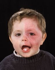
What does this kid have?
|
Sturge-Weber syndrome. Vascular malformation on the trigeminal nerve distribution. Also has an AV malformation on the ipsilateral side of the brain.
|
|
|
What is a spider telangiectasia?
|
Arteriovenous fistula, disappears when compressed. Associated with hyperestrinism (pregnancy, cirrhosis). In cirrhosis, you can't break down estrogen.
|
|
|
What is a capillary hemangioma?
|
Facial lesion in newborns that regress with age.
|
|
|
What is the organism associated with Kaposi sarcoma?
|
Human herpesvirus type 8.
|
|
|
What is the benign capillary proliferation involving skin and visceral organs in AIDS patients? (Looks like Kaposi's but it not!)
|
Bacillary angiomatosis. Caused by bartonella henselae (also causes cat-scratch disease). Treat it with a sulfa drug.
|
|
|
(VAT) Vinyl chloride (plastics), arsenic (pesticides), and thorium dioxide can cause what vascular tumor in the liver?
|
Liver angiosarcoma.
|
|
|
What is the most common benign tumor of the liver and spleen?
|
Cavernous hemangioma, which arise from the endothelial cells that line the blood vessels.
|
|
|
What is Von Hippel-Lindau syndrome? How will the patient present.
|
von Hippel-Lindau syndrome (VHL) is an autosomal dominant, inherited, neurocutaneous dysplasia complex. VHL is characterized by cavernous hemangiomas in cerebellum (ataxia) and retina (blindness). Increased incidence of pheochromocytoma and bilateral renal cell carcinomas.
|
|
|
What is a glomus tumor?
|
Glomus bodies are AV anastomosis that are involved in thermoregulation. A glomus tumor involves the SMC of glomus bodies and are commonly found under the fingernails and are very painful.
|
|
|
A large lesion is found on the neck of a child with Turner's. What is it?
|
Cystic hygroma. aka Lymphangioma.
|
|
|
95% of the time, small vessel vasculitis will be what type of hypersensitivity? What skin lesions are present?
|
Type III - immune complex depositing causing fibrinoid necrosis.
Skin lesion - palpable purpura. |
|
|
What is the most common cause of a myocardial infarction in children?
|
Kawasaki's disease (children < 4 years of age). Medium-sized vessel vasculitis. Presents with vessel thrombosis and infarction or aneurysm. Clinical features: desquamating rash, lymphadenopathy, swelling of hands/feet, oral erythema.
|
|
|
Name the medium-sized vessel vasculitis that is associated with HBsAg. It normally occurs in middle-aged men. Arteries at risk of infarction include renal, coronary, and mesenteric.
|
Polyarteritis nodosa.
|
|
|
Male, 52, presents with a headache, jaw claudiaction, tenderness of the scalp and unilateral blindness. Labs show an increase in ESR. What is the diagnosis?
|
Giant cell (temporal) arteritis. Granulomatous large vessel vasculitis involving superficial temporal and ophthalmic arteries (blindness); thrombi contain microabscesses.
|
|
|
Male, 28, presents with foot claudication, ulcerations on same foot, and his small toes appears to be falling off. History reveals he smokes a pack a day. What is the diagnosis?
|
Buerger's disease (thromboangiitis obliterans). Medium-sized vessel vasculitis with digital vessel thrombosis.
|
|
|
Female, 21, had fingers which turned white-blue-red in response to cold. What is the diagnosis?
|
Raynaud's disease.
|
|
|
Young boy presents with hematuria, polyarthritis, and palpable purpura of buttocks and lower extremeties.
|
Small vessel vasculiti (most common in children): Henoch-Schonlein purpura. IgA-anti-Iga immunocomplexes. Glomerulonephritis (RBC casts) and GI bleeding will also be present.
|
|
|
25 y/o male presents with a saddle nose deformity, sinus infection, and nodular masses in the lung. Labs show c-ANCA (anti-neutrophil cytoplasmic antibody) positive.
|
Wegener's granulomatosis: necrotizing medium/small sized vessel vasculitis involving upper respiratory tract, lung, and renal vessels. Necrotizing granulomas in upper respiratory tract (saddle nose).
|
|
|
Cyclophosphamide is used to treat Wegener's granulomatosis. What can cyclophsphamide cause? How do you prevent this?
|
Hemorrhagic cystitis. MESNA.
|
|
|
Vascular, red pedunculated mass that ulcerates and bleeds easily. What is it?
|
Pyogenic granuloma. Post-traumatic or associated with pregnancy (estrogen related).
|
|
|
Patient presents with several skin lesions that have been present since birth. You determine that the lesions are talengiectasia. Dx?
|
Hereditary hemorrhagic telangiectasia. aka Osler-Weber-Rendu disease. Autosomal dominant.
|
|
|
Young Asian women presents with pulseless extremities and blurry vision. What vasculitis could this be?
|
Takayasu's arteritis. Granulomatous large vessel vasculitis involving aortic arch vessels.
|
|
|
Young boy presents to your office with palpable purprua and hematuria. His mother asks why the penicillin you gave him last week made his condition worse. What caused the new symptoms?
|
Microscopic polyangiitis. Small vessel vasculitis involving skin, lung brain, GI tract, and postcapillary venules and glomerular capillaries. Often precipitated by drugs, infections, or immune disorders. Labs: p-ANCA.
|
|
|
What are some clinical findings in Churg-Strauss syndrome?
|
Small vessel vasculitis in children/adults involving lung, heart, skin. Allergic rhinitis, asthma. Labs: p-ANCA (labs) and eosinpholia.
|
|
|
Patient in Arizona presents with a rattle snake bite. He is given an antivenom but rapidly develops a fever and urticaria. What happened?
|
Serum sickness due to envenomation with horse-antivenom. Serum sickness causes small vessel vasculitis involving immunocomplex deposition in skin vessels.
|
|
|
Upon returning from a camping trip in Colorado, a boyscout presents with a fever and a rash on his palms that has began to spread to his trunk. What could be the diagnosis?
|
Rocky Mountain spotted fever. Ricketsia rickettsiae. Organisms invade endothelial cells causing a small vessel vasculitis
|
|
|
How does sodium cause hypertension?
|
Excess sodium increases plasma volume. Excess sodium produces vasoconstriction of TPR arterioles: sodium enters arteriole SMCs and opens Ca channel causing vasoconstriction.
|
|
|
Name the elastic blood vessels.
|
Aorta, braciocephalic, subclavian, common carotid, ileac.
|
|
|
Name the adrenal causes of secondary hypertension.
|
Cushings (increased mineralcorticoids). Pheochromocytoma (increaesd catecholamines). Neuroblastoma (increased catecholamines). 11-hdroxylase deficiency (increased mineralocorticoids). Conn syndrome (increased aldosterone).
|
|
|
What is the most common cause of hypertension in young women?
|
Oral contraceptives. They increase synthesis of angiotensinogen.
|
|
|
Does Graves disease cause hyper or hypotension? How?
|
Hypertension. Graves disease increases cardiac contraction via hyperthyroidism.
|
|
|
What is the most common complication in hypertension? What is the most common cause of death?
|
Left ventricular hypertrophy.
Acute myocardial infarction. |
|
|
What is the primary mechanism of essential hypertension in black Americans and the elderly?
|
Reduced renal sodium excretion.
|
|
|
What is the most common cause of secondary hypertension?
|
Renovascular hypertension: activation of RAS. The cause in elderly men is atherosclerotic plaques. The cause in young women is fibromuscular hyperplasia in the renal artery.
|
|
|
What are some inherited causes of DVT's?
|
Factor V Leiden, prothrombin mutant (20210A transition), deficiency in ATIII, protein C, or protein S. Homocysteinemia can be acquired or inherited.
|
|
|
What is Virchow's triad?
|
Endothelial injury, stasis/turbulence, and hypercoagulability.
|
|
|
What INR values do you want to achieve with a patient on long term warfarin treatments?
|
INR 2.0 - 3.0
|
|
|
What test are used to diagnose or rule out a DVT?
|
D-dimer can be used to rule out a DVT in patients with a low pre-test probability.
Ultrasonography to test the compressability of the vessel lumen. Venography. |
|
|
Compare a venous thrombus to an arterial thrombus.
|
Venous - due to stasis/hypercoagulability, dark red, and treated with heparin/warfarin.
Arterial - due to endothelial injury, gray/white with lines of Zahn (alternating fibrin and platelets), and treated with aspirin. |
|
|
Antiphospholipid syndrome (antibodies against phospholipids) can induce a hypercoagulable state. What would a PTT test show?
|
The PTT is paradoxically prolonged. This is an in vitro artifact caused by the interaction of the antibodies with the reagent used in the test.
|
|
|
Explain how a women with no sexual history can test positive for syphilis.
|
She has SLE. Anti-cardiolipin antibodies can cause a false-positive serologic test for syphilis because the antigen in the standard tests is embedded in cardiolipin.
|
|
|
Adult polycystic kidney disease is associated with: a) AV fistulas, b) atherosclerotic aneurysm, c) berry aneurysm, d) dissecting aneurysm
|
C) berry aneurysm
|
|
|
"Flea-bitten" kidney is seen in what condition.
|
Malignant hypertension
|
|
|
What is the significance of where a DVT is located in the lower extremity?
|
DVT's distal to the knee in the calf area are normally asymptomatic and do not cause PE. DVT's proximal to the knee normally form from propagation of distal DVT's. Half of patients with symptomatic proximal DVTs have a silent PE and 10% have symptomatic PE.
|
|
|
What are some ECG findings due to a pulmonary embolism?
|
Most commonly: sinus tachycardia and nonspecific anterior T wave inversion. Pathoneumonic and less common: S1, Q3 (pathological Q in lead III), T3 (inverted). Right axis deviation. RBBB.
|