Reading...
![]()
Play button
![]()
Play button
![]()
Use LEFT and RIGHT arrow keys to navigate between flashcards;
Use UP and DOWN arrow keys to flip the card;
H to show hint;
A reads text to speech;
149 Cards in this Set
- Front
- Back
|
Hyperplasia
|
Cells have increased in number—hyperplasia
|
|
|
Dysplasia
|
Abnormal proliferation of cells with loss of size, shape, and
orientation |
|
|
In Situ Carcinoma
|
* Neoplastic cells have not invaded basement membrane
• High nuclear/cytoplasmic ratio and clumped chromatin • Neoplastic cells encompass entire thickness • Tumor cells are monoclonal |
|
|
Invasive carcinoma
|
* Cells have invaded basement membrane using collagenases and hydrolases
• Can metastasize if they reach a blood or lymphatic vessel |
|
|
Metastases
|
Spread to distant organs
• Must survive immune attack • “Seed and soil” theory of metastasis • Seed = tumor embolus • Soil = target organ—liver, lungs, bone, brain . . . • Angiogenesis allows for tumor survival • decreased cadherin, increased laminin, integrin receptors |
|
|
Metaplasia
|
1 adult cell type is replaced by another. Often 2° to irritation and/or environmental exposure (e.g., squamous metaplasia in trachea and bronchi of smokers)
|
|
|
Dysplasia
|
Abnormal growth with loss of cellular orientation, shape, and size in comparison to normal tissue maturation; commonly preneoplastic
|
|
|
Name reversible cellular - plasias
|
Hyperplasia, metaplasia and dysplasia
|
|
|
Anaplasia
|
Irreversible change. Abnormal cells lacking differentiation; like primitive cells of same tissue, often equated with undifferentiated malignant neoplasms. Little or no resemblance to tissue
of origin. |
|
|
Neoplasia
|
A clonal proliferation of cells that is uncontrolled and excessive
|
|
|
Desmoplasia
|
Fibrous tissue formation in response to neoplasm.
|
|
|
Grade
|
Degree of cellular differentiation based on histologic appearance of tumor
(The character of the tumor) |
|
|
Stage
|
Degree of localization/spread based on site and size of 1° lesion, spread to regional lymph nodes
|
|
|
Tumor staging system (TNM)
|
T = size of Tumor
N = number of involved lymph nodes M = metastases |
|
|
What are benign and malignant tumors of bone?
|
Rhabdomyoma Rhabdomyosarcoma
|
|
|
Name malignancies associated with Down syndrome
|
ALL (we ALL fall Down),
and AML |
|
|
Name malignancies associated with Xeroderma pigmentosum, Albinism
|
Melanoma, basal cell
carcinoma, and especially squamous cell carcinomas of skin |
|
|
Name malignancies associated with chronic atrophic gastritis, pernicious anemia, postsurgical gastric remnants
|
Gastric carcinoma
|
|
|
Name malignancies associated with Tuberous sclerosis
|
Astrocytoma, angiomyoma, lipoma, and cardiac
rhabdomyoma |
|
|
Name malignancies associated with Barrett’s esophagus (chronic GI reflux)
|
Esophageal
adenocarcinoma |
|
|
Paget’s disease of bone associated with
|
2° osteosarcoma and
fibrosarcoma |
|
|
Autoimmune diseases (e.g., Hashimoto’s thyroiditis and myasthenia gravis are associated with
|
Lymphoma
|
|
|
Dysplastic nevus is commonly associated with what?
|
Malignant melanoma
|
|
|
Radiation exposure is associated with what types of malignancies
|
Sarcoma, papillary thyroid
cancer |
|
|
What are oncogenes?
|
Gain of function cancer genes. Need damage to only 1 allele to start malignant growth
|
|
|
CML is associated with mutation in which gene?
|
abl - mutated Tyrosine Kinase Protein
|
|
|
Burkitt's lymphoma is associated with mutation in which gene?
|
c-myc
|
|
|
Colon cancer is associated with mutation in which gene?
|
ras
|
|
|
Multiple endocrine neoplasia (MEN) types II and III is associated with what type of mutation?
|
tet
|
|
|
Rb gene mutation is associated with what malignancies?
|
Retinoblastoma, osteosarcoma
|
|
|
BRCA1 and BRCA2 are associated with which malignancies?
|
BRCA1 - Breast and ovarian cancer,
BRCA2 - breast cancer |
|
|
p53 mutation is associated with which malignancies?
|
Most human cancers, Li-Fraumeni syndrome
|
|
|
What type of malignancy is colon cancer associated with?
|
DCC—Deleted in
Colon Cancer. |
|
|
What type of malignancy is pancreatic cancer associated with?
|
DPC—Deleted in
Pancreatic Cancer. |
|
|
CEA is a tumor marker for which malignancies?
|
Carcinoembryonic antigen. Very nonspecific produced by 70% of colorectal and pacreatic cancers; also produced by gastric and breast
carcinomas |
|
|
alpha-fetoprotein is a tumor marker for which malignancies?
|
Normally made by fetus. Hepatocellular carcinomas.
Nonseminomatous germ cell tumors of the testis (e.g., yolk sac tumor). |
|
|
h-CGH
|
Hydatidiform moles, Choriocarcinomas, and
Gestational trophoblastic tumors. |
|
|
Alkaline Phosphatase
|
Metastases to bone, obstructive biliary disease, Paget’s disease of bone
|
|
|
Bombesin
|
Neuroblastoma, lung and gastric cancer
|
|
|
TRAP staining stains what?
|
Tartrate-resistant acid phosphatase. Hairy cell TRAP leukemia—a B-cell neoplasm
(TRAP the HAIRY animal) |
|
|
Describe the mechanism of DIC
|
Disseminated Intravascular Coagulopathy:
1. activation of coagulation sequenc e through trauma -- leads to disseminated thrombi 2. Consumption of coagulation factors and platelets to sub-hemostatic levels 3. Activation of fibronolysis 4. Tissue hypoxia, microinfarcts and hemorrhage |
|
|
Describe Lab Findings in DIC
|
Increased PT and PTT because most clotting factors have been used up
|
|
|
Name three diseases where we see shistocytes
|
1. Thrombic Thrombocytopenia Purpura 2. Thrombotic Microangiopathy
3. Disseminated Intravascular Coagulopathy |
|
|
Describe the mechanism of Thrombotic Thrombocytopenia Purpura?
|
The mechanism is the lack of a protease that cleaves vWF back to its normal size (hereditary or adapted through immune response). Ultra large vWF does not clot, but snags many platelets, this leads to thrombocytopenia without overwhelming consumption of clotting factors.
|
|
|
Describe the bleeding pattern for TTP?
|
Ptechiae bleeding because we are dealing with platelet disorder (thispattern is characteristic for thrombocytopenia)
|
|
|
Name the first line of treatment for DIC?
|
1. threat the underlying cause -frozen blood plasma since most blood clotting factors (including fibinogen).
Also, give heparin in patients with clinical thrmbosis 3. Can also give inhibitor concentrates - antithrombin, protein C |
|
|
What type of necrosis is gangrene?
|
Coagulative necrosis
|
|
|
Why would someone in DIC develop a gangrene?
|
Multple disseminated thombotic events take place in DIC. Thrombi lead to localyzed hypoxia and consequenty to necrosis in the areas deprived of oxygen
|
|
|
Define Hyperthrophy
|
An increase in the size of an organ or tissue due to an increase in the size of cells.
Eg. increase in skeletal muscle mass as a response to exercise |
|
|
Define Hyperplasia
|
increase in the size of an organ or tissue due to an increase in teh size of cells
Eg. glandular proliferation in breast during pregnancy |
|
|
Define Aplasia
|
Is a failure of cell production/
Ex. agenesis, or absence of an organ due to a failure of production in embryonic development |
|
|
Define Hypoplasia
|
Decrease in cell production that is less extreme than aplasia
Eg. partial lack of growth and maturation of gonadal structures in Turner syndrome |
|
|
Define Atrophy
|
Decrease in the size of an organ/tissue due to a decrease in the mass of preexisting cells. Eg. due to aging, denervation, lack of endocrine stimulation
|
|
|
Define Metaplasia
|
Replacement of one differentiated tissue by another.
Eg. Barrett's esophagus |
|
|
What is Barretts' Esophagus?
|
Long standing GERD (gastro-esophageal reflux disease) can cause adenocarcenoma. This leads to a columna metaplasia - change of pseudostratified columnar epithelium with cilia and goblet cells to columnar epthelium normally seen in stomach
|
|
|
What are the causes of hypoxic cell injury?
|
Normally results from hypoxia which can result from the following:
1. ischemia (obstrution of arterial blood flow) 2. anemia - reduction in RBC mass 3. CO poisoning 4. Decreased perfusion |
|
|
Name adaptive responses to cell injury
|
Hyperthrophy (increase in size)
Athrophy (decrease in size) Hyperplasia (increase in cell number) Metaplasia (change from one differentiated tissue type to another) |
|
|
Define involution
|
Decrease in the number of organ cells when the organ is no longer needed, for example, Thymus
|
|
|
Define Necrosis
|
The sum of degradative and inflammatory reactions occuring after tissue death caused by injury (hypoxia, exposure to toxic chemicals).
It occurs within living organisms |
|
|
Define Autolysis
|
Refers to degradative reactions in cells caused by intracellular enzymes idigenous to the cell. Postmortem autolysis occurs after the death of the whole organism and is NOT necrosis
|
|
|
Name four types of necrosis
|
1. Coagulative necrosis
2. Liquefactive necrosis 3. Caseous necrosis 4. Fat necrosis |
|
|
Describe Coagulative Necrosis
|
1. Results most often from cutting off a blood supply to an organ (ischemia).
Most prevalent in the heart and the kidney 3. General preservation of tissue architecture 4.Increased cytoplasmic eosinophilia due to protein denaturation and loss of cytoplasmic RNA |
|
|
Describe Liquefactive Necrosis
|
1. Most often caused by bacterial infections or an interraption of a blood supply
2. Frequent in brain and liver 3. Due to enzymatic digestion 4. Tissue no longer looks intact like in coagulative, but is actively digested |
|
|
Describe Coagulative Necrosis
|
|
|
Describe this type of cellular response to sublethal injury
|
These are hydropic changes in the hepatocypes. We see how some cells look more swollen and pale
|
|
|
Describe cell response to sublethal or reversible damage
|
1. Hydropic changes in the kidney
2. Fatty changes in the liver |
|
Describe this adaptive response to reversible injury
|
This is a hydropic change in the kidney. Decreased Na+ pump --> hydrophic changes --> cells swell and now look more pale and distended
|
|
Describe this cellular response to sublethal injury
|
This is fatty liver.
We see an enlargement of individual hepatocytes. Oily feel of the organ on a gross level. Yellowish coloration. Hepatocytes accumulate fate granules (steatosis). Cessation of alcohol consumption can cause cells to reverse these morphologic changes |
|
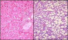
Describe this cellular respionse tosublethl injury
|

This is fatty liver on a microspic level. We see that hepatocypes accumulated fat.
Again, this is a reversible change |
|
|
Describe Caseous Necrosis
|
1. Shares features of both coagulative and liquefactive necrosis
2. Normally caused by TB (tubercolous granuloma) 3. Architecture is not preserved but tissue is not liquified. Gross appearance is soft and cheese like. 4. Histologic appearance is amorphous 5. Has increased affinity for eosinophilic dyes |
|
|
Define Fat Necrosis
|
Usually happens with liberation of pancretic enzymes with autodiestion of pancretic parenchyma or trauma to fat cells.
|
|
Understand the mechanisms of cellular responses to injury
|
none
|
|
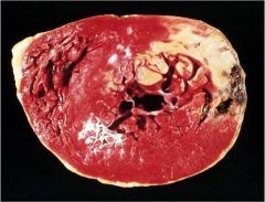
Identify the mechanism of necrosis
|
Coagulative necrosis (gross view)
|
|
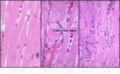
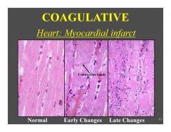
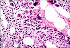
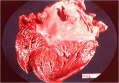
Indentify the type of necrosis in this cardiac tissue
|
This is coagulative necrosis. We see that cellular arcitecture is preserved here, but also can observe slow nuclear fragmentation (on the right we see nuclear fragements instead of cell nuclei)
|
|
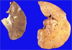
Name necrosis type
|
Coagulative (pale or anemic) necrosis. Renal arteries through renal hilum became occluded - resulting in a "pale" or "anemia" infarction
Again, we see no living tissue, yet the architecture is preserved. |
|
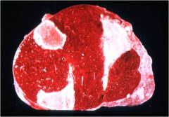
Name the type of necrosis
|
This is also coagulative necrosis. We see that the light area is dead due to hypoxia. The rest (darker area) is alive because it receives oxygen by diffusion
|
|
Name the type of necrosis
|
This is again coagulative necrosis due to adrenal infarct
|
|
Name the type of necrosis
|
Liquefactive necrosis due to a bacterial infection in the liver
|
|
Name the type of necrosis
|
Liquefactive necrosis in the brain
|
|
Name necrosis type
|
Liquefactive in the brain - can see stained bacteria in this slide
|
|
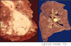
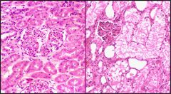
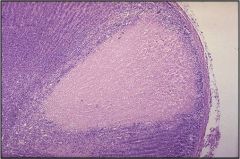
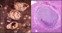
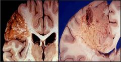
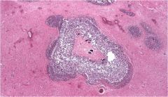
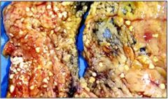
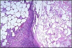
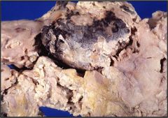
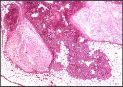
Name necrosis type
|
This is a caseous necrosis. Left: caseous necrosis in the lung due to TB infection. Right: in the lymph node, again due to a TB infection
|
|
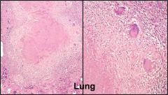
Name necrosis type
|
Caseous in the lung (go from small to large magnification)
|
|
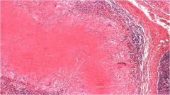
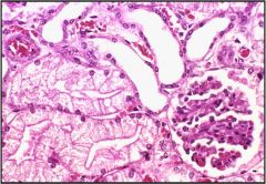
Name necrosis type
|
Caseous in the lymph node (high magnification) - we can see that architecture is not preserved but the tissue is not liquified. Increased eosinophilic staining
|
|
Name necrosis type
|
This fat necrosis in mesentery. We can see necrotic fat cells, inflammation
|
|
Name necrosis type
|
Again, this is a fat necrosis (high mag)
|
|
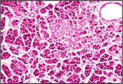
Name necrosis type
|
Fat necrosis in the pancreas
|
|
Name necrosis type
|
Fat necrosis in the pancreas
|
|
Name necrosis type
|
Fat necrosis in accute pancreatitis
|
|
Is this a necrosis? Why?
|
This is apoptosis, programmed cell death and not a necrosis.
|
|
Name this process - necrosis, apoptosis or autolysis?
|
This is autolysis.
Looks a bit like coagulative necrosis but no inflammation. Autolysis happens due to ingeneous enzymes as a cell death after death of the entireorganism or removal from the body. |
|
|
Describe hemoglobin structure
|
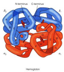
Hb is a multisubunit, plasma protein. It has
2 alpha and 2 beta globin chains: alpha2beta2 (adult) other globin subunit types are found in the fetus and newborns (zeta, epsilon, gamma, etc). Human body contains 750g of Hb which is replaced every ~120 days Hb also transports H+ and CO2 in addition to O2 |
|
|
Desribe Iron coordination in oxyhemoglobin
|
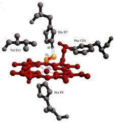
1. Fe2+ requires an octahedral coordination.
2. 4 ligands to Fe2+ are provided by nitrogens in the heme group. 3. A 5th ligand is provided by nitrogen from Histidine-93 in the globin chain, also known as the proximal His. A 6th ligand is provided by O2 which is sandwiched between the Fe2+ and nitrogen on Histidine-64 (the distal His) in the globin chain. |
|
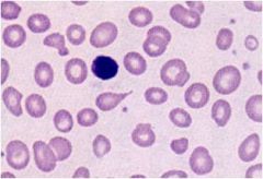
Define this type of anemia
|
Normocytic, normochromic anemia
|
|
Define this type of anemia
|
Microcytic hypochromic anemia
|
|
Define this type of anemia
|
Macrocytic normochromic
|
|
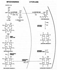
Describe the process of heme sythesis
|
Synthesis starts in the mitochondria, continues in the cytosol, and finishes in the mitochondria.
ALA-synthase and Ferrochelatase are two rate limiting enzymes in the entire heme synthesis pathway. |
|

Name the enzyme that upregulates heme synthesis when necessary (excess Fe present)
|
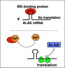
ALA-synthase (ALAS) is the rate limiting enzyme.
ALAS protein levels are upregulated in response to increased iron (Fe2+) in erythroid cells. the ALAS mRNA has a “cis-acting” iron response element (IRE) that modulates the rate of translation of the mRNA to protein. |
|
|
At the level of which enzymes is heme synthesis pathway regulated?
|
Feedback inhibition regulates heme synthesis at two levels:
-ALA-synthase -ferrochelatase |
|
|
Describe pathophysiology of Anemia of Lead Poisoning (Plumbism)
|
Lead exposure from: paint, gasoline fumes, lead pipes in plumbing, contaminated foods.
Pb targets many molecules vital to cell metabolism. Heme synthesis pathway especially sensitive to Pb. ALA-dehydratase and ferrochelatase most sensitive Pb targets in pathway -- Lead inhibits these enzymes directly as well as alters Iron uptake |
|
|
Describe Clinical Findings in Anemia of Lead Poisoning
|
Presenting symptoms: abdominal pain, motor neuropathy (palsy).
Hematology: often normocytic and normochromic. Microcytic and hypochromic with chronic exposure. Sometimes basophilic stippling due to iron induced aggregates of ribosomes. Treatment through removal of Pb source and/or chelation therapy with EDTA or dimercapto-succinate |
|
|
What is Congenital ALA-Synthase Deficiency?
|
An ALA-Synthase Deficiency causes anemia. Patients present with Sideroblastic anemia: Erythrocytes are hypochromic and microcytic.
|
|
|
What are Porphyrias?
|
Arise from specific inherited defects in enzymes of heme biosynthetic pathway.
Seven distinct porphyrias are described that correlate with deficiencies in each of the seven enzymes. |
|
|
Describe Acute Intermittent Porphyria (AIP)
|
1. Acute Intermittent Porphyria (AIP)
Manifests neurological and psychiatric illness, visceral pain. Most are due to accumulation of ALA-synthase enzyme. Treatment with analgesics for pain, maintain morale, alleviate fear. Infusion of hematin (Fe3+ heme). |
|
|
Name this type of anemia
|
Congenital Erythropoietic Porphyria (CEP)
Manifests cutaneous scarring due to photosensitivity. Accumulated isomers of Uro’gen and Copro’gen result from inhibition of dehydration. These compounds cause oxidative damage when exposed to light. treatment with spleenectomy or transfusion. |
|
|
Name four different orgains where Hb is produced during development
|
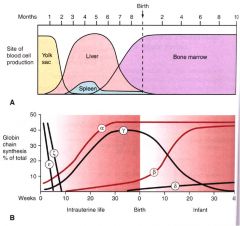
Changes or “switching” in globin chain composition correlate with sites of Hb synthesis during development.
1. Yolk sac 2. Spleen 3. Liver 4. Bone marrow |
|
|
Describe the pathophysiology of Thalassemia
|
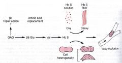
Sickle cell disease (HbS) results from mutations in the chain of Hb that promote self-aggregation of Hb into fibrils.
A Glu to Val amino acid change at position 6 in chain most common. Fibril formation precipitates from deoxy form of HbS. |
|
|
Describe two physiologic states of Hemoglobin
|
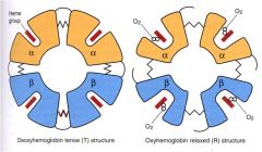
Hb exists in two general conformations: the T state and R state (Tight and Relaxed).
O2 binds preferentially to the R state: -OxyHb=R state -DeoxyHb=T state O2 binding invokes structural changes in heme: -Fe2+ is displaced to be in same plane as porphyrin ring. |
|
|
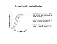
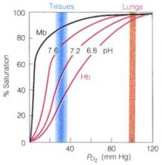
Describe Oxygen binding kinetics of hemoglobin
|
|
|
|
Compare Fetal and Adult Hemoglobin
|
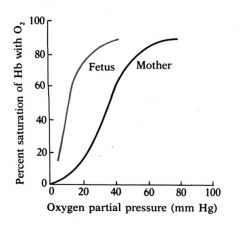
Fetal circulation and maternal circulation come in close contact however, oxygen transport relies on exchange of O2, not RBCs.
The pO2 in the placenta is well below that in the maternal circulation (e.g.<20Torr). Fetal Hb (HbF) must have a higher affinity for O2 than maternal (adult) Hb (HbA) in order to “extract” O2 from the maternal circulation. |
|
|
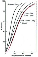
Describe the effect of 2,3-BPG on Hemoglobin
|
2,3-bisphosphoglycerate (BPG):
Is an allosteric inhibitor of Hb-O2 binding. Promotes release of O2 from Hb. |
|
|
Provide physiological exoplanation for the higher affinity of Fetal Hb to Oxygen
|
The physiological basis for the ability of the fetus to withdraw O2 from the mothers circulation relies on the property that, in vivo, HbF has a higher affinity for O2 than HbA.
Adult = HbA = alpha2beta2 Fetal = HbF = alpha2gamma2 |
|
|
Describe the effect of pH on Hemoglobin
|
H+ is displaced upon O2 binding to Hb. Therefore, increase in H+ drives the reaction backwards.
Lower pH (higher H+ concentration) causes release of O2. This is known as the Bohr effect. This is a good thing! Working muscles, which demand more O2, propagate acidic conditions e.g. lactic acid. |
|
|
Describe the effect of Temperature on Hemoglobin
|
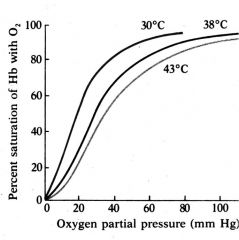
The heme-O2 binding reaction is exothermic. Therefore, oxygen affinity of Hb drops with elevated temperature.
Again, this is a good thing! Working muscles, which demand more O2, are elevated in temperature. |
|
|
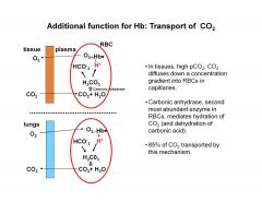
Describe the effect of CO2 on hemoglobin oxygen dissosiation curve
|
CO2, a major product of glycolysis, is generated rapidly in tissues,especially in working muscles.
CO2 reacts with H20 to form carbonic acid. Dissociated carbonic acid lowers pH and promotes O2 dissociation from Hb via the Bohr effect. |
|
|
How is CO2 transported in the blood?
|
|
|
|
What is Carboxyhemoglobinemia?
|
CO (carbonmonoxide) will bind in the heme pocket in place of O2 with about 250 times greater affinity as O2.
|
|
|
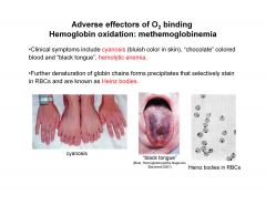
What is Methemoglobinemia?
|
Elevated levels of methemoglobin in the blood are caused when the mechanisms that defend against oxidative stress within the red blood cell are overwhelmed and the oxygen carrying ferrous ion (Fe2+) of the heme group of the hemoglobin molecule is oxidized to the ferric state (Fe3+).
MetHb is unable to bind and release O2. Presence of metHb shifts O2 dissociation curve to the left. |
|
|
Describe clinical symptoms of methemoglobinuria
|
|
|
|
Describe Glucose metabolism in erythrocytes
|
RBCs are devoid of nucleus, so most of their metabolic funsction is directed at maintenance. Glucose metabolism proceeds through anaerobic glycolysis which yields 2 ATP for each glucose.
Glycolysis occurs in the cytoplasm. RBC longevity is reliant upon ATP production which is very sensitive to the “fitness” of the enzymes required for glucose catabolism. |
|
|
Name three most critical enzymes in metabolism
|
Glycolysis requires eleven enzymes. Three critical enzymes are:
-hexokinase (HK) -phosphofructokinase (PFK) -pyruvate kinase (PK). |
|
|
What is methemoglobin reductase pathway?
|
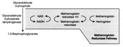
NADH produced from G3PDH acts as a reducing cofactor for the metHb-reductase enzyme.
This is the major pathway for reducing metHb back to functional Hb. Non-enzymatic mechanism of metHb reduction require ascorbic acid or methylene blue, which can be applied clinically. |
|
|
Name three most common ROS (reactive oxygen species)
|
O2- is most common in the process of metHb formation from Hb. Also promotes cell lysis via membrane lipid oxidation.
H2O2 is more stable than OH yet both act to oxidize and denature Hb. |
|
|
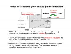
Describe Hexose monophosphate (HMP) pathway: glutathione reduction
|
|
|
|
What is Glucose-6-Phosphate Dehydrogenase Deficiency?
|
The most common RBC metabolic disorder. >2 million people affected.
The G6PD gene is on the X chromosome and demonstrates a recessive X-linked inheritance. i.e. males are most affected. Deficiencies compromise the RBC defense of oxidation due to the inability to maintain reduced form of GSH. Two Mutations are most clinically relevant:G6PD A- (in African Americans) and G6PD Mediterranean, both generally decrease the half-life of enzyme. |
|
|
What is Pyruvate Kinase Deficiency?
|
PK deficiency is prevalent in the US, Europe and Japan. ~1% of US population is heterozygous. Overall decrease in ATP compromises repair processes resulting in hemolytic anemia. No effective therapy, treatments include RBC transfusions.
PK deficiency shows autosomal recessive inheritance and is the most common genetic disorder of the glycolytic pathway. |
|
|
Commen on the iron content of a typical diet and how much is usually absorbed/day
|
A typical diet consists of 10-20 mg/day iron, of which ~10% is adsorbed.
A balance is maintained: roughly 1-2 mg iron is lost daily through loss of epithelia. Under higher demand for iron adsorption increases to 20% of dietary intake. |
|
|
Names two forms in which iron is absorbed
|
Inorganic (Fe2+, Fe3+ salts) or organic (heme). Heme iron is rapidly adsorbed whereas Fe3+ is not very soluble and poorly adsorbed. Fe2+ is more soluble than Fe3+.
Heme is absorbed by machinery completely different to that of inorganic iron. The process is more efficient and is independent of duodenal pH . Consequently meats are excellent nutrient sources of iron. |
|
|
Name three major compartments of iron in the body
|
-Functional
-Transport -Storage RBCs contain 70% of body iron. Approx 25% is in storage in ferritin and hemosiderin |
|
|
What is Ferritin?
|
Ferritin is a huge spherical protein (Mw=440,000 Da) consisting of 24 subunits.
It is common to all cells. High levels occur in erythroid precursors. Forms a “shell” that can encompase >4500 iron atoms. Iron can reversibly pass through “channels” in the shell. |
|
|
What is Hemosiderin?
|
less-well characterized complex of iron in a ferric hydroxyphosphate form and protein.
It is a storage compartment when ferritin capacity is exceeded. |
|
|
What is Transferrin?
|
Transferrin is a medium-large soluble protein (MW=79,000 Da) that mediates iron transport between tissues.
It is present in plasma and extracellular fluid. It binds IRON IN 3+ STATE |
|
|
Describe the mechanism of Iron delivery inside the cells
|
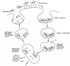
1. Uptake into cells is mediate by the transferrin receptor.
2. Apotransferrin has low affinity for the receptor; iron-bound transferrin binds tightly to the receptor. 3. Binding promotes receptor clustering and endocytosis of transferrrin-receptor complexes into an endosome. 4.Acidification of endosome causes iron dissociation (but not transferrin dissociation from the receptor!). 5. Transferrin is recycled and |
|
|
Describe lab values that measure iron status
|
Iron status is determined by laboratory methods that measure ferritin and transferrin-associated iron levels in serum.
Serum iron is characterized by the total iron binding capacity (TIBC) and unsaturated iron binding capacity (UIBC). |
|
|
Define Thrombus
|
Solidified blood
Inside the vascular space Of a living organism |
|
|
Where intravascular thrombi are usually located?
|
Intravascular, by definition:
Arteries Veins Heart chambers Heart valves |
|
|
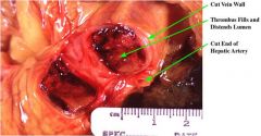
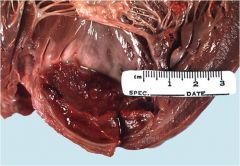
Name this
|
Aortic valve thrombus
|
|
|
Name potential fates of thrombi
|
Resolve (heal without a trace)
Propagate Embolize Organize Recanalize Calcify (phlebolith) |
|
|
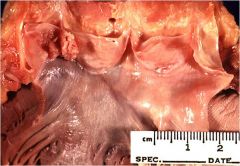
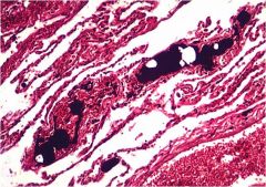
Name this stage of thrombus formation
|
Recanalization
|
|
|
Name three types of emboli
|
Gas: air, nitrogen (the “bends”)
Liquid: amniotic fluid, contrast material, fat after soft tissue trauma or fracture, injectables Solid: Thrombus is most common; also foreign bodies (bullet, catheter tip), atheroembolism, bone marrow Thromboemboli can be infected (septic) |
|
|
What are common origins/sites of thromboembolism?
|
Venous (systemic veins to pulmonary arteries)
Arterial (heart or aorta to systemic circulation Paradoxic (systemic vein, through a septal defect in the heart, to the systemic circulation) |
|
|
Describe this type of embolus
|

This is a saddle embolism
|
|
|
Name this type of embolus
|
Fat embolus
|
|
|
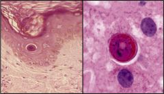
Name this type of embolus
|
Paradoxic embolus in foramen ovale
|
|
|
Define infarct
|
Localized area of ischemic necrosis
Usually caused by vascular blockage Note: Takes time (4-6 hours) to be seen microscopically |
|
|
Define Infarction
|
The process of production of an infarct
|
|
|
Describe two common morphologies of infarct
|
Varies with different tissues and durations:
1. Pale, roughly wedge-shaped in solid organs with single blood supply (e.g., kidneys and spleen) 2. Hemorrhagic, in soft organs with a dual blood supply (e.g., lungs, bowel) |
|
|
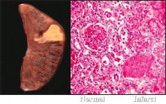
What kind of infarct is this?
|
Ischemic splenic infarct
|
|
|
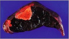
What kind of infarct is this?
|
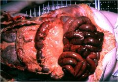
Hemorrhagic infarct of bowel
|
|
|
Define ischemia
|
Decreased blood flow to an organ or tissue
Often distinguished from infarction clinically. |