![]()
![]()
![]()
Use LEFT and RIGHT arrow keys to navigate between flashcards;
Use UP and DOWN arrow keys to flip the card;
H to show hint;
A reads text to speech;
68 Cards in this Set
- Front
- Back

This is an image of a kidney. What is A? |
Cortex |
|

What is B? |
Medulla
|
|

What is C? |
Papilla |
|

What is D? |
Fibrous Capsule |
|
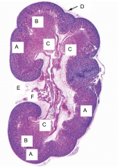
What is E? |
Hilum |
|

What is F? |
Ureter |
|

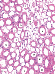
This is an image of the renal cortex. What is A? |
Vein |
|

What is B? |
Interlobular Artery
|
|

What is C? |
Glomeruli
|
|

What is D? |
Tubules |
|
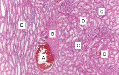
What is E? |
Medullary Ray |
|

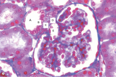
This is an image of the Kidney. What is this structure called? |
Renal Corpuscle |
|

This is an image of the Kidney. In which part of the kidney is this structure usually found? |
Renal Cortex |
|

What is A? |
Proximal Convoluted Tubule |
|

What is B? |
Renal Interstitium |
|

What is C? |
Parietal Epithelial Cells |
|

What is D? |
Endothelial Cells |
|
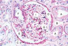
What is E? |
Nuclei of the Mesangial Cells |
|

What is F? |
Afferent Arteriole |
|

What is G? |
Mesangium |
|

What is H? |
Glomerular Capillaries |
|

What is I? |
Glomerular Basement Membrane |
|
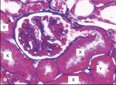
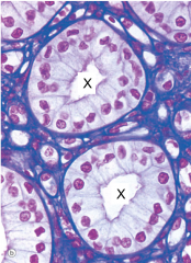
This is an image of the renal cortex. What is X? |
Proximal Convoluted Tubule |
|

What is Y? |
Capillary |
|

This is an image of the renal cortex. What is this structure called? |
Proximal Convoluted Tubule |
|
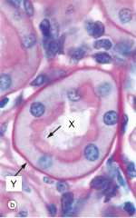
What is X? |
Brush Border |
|

What is Y? |
Basement Membrane |
|
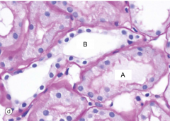
This is an image of there renal cortex. What is A? |
Distal Convoluted Tubule
|
|

What is B? |
Proximal Convoluted Tubule |
|

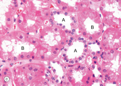
This is an image of the kidney. What is A? |
Proximal Convoluted Tubule |
|
What is B? |
Distal Convoluted Tubule |
|
This is an image of a structure found within the renal cortex. What is this structure called? |
Juxtaglomerular Apparatus |
|

What is A? |
Distal Convoluted Tubule |
|

What is B? |
Macula Densa |
|

What is C? |
Lacis Cells |
|

What is D? |
Juxtaglomerular Cells |
|

What is E? |
Afferent Arterioles |
|

This is an image of the Loop of Henle. What is A? |
Collecting Tubule |
|

What is B? |
Vasa Recta
|
|
What is C? |
Thin Limbs
|
|

What is D? |
Thick Ascending Limbs
|
|

What is E? |
Collecting Duct |
|

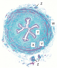
This is an image of the Loop of Henle. What is W? |
Thin limbs |
|
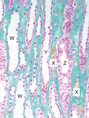
What is X? |
Vasa Recta
|
|

What is Y? |
Thick Ascending Limb
|
|

What is Z? |
Collecting Duct |
|

This is an image of collecting tubules and ducts. What is A? |
Intercalated Cells |
|

What is B? |
Supporting Tissue
|
|
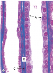
What is C? |
Basement Membrane |
|
This is an image of a structure found within the renal medulla. What is A? |
Collecting Ducts |
|

This is an image of the renal papillae. What is A? |
Ducts of Bellini |
|

What is B? |
Pelvicalyceal System |
|

What is C? |
Ureter |
|

What is D? |
Transitional (Urinary) Epithelium |
|
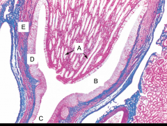
What is E? |
Smooth Muscle |
|

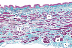
What part of the urinary system does this image show? |
Ureter |
|

What is W? |
Collagenous Adventitia |
|

What is X? |
Outer Circular Muscle Layer |
|

What is Y? |
Longitudinal Muscle Layer
|
|
What is Z? |
Blood Vessels |
|
What part of the urinary system does this image show? |
Bladder |
|

What is W? |
Outer Longitudinal Muscle Layer |
|

What is X? |
Adventitia
|
|

What is Y? |
Inner Longitudinal Muscle Layer |
|

What is Z? |
Outer Circular Muscle Layer |
|
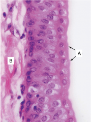
This is an image of the epithelium of the urinary system. What type of epithelium is this? |
Transitional (Urinary) Epithelium |
|

What is A? |
Umbrella Cells |
|

What is B? |
Lamina Propria |