Reading...
![]()
Play button
![]()
Play button
![]()
Use LEFT and RIGHT arrow keys to navigate between flashcards;
Use UP and DOWN arrow keys to flip the card;
H to show hint;
A reads text to speech;
13 Cards in this Set
- Front
- Back
|
A 50 female presents with urinary frequency, urgency, suprapelvic pain and hematuria. A culture of the urine is negative. What is the likely cause.
A. Fungal cystitis B. Bacterial cystitis C. Interstitial cystitis D. Cystitis cystica E. hemorrhagic cystits |
C. Interstitial cystitis is otherwise known as Hunner's cystitis. It presents just like in this case (same symptoms, middle-aged women). Histology often shows mast cell present, however this finding is inconsistent and non-specific. Diagnosis is rendered clinically by the presence of mucosal tears and hemorrhage after bladder distension. The pathologist role is mostly to r/o CIS since it can present similarly.
|
|
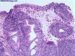
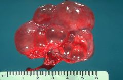
The lesion shown here..
A. Can transform to carcinoma B. Is associated with urinary fistulas C. Is always limited to the lamina propria D. Is most likely from a child's bladder |
This is cystitis cystica/glandularis. Cystica when the lumen is extending; glandularis when not. Two types are described, common type and intestinal type. The photo was of common type. Intestinal type has goblet cells and resembles intestinal epithelium. Mucin extravasation can be marked with dissecting pools of mucin. The key is the lack of other features of carcinoma. Only extensive intestinal metaplasia infers a higher risk of adenocarcinoma. The above photo shows a mixed common and intestinal type.
|
|
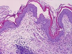
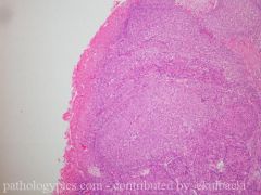
This is a bladder biopsy. What is not true about this entity?
A. Highest incidence occurs in people with Schistosoma haematobium. B. It is most common in women at the trigone. C. Is associated with neurogenic bladders and indwelling catheters. D. Infers an increased risk of carcinoma. |
B. This is squamous metaplasia in the bladder. It is associated with irritants and infers a risk of carcinoma if the irritant exposure is long. Uniquely, women can get non-keratinizing squamous metaplasia which does occur at the trigone and is not associated with carcinoma.
|
|
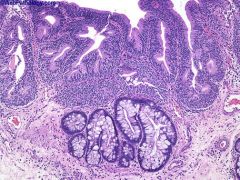
The image depicted was a biopsy from the bladder. What is it?
A. Nephrogenic adenoma B. Urethelial papilloma C. PUNLMP D. Low grade papillary transitional carcinoma E. Inverted papilloma |
A. Nephogenic adenoma has a single layer of cells without cytologic atypia. They are usually papillary and lined by cuboidal cells with a hobnail pattern. It a metaplastic reaction to host injury where the bladder seems to be creating immature epithelium. It is not a precursor lesion but 35% recur.
|
|
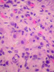
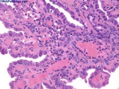
What is the likely cause of the structures at the tip of the arrows?
A. Adenovirus B. Parvovirus C. UTI D. Foreign bodies E. Radiation treatment |
C. The arrows point at michaelis-Gutmann bodies which are often present in malakoplakia, a chronic granulomatous reaction that can be seen in any organ but is most often seen in the bladder. The adjacent areas show histiocytes and inflammation. The causitive agent is usually E. Coli or Proteus and the reaction is an inability to process bacteria. It is most common in HIV patients.
|
|
The image depicted was a biopsy from the bladder. What is it?
A. Nephrogenic adenoma B. Urethelial papilloma C. PUNLMP D. Low grade papillary transitional carcinoma E. Inverted papilloma |
E. This is an inverted papilloma. They are most common in males with a median age of 55. They are characterized by anastomosing cords or islands of urothelium with minimal cytologic atypia.
|
|
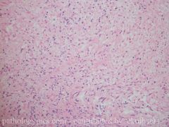
Based on the image shown, what is the most likely diagnosis of this bladder lesion in a patient with no significant past medical history.
A. Postoperative spindle cell nodule B. Pseudosarcomatous fibromyxoid tumor C. Carcinosarcoma D. Leiomyosarcoma |
B. Most inflammatory pseudotumors (AKA pseudosarcomatous fibromyxoid tumor) of the bladder are pedunculated and extend into the bladder lumen. They are can be large and are composed of spindle cells in a collagenous or myxoid background. A prominent network of thin wall vessels is common. Mitosis can be seen but are not atypical. Excision is curative. Post operative spindle cell neoplasm are smaller, <1cm, and occur after surgery.
|
|
|
What is the single most important risk factor for development of renal cell carcinoma?
A. Obesity B. Male gender C. Smoking D. NSAIDs E. Viral infections |
C. Smoking is the single most important risk factor. Obesity in females and various chemical agents have also be implicated. Of note is long term use of phenacetin and transitional cell carcinoma of the renal pelvis.
|
|
|
Phenacetin is associated with the development of what tumor?
A. Papillary renal cell carcinoma B. Metanephric adenoma C. Urethral condyloma D. Renal pelvis transitional cell carcinoma E. Neuroendocrine tumors of the kidney |
D. Renal pelvis transitional cell carcinoma. A cell strong association is seen with acetaminophen.
|
|
|
The vacuolization of the cells of the proximal tubules in cyclosporin toxicity...
A. Are a result of dilatation of endoplasmic reticulum on EM. B. Are often the only tissue manifestation. C. Are specific to cyclosporin toxicity. D. are irreversible. |
A. In addition to the vacuolization seen in acute cyclosponin toxicity, patients can have thrombotic microangiopathy, with platelet and fibrin thrombi in the glomeruli and vessels. The above findings are also seen in Tacrolimus toxicity. The microangiopathy by itself is also non-specific as it is seen in HUS. The nephropathy is usually dose dependent and reversible.
|
|
|
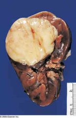
What is the most common cause of an abdominal mass in a newborn?
A. Multicystic nephroma B. Nephroblastoma C. Neuroblastoma D. Multicystic renal dysplasia E. Mesoblastic nephroma |
D. Multicystic renal dysplasia
|
|
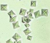
Which statement about the crystals seen here is false?
A. The majority of kidney stones are composed of this mineral. B. Formation of this crystal is one of the toxic effects of ethylene glycol. C. The crystals are deposited in the interstitium, tubules, and rarely, the glomeruli of the kidney. D. Lesch-Nyhan syndrome patients commonly have these crystals in there urine. |
D. Calcium oxylate and calcium oxylate/calcium phosphate stones account for 75% of kidney stones. Calcium oxylate looks like an envelope. Struvite stones (15%) are composed of magnesium ammonium phosphate (rectangular) and are associated with urea-slitting bacterial. Uric acid stones form in acidic urine and can be a result of gour or Lesch-Nyhan syndrome. Cystine crystals are flat hexagons.
|
|
Which statement if not true regarding clear cell renal sarcoma?
A. Metastases are often found >5 years after resection of the tumor. B. Bone metastases are common. C. A clear cytoplasm is a prominent feature in only 20% of cases. D. Like Wilm's, it is insulin-like growth factor positive and WT1 positive. |
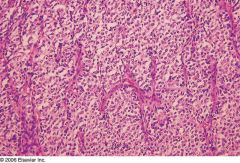
Clear cell sarcoma is also known as bone-metastasizing renal tumor. Despite its name, a clear cytoplasm is a prominent feature in only 20% of cases. It used to be considered a variant of Wilm's but has a different natural history (bone mets) and does not stain with insulin-like growth factor. It does stain for WT1. Histology shows a diffuse growth of small cells with round nuclei, nuclear grooves, inconspicuous nucleoli and a low mitotic rate.
|