![]()
![]()
![]()
Use LEFT and RIGHT arrow keys to navigate between flashcards;
Use UP and DOWN arrow keys to flip the card;
H to show hint;
A reads text to speech;
55 Cards in this Set
- Front
- Back
|
Fn of synovial membrane |
secretes synovial fluid |
|
|
Fn of synovial fluid |
-provides nutrients -decreases friction -shock absorption |
|
|
Remember joint pain is coming from... |
damage to well-innervated joint, capsule, ligaments, and bone (cartilage and synovial membranes poorly innervated) |
|
|
Hilton's law |
sensory nerve supply to joints is by the nerves that supply the muscles that act on it |
|
|
3 factors responsible for joint stability |
1. shape of articulating surfaces 2. ligaments 3. muscles |
|
|
3 main pectoral girdle joints and the 4th conceptual joint |
1. sternoclavicular 2. acromioclavicular 3. glenohumeral 4. scapulothoracic |
|
|
bursae |
Synovial fluid sac around many joints that reduce friction -- subject to inflammation (bursitis) |
|
|
Fn of supporting structures like the ligaments |
-increase stability -limits range of motion ( abnormal range would indicate damage) |
|
|
What type is the sternoclavicular joint |
Saddle synovial joint (but acts like ball-and-socket joint ) |
|
|
Major structural components of the sternoclavicular joint |
1. Anterior & Posterior sternoclavicular ligament 2. Interclavicular ligament 3. Costoclavicular ligament |
|
|
Sternoclavicular joint dislocation |
shoulder displacement 90% anterior displacement- benign Posterior displacement- can poke into the lungs (SOB) |
|
|
Subluxation of the SC joint |
1 of 2 ligament is torn (either a. or p. sternoclavicular ligament or costoclavicular) |
|
|
Dislocation of the SC joint |
When both sternoclavicular and costoclavicular ligaments are torn |
|
|
Are the ligaments more likely to be torn? |
No, they are strong. Therefore you're more likely to get an avulsion facture |
|
|
What type of joint is the acromioclavicular joint? |
Plane type of synovial joint, uniaxial |
|
|
Important ligaments in the acromioclavicular joint |
-Acromioclavicular ligament -Coracoclavicular ligament 1. Trapezoid (horizontal; lateral) 2. Conoid (vertical; medial) |
|
|
AC subluxation Grade II |
elbow injury that travels up and ruptures acromioclavicular ligament only |
|
|
AC Dislocation Grade III |
direct trauma to shoulder rupturing acromioclavicular and coracoclavicular ligament |
|
|
Shoulder separation (falling shoulder) Loss of ligaments in AC joint -gravity pulls it downward |
|
|
Glenohumeral joint is what kind of joint? |
Ball & Socket synovial joint |
|
|
Ligaments for glenohumeral joint |
1. Glenohumeral ligaments (superior, middle, inferior) 2. Coracoacromial ligament 3. Coracohumeral ligament 4. Transverse humeral ligament |
|
|
Mobility of the glenohumeral joint is enhanced by what? |
1. Poor articular congruency 2. Several bursae 3. lots of articular cartilage 4. Lax Joint capsule |
|
|
Stability of the glenohumeral joint is enhanced by what? |
1. glenoid labrum 2. musculotendinous rotator cuff 3. ligaments |
|
|
What is the axillary recess? |
The bottom of glenoid cavity that is prone to injury |
|
|
Main Arterial supply to glenohumeral joint |
Suprascapular artery Anterior & Posterior humeral circumflex arteries |
|
|
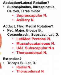
Innervation to the glenohumeral joint? |
|
|
|
Shoulder dislocation by glenohumeral joint |
-most frequent dislocated joint -90% anterior -risk of axillary n and a damage |
|
|
Impingement syndrome |
-supraspinatus tendon becomes impinged in the coracoacromial arch -leads to tendonitis and thickening of tendon |
|
|
Shoulder instability |
Chronic overuse can stretch glenohumeral stabilizers -increases stress and weakens rotator cuffs -leads to humeral head subluxation; secondary impingement |
|
|
Coracoacromial arch is so strong... |
it would sooner fracture humerus or clavicle before fracturing the arch |
|
|
Diagnosis of shoulder instability |
Apprehension test |
|
|
Type of joint at the elbow |
hinge type of synovial joint |
|
|
Two articulations of the elbow |
1. Humeroradial 2. Humeroulnar |
|
|
The elbow joint is weak ________ but reinforced ___________ by ________ |
The elbow joint is weak anterior and posteriorly but reinforced medially and laterally by collateral ligaments |
|
|
Elbow's prominent bursae that help to facilitate movement |
subcutaneous olecranon bursae biceps bursa |
|
|
Ligaments found in the elbow |
1. Ulnar collateral ligament 2. Radial collateral Ligament |
|
|
Ulnar collateral ligament parts |
Anterior (strongest) Posterior (weakest) Oblique |
|
|
What is reducing friction for the olecranon |
subcutaneous olecranon bursa |
|
|
Blood supply to the elbow joint |
Radial collateral a. ANASTOMOSES with radial recurrent a. Superior ulnar collateral a ANASTOMOSES with Posterior Ulnar Recurrent A Inferior Ulnar collateral a ANASTOMOSES with anterior ulnar recurrent a |
|
|
Innervation of the elbow joint |
musculocutaneous n. median n. radial n. ulnar n. |
|
|
elbow dislocations |
mostly posteriorly ulnar n at risk |
|
|
Tommy John |
Ulnar collateral ligament replacement surgery -use palmaris longus, donor tendon |
|
|
Radioulnar joints |
Proximal and Distal (synovial pivot joints) Intermediate (Syndesmosis joint ocnnect via interosseous membrane) |
|
|
Movement of radioulnar joint |
supination and pronation |
|
|
Proximal Radioulnar joint |
Articulation of head of radius & radial notch of ulna -located w/in loose fibrous capsule/synovial membrane -anular ligaments allow rotation of radial head |
|
|
Proximal Radioulnar joint blood supply and innervation |
blood supply: Radial collateral a. ANASTOMOSES with radial recurrent a. innervation: musculocutaneous n. median n. radial n. |
|
|
subluxation of the proximal radio-ulnar joint |
Nursemaid's elbow |
|
|
Distal radioulnar joint |
Pivot joint articulation: head of ulna, ulnar notch of radius ligaments: anterior radioulnar and posterior radioulnar ligaments (WEAK) Has an articular disc |
|
|
Triangular Fibrocartilage complex (TFCC) |
Distal radioulnar joint: Articular disc -distributes forces during supination and pronation -permits mvmt -increases joint stability |
|
|
Distal radioulnar joint blood supply and innervation |
blood supply: anterior and posterior interosseous arteries innervation: anterior and posterior interosseous nerves |
|
|
Ulnar sided wrist trauma |
affects TFCC |
|
|
Wrist Joint |
Ulna doesn't participate in the wrist joint Radiocarpal joint (Synovial: Condyloid) -distal end of the radius with the proximal row of carpal bones except pisiform (ie. scaphoid, lunate, triquetrum) |
|
|
Ligaments of wrist joint |
1. Palmar Radiocarpal Ligament 2. Dorsal Radiocarpal Ligament 3. Radial Collateral Ligament of the wrist 4. Ulnar Collateral ligament of the wrist |
|
|
Wrist sprain |
ligaments in wrist torn/stretched -scapholunate ligament most commonly injured (lunate is out of place and leads to carpal tunnel syndrome--median n compression) |
|
|
Wrist joint blood supply and innervation |
blood supply: dorsal and palmar carpal arches innervation: anterior interosseous branch of the median nerve posterior interosseous branch of the radial nerve dorsal and deep branches of ulnar nerve |