Reading...
![]()
Play button
![]()
Play button
![]()
Use LEFT and RIGHT arrow keys to navigate between flashcards;
Use UP and DOWN arrow keys to flip the card;
H to show hint;
A reads text to speech;
16 Cards in this Set
- Front
- Back
|
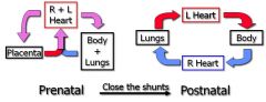
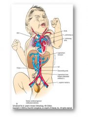
Difference between pre and post natal heart?
|
|
|
|
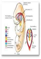
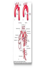
Each pharyngeal arch has an artery (1-4 and 6); forms a basket of arterial arches that surround the foregut
|
|
Remember arches 4 and 6
|

Arch 4 persists on both sides but become diff structures; R = sbclavian, L = aortic arch
Arch 6 L = pulmonary trunk and ductus arteriosus, R = regresses Recurrent laryngeal nerve originally trapped below 6 is released on the right so it lodges under the right subclavian, on the left it remains trapped under the ligamentum arteriosum (ductus arteriosus in the fetus) |
|
What happens if the right dorsal aorta fails to regress?
|
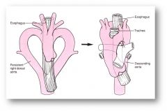
Double Aorta that entraps the esophagus
|
|
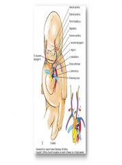
Caval System After Remodeling
|

IVC dervies from all three cardinal sets (cardinal, subcardinal, supracardinal veins) while the SVC is derived from the cardinal vein only
Azygos, hemiazygos and accesory hemiazygos arise from the supracardinal veins |
|
From where does the hepatic portal system develop?
|
Vitelline Veins; note that the cardinal and vitelline veins tend to regress on the left and persist on the right
|
|

How do the umbilical veins develop?
|
The umbilical veins, which drain the placenta, also remodel except the right umbilical vein regresses and the left persists
|
|
|
Describe the 3 important shunts in fetal circulation
|
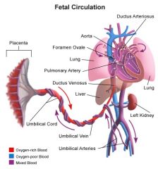
1. ductus venosus (bypass hepatic circulation)
2. foramen ovale (bypass lungs to pump already oxygenated blood from umbilical vein to heart and body) 3. ductus arteriosus (bypass lungs, pumps deoxygenated blood to lower body for maternal circulation) |
|
Describe what happens at birth to change fetal circulation
|
Infant takes breath -> decreased pulmonary resistance = increased left atrial pressure vs right atrial pressure -> foraman ovale closes (now called fossa ovalis) -> increase in 02 leads to decrease in prostaglandins, causing closure of ductus arteriosus
|
|
|
Aortic Coarctation; happens when the ductus arteriosus contracts after birth and the lumen of the aorta becomes constricted
|
|
|
Aortic Coarctation; occurs near the ductus arteriosus, but it can be preductal, ductal or postductal
|
|
three types of atrial septal defects?
|
Sinus Venosus (5%) (roof)
Secundum (90%) (center/middle); results when the foramen ovale and foramen (ostium) secundum overlap Primum (5%) (base) |
|
|
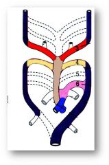
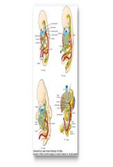
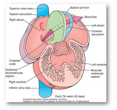
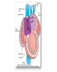
The smooth walls of the atria form by the incorporation of veins into the walls (referred to as intussusception). The colored zones in the figure show where the vena cavae and pulmonary veins end up in the atrial walls. If too much intussusception occurs then the venous entry points can actually overlap the atrial septum (which you have to imagine will form soon in the primitive atrium shown in this figure).
|
|
|
Ventricular Septal Defect
|
|
|
What keeps ductus arteriosus open?
|
Prostalgnadins (from placenta)
Low p02 |
|
|
What closes the ductus arteriosus?
|
Cutting the umbilical cord; prostaglandin synthesis inhibitor
Increase p02 (first breath) |