Reading...
![]()
Play button
![]()
Play button
![]()
Use LEFT and RIGHT arrow keys to navigate between flashcards;
Use UP and DOWN arrow keys to flip the card;
H to show hint;
A reads text to speech;
22 Cards in this Set
- Front
- Back

|
Left coronary Artery And its Branches
LCA (red) LAD (purple) Circumflex (Burgendy) |
|
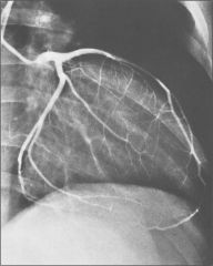
+ angina pectoris
|
Note atherosclerotic plaque in LAD
|
|
|
|
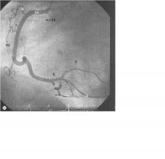
right coronary artery
24 = SA nodal artery |
|
|
right coronary artery
24 = SA nodal artery |
|
|
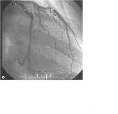
Left Coronary Artery
14 = LAD 7 = circumflex |
|
|
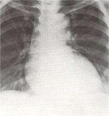
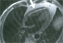
LV Enlargement; often ude to aortic stenosis
|
|
|
Double Density sign of left atrial enlargement; note the right border has a double border
|
|
|
Right ventricular hypertrophy in a man w pulmonary hypertension
|
|
|
right atrial enlargement
|
|
|
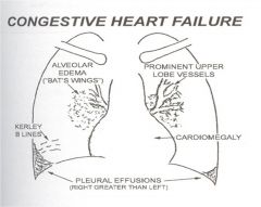
CHF
|
|

|
CHF w left ventricular hypertrophy following chronic rheumatic aortic stenosis
note engorgement of pulmonary arteries and veins |
|
|
Constrictive pericarditis in a 45 yo man with dyspnea and chest pain
|
|
|
Acute Pericarditis; bright outlining of the pericardial sac
|
|

|
Normal Aorta
|
|

|
Saccular aneurysm of the aortic arch
|
|
|
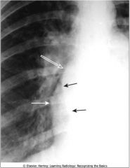
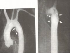
Aortic Dissection; black arrows = intimal flap
|
|
|
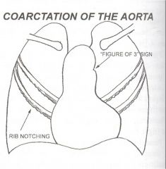
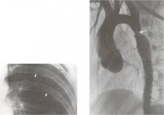
Coarctation of the aorta
|
|

|
Coarctation 4
|
|
|
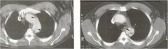
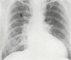
Coarctation; note dilation proximal to the coarctation; radial pulses will be much stronger than foot pulses
Note notching of the ribs |
|
|
Notching of ribs in pt with coarctation of the aorta (sign of 3)
|
|
|
Thoracic aortogram post high speed MV accident; traumatic aortic ruptures occur just beyond the origin of the left subclavian artery
|
|
|
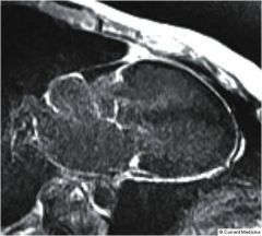
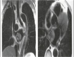
Left atrial Myxoma; (this view is from behind the pt);
|