![]()
![]()
![]()
Use LEFT and RIGHT arrow keys to navigate between flashcards;
Use UP and DOWN arrow keys to flip the card;
H to show hint;
A reads text to speech;
473 Cards in this Set
- Front
- Back

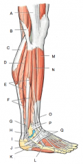
A |
Phalanges |
|

B |
Metatarsals |
|

C |
Tarsals |
|

D |
Distal Phalanx |
|

E |
Middle Phalanx |
|

F |
Proximal Phalanx |
|

G |
Lateral Cuneiform |
|

H |
Cuboid |
|

I |
Calcaneus |
|

J |
Hallux |
|

K |
Medial Cuneiform |
|
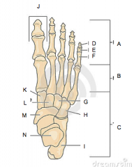
L |
Intermediate Cuneiform |
|

M |
Navicular |
|

N |
Talus |
|

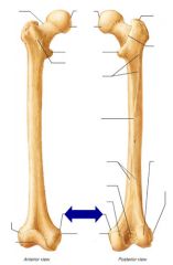
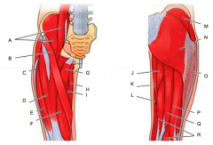
C |
Head of fibula |
|

Name the bone D |
Fibula |
|

E |
Lateral malleolus |
|

F |
Medial condyle |
|

G |
Tibial tuberosity |
|

H |
Interosseous membrane |
|

I |
Anterior boarder of tibia |
|

Name the bone J |
Tibia |
|

K |
Medial malleolus |
|

L |
Articular surface |
|

M |
Intercondylar eminence |
|

N |
Lateral condyle |
|

O |
Head of fibula |
|

Name the joint P |
Proximal tibiofibular joint |
|
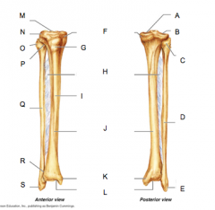
Name the bone Q |
Fibula |
|

Name the joint R |
Distal tibiofibular joint |
|

S |
Lateral malleolus |
|
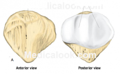
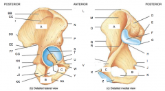
A |
Apex |
|

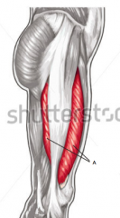
A |
Gluteal tuberosity |
|

B |
Linea aspera |
|

C |
Lateral condyle |
|

D |
Patellar surface |
|

E |
Intercondylar fossa |
|

F and G |
Medial epicondyle |
|

H |
Medial condyle |
|

I and J |
Lesser trochanter |
|

K |
Head of femur |
|

L |
Neck |
|

M |
Fovea capitis |
|

N |
Greater trochanter |
|
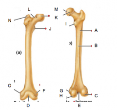
O |
Lateral epicondyle |
|
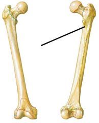
|
Pectineal line |
|
|
Adductor tubercle |
|

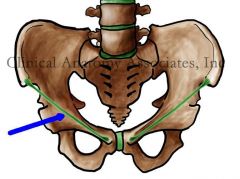
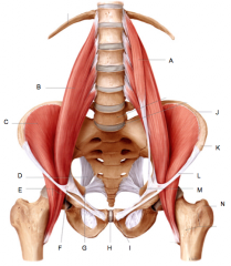
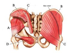
Name the bone A |
Ilium |
|

Name the bone B |
Ischium |
|

Name the bone C |
Pubis |
|

E |
Posterior superior iliac spine |
|

F |
Articular surface |
|

G |
Posterior inferior iliac spine |
|

H |
Greater sciatic notch |
|

I |
Ischial spine |
|

J |
Lesser sciatic notch |
|

K and KK |
Obturator foramen |
|

L |
Iliac crest |
|

M |
Iliac fossa |
|

N and O |
Anterior superior iliac spine |
|

P and Q |
Anterior inferior iliac spine |
|

R |
Arcuate line |
|

S |
Acetabulum |
|

T |
Body of pubis |
|

U |
Pectineal line of pubis |
|

V |
Superior pubic ramus |
|

W |
Pubic tubercle |
|

Z |
Inferior pubic ramus |
|

AA |
Ischial ramus |
|

BB |
Ala |
|

CC |
Gluteal lines |
|

DD |
Posterior superior iliac spine |
|

EE |
Posterior inferior iliac spine |
|

FF |
Greater sciatic notch |
|

GG |
Body of ischium |
|
HH |
Ischial spine |
|

II |
Lesser sciatic notch |
|

JJ |
Ischial tuberosity |
|
|
What is the iliopubic eminence |
Raised knob between pectineal line of pubis and arcuate life of ilium |
|
|
Inguinal ligament |
|
|
Sacrospinous ligament |
|

B |
Sacral canal |
|

C |
Body |
|

D |
Superior articular surface |
|

E |
Auricular surface |
|

F and Form _____ |
Spinous tubercles; median sacral crest |
|

G |
Anterior sacral foramen |
|

H |
Posterior sacral foramen |
|
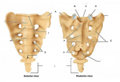
I |
Coccyx |
|

J |
Sacral promontory |
|

A |
Sacral promontary |
|

B |
Symphysis pubis |
|
C |
Sacroiliac joint |
|

D |
Body of sacrum |
|

Name the bone E |
Pubis |
|

Name the bone F |
Ischium |
|

Name the bone G |
Ilium |
|

A |
False pelvis |
|
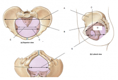
B |
Pelvic brim |
|

C |
Pelvic inlet |
|

D |
True pelvis |
|

E |
Pelvic outlet |
|

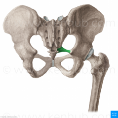
A |
Adductor longus |
|
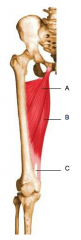
B |
Adductor magnus |
|

C |
Adductor hiatus |
|

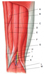
A |
Patella |
|

B |
Tibial tuberosity |
|

D |
Gastrocnemius |
|

E |
Fibularis longus |
|
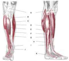
F |
Soleus |
|

G |
Tibialis anterior |
|

H |
Fibularis brevis |
|

I |
Extensor digitorum longus |
|

J |
Extensor hallucis longus |
|

A |
Pectineus |
|
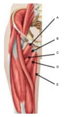
B |
Adductor magnus |
|

C |
Adductor brevis |
|

D |
Adductor longus |
|

E |
Gracilis |
|

A |
Piriformis |
|
B |
Gemellus superior |
|

C |
Obturator internus |
|

D |
Gemellus inferior |
|

E |
Quadratus femoris |
|

A |
Iliotibial tract |
|

B |
Biceps femoris |
|

C |
Gastrocnemius |
|

D |
Soleus |
|

E |
Fibularis longus |
|

F |
Fibularis brevis |
|

G |
Fibularis longus |
|

H |
Superior fibular retinaculum |
|

I |
Inferior fibular retinaculum |
|

J |
Fibularis tertius |
|
K |
Fibularis brevis |
|

L |
Extensor digitorum brevis |
|

M |
Tibialis anterior |
|

N |
Extensor digitorum longus |
|

O |
Extensor hallucis longus |
|

P |
Superior extensor retinaculum |
|

Q |
Inferior extensor retinaculum |
|

A |
Iliopsoas |
|

B |
Sartorious |
|

C |
Iliotibial tract |
|

D |
Rectus femoris |
|

E |
Vastus lateralis |
|

F |
Vastus medialis |
|

G |
Adductor longus |
|

H |
Gracilis |
|

I |
Adductor magnus |
|

J |
Adductor magnus |
|

K |
Gracilis |
|

L |
Sartorius |
|
M |
Gluteus medius |
|

N |
Gluteus maximus |
|

O |
Iliotibial tract |
|

P |
Biceps femoris |
|

Q |
Semitendinosus |
|

R |
Semimembtanosus |
|

A |
Adductor magnus |
|

B |
Biceps femoris, long head |
|

C |
Gracilis |
|

D |
Semitendinosus |
|
E |
Semimembranosus |
|

F |
Iliotibial tract |
|

G |
Biceps femoris, short head |
|

H |
Sartorius |
|

I |
Semimembranosus |
|

L |
Gastrocnemius |
|

M |
Gastrocnemius |
|
|
Plantaris |
|

A |
Gastrocnemius |
|
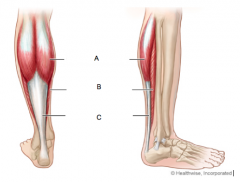
B |
Soleus |
|

C |
Calcaneal tendon |
|

A |
Psoas minor |
|

B |
Psoas major |
|

C |
Iliacus |
|
E |
Iliopsoas |
|

G |
Ischial spine |
|

H |
Pubic symphysis |
|

I |
Pubic tubercle |
|

J |
Iliac crest |
|

K |
Anterior superior iliac spine |
|

L |
Inguinal ligament |
|

N |
Greater trochanter of femur |
|
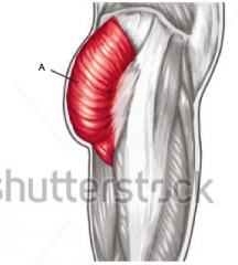
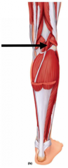
A |
Gluteus maximus |
|
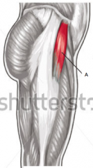
A |
Tensor fasciae latae |
|
A |
Vastus intermedius |
|

A |
Tibialis anterior |
|

B |
Fibularis longus |
|

C |
Extensor digitorum longus |
|

D |
Fibularis brevis |
|

E |
Extensor hallucis longus |
|

F |
Fibularis tertius |
|

G |
Superior extensor retinaculum |
|

H |
Inferior extensor retinaculum |
|

I |
Gastrocnemius |
|

J |
Gastrocnemius |
|

K |
Plantaris |
|

L |
Soleus |
|

M |
Calcaneal tendon |
|

N |
Popliteus |
|

O |
Soleus |
|

P |
Fibularis longus |
|
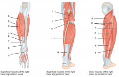
Q |
Tibialis posterior |
|

R |
Flexor digitorum longus |
|

S |
Flexor hallucis longus |
|

T |
Fibularis brevis |
|

A |
Gluteus minimus |
|

B |
Gluteus medius |
|

C |
Gluteus maximus |
|
D |
Obturator internus |
|

A |
Posterior ramus |
|

B |
Spinal nerve |
|

C |
Posterior root |
|

D |
White matter |
|
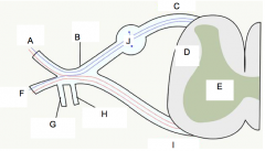
E |
Gray matter |
|

F |
Anterior Ramus |
|

I |
Anterior root |
|

J |
Posterior root ganglion |
|

A |
Posterior median sulcus |
|

B |
Gray commissure |
|

C |
Posterior horn |
|

D |
Anterior horn |
|

E |
Lateral horn |
|

F |
Gray matter |
|
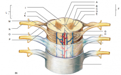
G |
Central Canal |
|

H |
Anterior median fissure |
|

I |
Posterior funiculus |
|

J |
Anterior funiculus |
|

K |
Lateral funiculus |
|

L |
White matter |
|

M |
Posterior root ganglion |
|

N |
Spinal nerve |
|

O |
Posterior root |
|

P |
Anterior root |
|

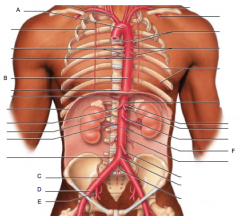
Name the artery A |
Common iliac artery |
|

Name the artery B |
Internal iliac artery |
|

Name the artery C |
External iliac artery |
|

Name the nerve D |
Fermoral nerve |
|

Name the nerve E |
Obturator nerve |
|

Name the artery F |
Femoral artery |
|

Name the nerve G |
Superior gluteal nerve |
|

Name the nerve H |
Inferior gluteal nerve |
|

Name the nerve I |
Sciatic nerve |
|

Name the nerve J |
Tibial nerve |
|
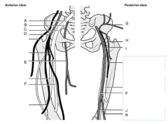
Name the nerve K |
Common fibular nerve |
|

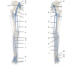
Name the nerve A |
Common fibular nerve |
|

Name the nerve B |
Deep fibular nerve |
|

Name the nerve C |
Superficial fibular nerve |
|

Name the artery D |
Anterior tibial artery |
|

Name the artery E |
Dorsalis pedis artery |
|

Name the artery F |
Popliteal artery |
|

Name the artery not labeled above D on the right |
Posterior tibial artery |
|

Name the artery G |
Medial plantar artery |
|
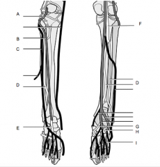
Name the artery H |
Lateral plantar artery |
|

Name the artery I |
Plantar arch |
|

A |
Ascending aorta |
|

B |
Superior vena cava |
|

C |
Pulmonary artery |
|

D |
Pulmonary veins |
|
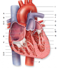
E |
Right atrium |
|

F |
Right atrioventricular valve (Tricuspid) |
|

G |
Right ventricle |
|

H |
Inferior vena cava |
|

I |
Aortic arch |
|

J |
Pulmonary trunk |
|

K |
Left atrium |
|

L |
Aortic semilunar valve |
|

M |
Left atrioventricular valve (bicuspid) |
|

N |
Pulmonary semilunar valve |
|

O |
Left ventricle |
|

P |
Descending aorta |
|

A |
Aortic arch |
|

B |
Thoracic aorta |
|
C |
Common iliac artery |
|

D |
External iliac artery |
|

E |
Internal iliac artery |
|

F |
Abdominal aorta |
|

A |
External iliac vein |
|

B |
Common iliac vein |
|

C |
Internal iliac vein |
|

D |
Femoral vein |
|

E |
Great saphenous vein |
|

F |
Popliteal vein |
|

G |
Lesser saphenous vein |
|

H |
Anterior tibial vein |
|

I |
Posterior tibial vein |
|

J |
Fibular vein |
|
K |
Dorsal venous arch |
|

L |
Digital vein |
|

M |
Lateral plantar vein |
|

N |
Medial plantar vein |
|

O |
Plantar venous arch |
|

A |
Dorsal arch |
|
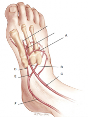
B |
Dorsalis pedis artery |
|

C |
Anterior tibial artery |
|

D |
Medial plantar artery |
|

E |
Lateral plantar artery |
|

F |
Posterior tibial artery |
|
|
The Right atrium, Right ventricle, Pulmonary arteries, and Pulmonary veins are all part of the _____________ circulation system. |
Pulmonary |
|
|
The Left atrium, Left ventricle, Aorta and systemic arteries, and Systemic veins are all part of the _______________ circulation system. |
Systemic |
|
|
From the superior and inferior vena cava blood goes to the _________. |
Right atrium |
|
|
From the right atrium blood goes to the ________. |
Right atrioventricular valve (tricuspid) |
|
|
From the right atrioventricular valve blood goes to the ________. |
Right ventricle |
|
|
From the right ventricle blood goes to the _______. |
Pulmonary semilunar valve |
|
|
From the pulmonary semilunar valve blood goes to the _______. |
Pulmonary trunk |
|
|
From the pulmonary trunk blood goes to the _______. |
Pulmonary arteries |
|
|
From the pulmonary arteries blood goes to the ________. |
Lungs |
|
|
From the lungs blood goes to the ________. |
Pulmonary veins |
|
|
From the pulmonary veins blood goes to the ______. |
Left atrium |
|
|
From the left atrium blood goes to the _______. |
Left atrioventricular valve (bicuspid) |
|
|
From the left atrioventricular valve blood goes to the _____. |
Left ventricle |
|
|
From the left ventricle blood goes to the ______. |
Aortic semilunar valve |
|
|
From the aortic semilunar valve blood goes to the _______. |
Ascending aorta |
|
|
From the ascending aorta blood goes to the _____. |
Aortic arch |
|
|
From the aortic arch blood goes to the ______. |
Descending aorta |
|
|
From the descending aorta blood goes to the _____. |
Common Iliac artery |
|
|
From the common iliac artery blood goes to the _________ and ___________. |
External iliac artery; internal iliac artery |
|
|
From the external iliac artery blood goes to the ______. |
Femoral artery |
|
|
From the femoral artery blood goes to the _______. |
Popliteal artery |
|
|
From the popliteal artery blood goes to the ________ and _______. |
Anterior tibial artery; posterior tibial artery |
|
|
From the anterior tibial artery blood goes to the _______. |
Dorsalis pedis artery |
|
|
From the dorsalis pedis artery blood goes to the _______ |
Dorsal arch |
|
|
From the dorsal arch blood goes to the _______. |
Plantar arch |
|
|
From the plantar arch artery blood goes to the _________. |
Digital arteries |
|
|
From the posterior tibial artery blood goes to the _________, __________, and _________. |
Fibular artery; Lateral plantar artery; Medial plantar artery |
|
|
From the lateral plantar artery blood goes to the ________. |
Plantar arch |
|
|
From the medial plantar artery blood goes to the _________. |
Plantar arch |
|
|
From the digital arteries blood goes to the ____. |
Digital veins |
|
|
From the digital veins blood goes to the ______ and _______. |
Dorsal venous arch; plantar venous arch |
|
|
From the dorsal venous arch blood goes to the _____ and _____. |
Lesser saphenous vein; greater saphenous vein |
|
|
From the lesser saphenous vein blood goes to the _____. |
Popliteal vein |
|
|
From the popliteal vein blood goes to the _____. |
Femoral vein |
|
|
From the femoral vein blood goes to the _____. |
External iliac vein |
|
|
Fro the external iliac vein blood goes to the ____. |
Common iliac vein |
|
|
From the greater saphenous vein blood goes to the _____. |
Femoral vein |
|
|
From the plantar venous arch vein blood goes to the ______ and ______. |
Medial plantar vein; lateral plantar vein |
|
|
From the medial plantar vein blood goes to the ______. |
Posterior tibial vein |
|
|
From the posterior tibial vein blood goes to the _______. |
Popliteal vein |
|
|
From the Lateral plantar vein blood goes to the _____. |
Posterior tibial vein |
|
|
Blood goes from the fibular vein to the _____. |
Posterior tibial vein |
|
|
Blood goes from the anterior tibial vein to the _____. |
Popliteal vein |
|
|
Blood goes from the internal iliac vein to the _____. |
Common iliac vein |
|
|
From the common iliac vein blood goes to the _____. |
Inferior vena cava |
|
|
From the inferior vena cava and the superior vena cava blood goes to the _____. |
Right atrium |
|
|
Muscle(s) innervated by the Inferior Gluteal N. |
Gluteus Maximus |
|
|
Muscle(s) innervated by the Superior Gluteal N. |
Gluteus Medius Gluteus Minimus Tensor Fasciar Latae |
|
|
Muscle(s) innervated by the Femoral N. |
Iliacus Sartorius Rectus Femoris Vastus Lateralis Vastus Medialis Vastus Intermedius Pectineus |
|
|
Muscle(s) innervated by the Obturator N. |
Gracilis Adductor Longus Adductor Magnus Adductor Brevis |
|
|
Muscle(s) innervated by the Tibial N. |
Biceps Femoris, Long Head Semitendinosus Semimembranosus Gastrocnemius Soleus Plantaris Popliteus Flexor Hallucis Longus Flexor Digitorum Longus Tibialis posterior
|
|
|
Muscle(s) innervated by the Common Fibular N. |
Biceps Femoris, Short Head |
|
|
Muscle(s) innervated by the Deep Fibular N. |
Tibialis Anterior Extensor Digitorum Longus Extensor Hallucis Longus Fibularis Tertius |
|
|
Muscle(s) innervated by the Superficial Fibular N. |
Fibularis Longus Fibularis Brevis |
|
|
Muscle(s) that extend the hip joint |
Gluteus Maximus Adductor Magnus Biceps Femoris, Long Head Semitendinosus Semimenbranosus
|
|
|
Muscle(s) that laterally rotate the hip joint |
Gluteus Maximus Piriformis Gemellus Superior Obturator Internus Gemellus Inferior Quadratus Femoris Sartorius
|
|
|
Muscle(s) that abduct the hip joint |
Gluteus Medius Gluteus Minimus Tensor Fasciae Latae |
|
|
Muscle(s) that flex the hip joint |
Tensor Fasciae Latae Psoas Major Iliacus Iliopsoas Sartorius Rectus Femoris Pectineus Adductor Longus Adductor Brevis |
|
|
Muscle(s) that medially rotate the hip joint |
Tensor Fasciae Latae |
|
|
Muscle(s) that flex the knee joint |
Sartorius Gracilis Biceps Femoris, Long Head Biceps Femoris, Short Head Semitendinosus Semimembranosus Gastrocnemius Plantaris Popliteus
|
|
|
Muscle(s) that extend the knee joint |
Rectus Femoris Vastus Lateralis Vastus Medialis Vastus Intermedius
|
|
|
Muscle(s) that adduct the hip joint |
Gracilis Pectineus Adductor Longus Adductor Brevis Adductor Magnus |
|
|
Muscle(s) that dorsiflex the foot |
Tibialis Anterior Extensor Digitorum Longus Extensor Hallucis Longus Fibularis Tertius |
|
|
Muscle(s) that invert the foot |
Tibialis Anterior Tibialis Posterior |
|
|
Muscle(s) that extend digits #2-5 |
Extensor Digitorum Longus |
|
|
Muscle(s) that extend the hallux |
Extensor Hallucis Longus |
|
|
Muscle(s) that evert the foot |
Fibularis Tertius Fibularis Longus Fibularis Brevis
|
|
|
Muscle(s) that plantar flex foot |
Fibularis Longus Fibularis Brevis Gastrocnemius Soleus Plantaris Flexor Hallucis Longus Flexor Digitorum Longus Tibialis Posterior |
|
|
Muscle(s) that medially rotate the tibia |
Polpiteus |
|
|
Muscle(s) that flex the hallux |
Flexor Hallucis Longus |
|
|
Muscle(s) that flex digits #2-5 |
Flexor Digitorum Longus |
|
|
Patellofemoral joint structural classification and type |
Synovial, hinge and planar |
|
|
Patellofemoral joint functional classification |
Diarthrosis |
|
|
Superior tibiofibular joint structural classification and type |
Synovial, planar |
|
|
Superior tibiofibular joint functional classification |
Amphiarthrosis |
|
|
Inferior tibiofibular joint structural classification and type |
Fibrous, syndesmosis |
|
|
Inferior tibiofibular joint functional classification |
Amphiarthrosis |
|
|
Interphalangeal joint structural classification and type |
Synovial, hinge |
|
|
Interphalangeal joint functional classification |
Diarthrosis |
|
|
Metatarsophalangeal joint structural classification and type |
Synovial, condylar |
|
|
Metatarsophalangeal joint functional classification |
Diarthrosis |
|
|
Talocrural joint structural classification and type |
Synovial, hinge |
|
|
Talocrural joint functional classification |
Diarthrosis |
|
|
Intertarsal joint structural classification and type |
Synovial, planar |
|
|
Intertarsal joint functional classification |
Diarthrosis |
|
|
Coxal joint structural classification and type |
Synovial, ball-and-socket |
|
|
Coxal joint functional classification |
Diarthrosis |
|
|
Symphysis pubis structural classification and type |
Cartilagious, symphysis |
|
|
Symphysis pubis functional classification |
Amphiarthrosis |
|
|
Sacroiliac joint structural classification and type |
Synovial, planar |
|
|
Sacroiliac joint functional classification |
Diarthrosis |
|
|
Tibiofemoral joint structural classification and type |
Synovial, hinge |
|
|
Tibiofemoral joint functional classification |
Diarthrosis |
|
|
Tarsometatarsal joint structural classification and type |
Synovial, planar |
|
|
Tarsometatarsal joint functional classification |
Diarthrosis |
|
|
Origin of Gluteus maximus |
Ilium, posterior sacrum and coccyx |
|
|
Insertion of Gluetus maximus |
Gluteal tuberosity of femur, iliotibial tract of fasciae latae |
|
|
Origin of Gluteus medius |
Gluteal lines of ilium, iliac crest |
|
|
Insertion of Gluteus medius |
Greater trochanter of femur |
|
|
Origin of Gluteus minimus |
Gluteal lines of ilium |
|
|
Insertion of Gluteus minimi |
Greater trochanter of femur |
|
|
Origin of Tensor fasciae latae |
Anterior superior iliac spine |
|
|
Insertion of Tensor fasciae latae |
Iliotibial tract of fascia |
|
|
Origins on Piriformis |
Anterior sacrum |
|
|
Insertion of Piriformis |
Greater trochanter of femur |
|
|
Origin on Gemellus superior |
Ischial spine |
|
|
Insertion of Gemellus superior |
Greater trochanter of femur |
|
|
Origin on Obturator internus |
Medial margin of obturator foramen |
|
|
Insertion of Obturator internus |
Greater trochanter of femur |
|
|
Origin of Gemellus inferior |
Ischial tuberosity |
|
|
Insertion of Gemellus inferior |
Greater trochanter of femur |
|
|
Origin of Psoas major |
Vertebrae T12-L5 |
|
|
Insertion of Psoas major |
Lesser trochanter of femur |
|
|
Origin of Psoas minor |
Vertebrae T12-L1 |
|
|
Insertion of Psoas minor |
Iliopubic eminence |
|
|
Origin of Iliacus |
Iliac fossa |
|
|
Insertion of Iliacus |
Lesser trochanter of femur |
|
|
Origin of Iliopsoas |
Conversion of iliac and posts major once they pass under the inguinal ligament |
|
|
Insertion of Iliopsoas |
Lesser trochanter of femur |
|
|
Origin of Sartorius |
Anterior superior iliac spine |
|
|
Insertion of Sartorius |
Proximal medial tibia |
|
|
Origin of Rectus femoris |
Anterior inferior iliac spine |
|
|
Insertion of Rectus femoris |
Tibial tuberosity |
|
|
Origin of Vastus lateralis |
Linea aspera, lateral lip |
|
|
Insertion of Vastus lateralis |
Tibial tuberosity |
|
|
Origin of Vastus medialis |
Linea aspera, medial lip |
|
|
Insertion of Vastus medialis |
Tibial tuberosity |
|
|
Origin of Vastus intermedius |
Anterior upper femur |
|
|
Insertion of Vastus intermedius |
Tibial tuberosity |
|
|
Origin of Gracilis |
Inferior ramus of pubis |
|
|
Insertion of Gracilis |
Proximal medial tibia |
|
|
Origin of Pectineus |
Pectineal line of pubis |
|
|
Insertion of Pectineus |
Pectineal line of femur |
|
|
Origin of Adductor longus |
Pubis |
|
|
Insertion of Adductor longus |
Linea aspera of femur |
|
|
Origin of Adductor brevis |
Inferior ramus of pubis |
|
|
Insertion of Adductor brevis |
Linea aspera of femur |
|
|
Origin on Adductor magnus |
Inferior rams of pubis, ischial tuberosity |
|
|
Insertion of Adductor magnus |
Linea aspera, adductor tubercle of femur |
|
|
Origin of Biceps femoris, long head |
Ischial tuberosity |
|
|
Insertion of Biceps femurs, long head |
Head of fibula |
|
|
Origin of Biceps femoris, short head |
Linea aspera of femur |
|
|
Insertion of Biceps femoris, short head |
Head of fibula |
|
|
Origin of Semitendinosus |
Ischial tuberosity |
|
|
Insertion of Semitendinosus |
Proximal medial tibia |
|
|
Origin of Semimembranosus |
Ischial tuberosity |
|
|
Insertion of Semimembranosus |
Proximal posterior tibia |
|
|
Origin of Tibialis anterior |
Lateral condyle and proximal tibia, interosseous membrane |
|
|
Insertion of Tibialis anterior |
Medial cuneiform, metatarsal I |
|
|
Origin of Extensor digitorum longus |
Lateral condyle of tibia, anterior fibula, interosseous membrane |
|
|
Insertion of Extensor digitorum longus |
Distal phalanges digits 2-5, dorsal surface |
|
|
Origin of Extensor hallucis longus |
Anterior fibula, interosseous membrane |
|
|
Insertion of Extensor hallucis longus |
Distal phalanx of hallux, dorsal surface |
|
|
Origin of Fibularis tertius |
Distal fibula, interosseous membrane |
|
|
Insertion of Fibularis tertius |
Base of metatarsal V |
|
|
Origin of Fibularis longus |
Head and proximal lateral fibula, lateral condyle of tibia |
|
|
Insertion of Fibularis longus |
Base of metatarsal I, medial cuneiform |
|
|
Origin of Fibularis brevis |
Lateral fibula |
|
|
Insertion of Fibularis brevis |
Base of metatarsal V |
|
|
Origin of Gastrocnemius |
Medial and lateral condyles of femur |
|
|
Insertion of Gastrocnemius |
Calcaneus |
|
|
Origin of Soleus |
Proximal fibula, soleal line of tibia |
|
|
Insertion of Soleus |
Calcaneus |
|
|
Origin of Plantaris |
Distal lateral femur |
|
|
Insertion of Plantaris |
Calcaneus |
|
|
Origin of Popliteus |
Lateral condyle of femur |
|
|
Insertion of Popliteus |
Posterior proximal tibia |
|
|
Origin of Flexor hallucis longus |
Posterior distal fibula |
|
|
Insertion of Flexor hallucis longus |
Distal phalanx of hallux |
|
|
Origin of Flexor digitorum longus |
Posterior tibia |
|
|
Insertion of Flexor digitorum longus |
Distal phalanx of digits 2-5 |
|
|
Origin of Tibialis posterior |
Tibia, fibula, posterior interosseous membrane |
|
|
Insertion of Tibialis posterior |
Navicular, cuneiforms, cuboid, metatarsals II-IV |