![]()
![]()
![]()
Use LEFT and RIGHT arrow keys to navigate between flashcards;
Use UP and DOWN arrow keys to flip the card;
H to show hint;
A reads text to speech;
26 Cards in this Set
- Front
- Back
|
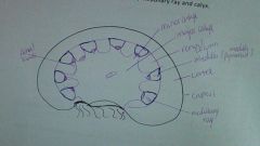
draw and label a diagram showing a low power image of the kidney section |
|
|
|
1. glomerulus 2. bowman's capsule 3. collecting duct 4. distal tubule 5. loop of henle 6. capillary network 7. proximal tubule 8. arteriole from glomerulus 9. arteriole from renal artery |
|
|
approximately how many nephrons are in each kidney? |
800,000 - 1.5 million |
|
|
parts of the nephron in the renal cortex |
1. renal corpuscle - glomerulus - bowman's capsule - urinary space 2. convoluted tubules - proximal tubule -distal tubule |
|
|
parts of the nephron in the medulla |
1. loop of henle 2. collecting duct |
|
|
parts of nephron in the medullary ray |
1. loop of henle 2. collecting tubules |
|
|
are the medullary rays longitudinal or transverse? why? |
longitudinal. tubules run parallel to each other and perpendicular to the capsule |
|
|
why do the descending and ascending tubules sit close and parallel to each other? |
descending part reabsorbs water back into the blood - permeable ascending reabsorbs ions (Na+, Cl-, Ca2+, K+) - impermeable |
|
|
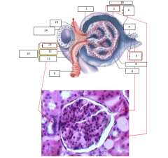
1. vascular pole 2. parietal epithelium 3. visceral epithelum (podocyte) 4. tubular pole 5. proximal convoluted tubule 6. capsular space 8. glomerular capillary 9. afferent arteriole (arteriole from renal artery) 10. juxtaglomerular complex 11. extraglomerular mesangial cells 12. juxtaglomerular cells 13. macula densa 14. distal convulated tubule 15. efferent arteriole (arteriole from glomerulus) 16. glomerular capsule |
|
|
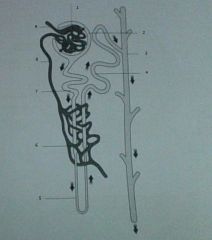
1. renal corpuscle 2. renal capsule 3. renal space 4. proximal convoluted tubule 5. urinary/tubular pole 6. glomerulus 7. vascular pole 8. distal convoluted tubule |
|
|
histologically, how might you differentiate between the afferent and efferent arterioles? |
afferent has smooth muscle cells in its wall (juxtaglomerular cells) |
|
|
how would you recognize macula densa cells |
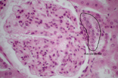
cluster of cells in the juxtaglomerular complex near the DCT |
|
|
where would you expect to locate juxtaglomerular mesangial cells |
modified smooth muscle cells of the afferent arteriole |
|
|
where would you expect to locate extraglomerular mesangial cells |
outside glomerulus near the arterioles |
|
|
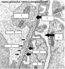
1. endothelial cell cytoplasm 2. fused basal lamina 3. urinary space 4. pedicel 5. filtration slits 6. podocyte 7. fenestrations 8. capillary lumen |
|
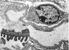
use the previous EM view to help you label the same features on this EM |
|
|
|
list the possible function of mesangial cells |
provide structural support for and regulate blood flow of the glomerular capillaries by their contractile activity specialized cells around blood vessels in thekidneys, at the mesangium. Primary function of mesangial cells is to remove trapped residues and aggregated protein from the basement membrane thus keeping the filter free of debris |
|
|
what might be one effect on renal function if, in a kidney disease, there is a proliferation of mesangial cells? |
debris would accumulate in the basement membrane of kidney cells --> obstruction to nearby blood vessels and blocked filtration/reabsorption into the blood |
|
|
what might be one effect on renal function if, in a kidney disease, there is damage to the basement membrane underlying the podocytes and endothelial cells in the renal corpuscle? |
capillaries become inflamed disruption in filtration |
|
|
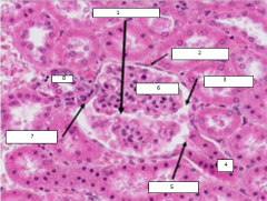
how can you differentiate between the proximal and distal convoluted tubules (histology)? |
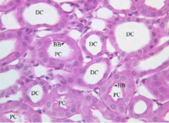
proximal convoluted - lined with cuboidal epithelial; basal striations; microvilli on apical surface distal convoluted - few microvilli; cuboidal epithelium;basal striations |
|
|
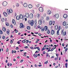
what is unique about transitional epithelium? how does its structure relate to its function? |
can stretch in order to accommodate changes in volume the cells of transitional epithelium are connected by tight junctions, or virtually impenetrable junctions that seal together the cellular membranes of neighboring cells. This barrier prevents reabsorption of toxic wastes and pathogens by the bloodstream. When the organ or tube is stretched (e.g. when the bladder is filled with urine), the tissue compresses and the cells become stretched. When this happens, the cells flatten, and they appear to be squamous and irregular. |
|
|
transitional epithelium (top portion) basal layer is cuboidal columnar cells at superficial layer |
|
|
3 histological features of the wall of the urinary bladder |
1. transitional epithelium (innermost layer) + folds 2. lamina propria 3. smooth muscle 4. adipose tissue |
|
|
What are the cells of the visceral layer of Bowman’s Capsule called |
podocytes podocytes are epithelial cells that have extensive cytoplasmic processes and numerous secondary processes called foot processes |
|
|
Which one of the following can be used to differentiate proximal from distal convoluted tubules? |
Presence/absence of a brush border proximal convoluted tubules have a brush border of Microvilli |
|
|
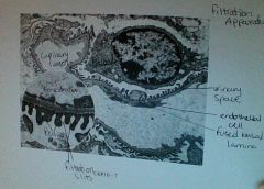
In life, what would be found in the true space marked X? |
Urinary filtrate (urine) this space is called the urinary or bowman’s space. It is lined by the visceral and parietal layers of the bowman’s capsule. The glomerular filtration apparatus produces the urinary filtrate |