![]()
![]()
![]()
Use LEFT and RIGHT arrow keys to navigate between flashcards;
Use UP and DOWN arrow keys to flip the card;
H to show hint;
A reads text to speech;
21 Cards in this Set
- Front
- Back
|
Notes: |
Sarcomere ---Structure ------Actins -- thin filaments ---------G-Actin ---------F-Actin ------Myosins -- thick filaments ------Arrangement of Actin and Myosin ---------Regular, repetitive ---------2 Actin:1 Myosin ---------Midpoint of sarcomere ------------Actin deficient ------Regions of a sarcomere ---------I-Band ---------A-Band ---------H-Zone (Middle of A-Band) ------Details of myofilament ---------Thin--Actin ------------Binding Site (Receptor) for myosin heads ------------Tropomyosin filamentous ------------Troponin ---------Thick ------------2 heads ---------------Actin binding site on each neck for each head ------------Neck for each head ---------------Flexible ------------No Myosin Heads ---------------@ M-line (H-Zone) |
|
|
Notes: |
Sarcomere (cont'd) ---Other Organelles ------Mitochondria ------Smooth ER ---------Sarcoplasmic Reticulum (SR) ------------Ca2+ reservoir ------Transverse Tubule ---------T-Tubule ---------Triad Sliding Filament Theory |
|
|
NOTES: |
NERVE SIGNAL ---GENERATION OF MEMBRANE POTENTIAL |
|
|
1. Striations seen between different muscle types represent a similar ________ between those different muscle types 2. Muscles don't contract on their own; they respond to a signal from the ________ 3. Tendon attaches to the ________ 4. the nuclei are placed ________ on the muscle cell 5. the ________ is the term that we give to the plasma membrane of the muscle cell 6. the ________ is immediately surrounding the previous term Figure 9.1 on PAGE 279 |
1. contractile mechanism 2. nervous system 3. periosteum 4. peripherally 5. sarcolemma 6. endomysium |
|
|
1. ________ fibers connect the different connective tissue sheaths 2. there is a fairly high density of ________ organelles in the muscle cells; this means there is a lot of energy in the form of ________ being used up 3. the reason muscle cells are striated, is because the ________ are striated; the greater the number of them, the more easily the striations are to see 4. the name for the sets of 3 dark lines circumscribing the myofibrils are ________ lines 5. ________ are the basic unit of structure and function of striated muscle |
1. collagen 2. mitochondria, ATP 3. myofibrils 4. M lines 5. sarcomeres; to be the functional part, it requires ATP and neural stimulation |
|
|
1. what is a sarcomere, with respect to myofibrils? 2. what is a myofibril, with respect to sarcomeres? 3. What is the most abundant protein found in striated muscle cells? 4. amino acids are held together by ________ bonds 5. G-Actins form long filaments known as ________ actin |
1. cylindrically shaped subset of a myofibril; 2. many sarcomeres lined up in sequence 3. actin; protein making up the cytoskeleton of the cell; tend to be clustered around the outside edge of cell attached to intermediate filaments; most common intracellular proteins 4. peptide 5. filamentous (F-Actin) |
|
|
1. The thin blue lines on figure 9.1 (Page 280) represent ________ proteins 2. the second most common protein are ________ 3. what is a multimer? 4. The 2 most common proteins in muscle cells are ________ and ________ 5. actins are referred to as ________ filaments, and myosins as ________ filaments 6. the proteins that form the M line help keep thick filaments from ________ |
1. actin 2. myosins 3. many molecules making up an even greater single piece 4. actins, myosins 5. thin, thick 6. slipping out of register |
|
|
1. actins and myosins ________ with each other 2. ________ proteins attach thick filaments at distal ends to zig-zag lines that form the end of the sarcomere 3. actin and myosin are arranged ________ to the long axis of the muscle cell 4. T/F. There are other proteins that are integral to the function of the sarcomere that are not actin and myosin. 5. ________ are the terminal structures of the sarcomere at both ends |
1. interact 2. Elastic (titin) 3. parallel 4. True 5. Z discs |
|
|
1. Z discs directly attach to ________ filaments, and indirectly to ________ filaments 2. thick filaments do not extend to the ________ of the sarcomere 3. the purpose of muscle is ________, and as it contracts the length of the muscle ________ 4. during muscle contraction, the length of the sarcomere ________ 5. if a muscle shortens during contraction, it's because all of its ________ are shortening |
1. thin actin, thick myosin 2. center 3. mobilization, shortens 4. shortens 5. sarcomeres |
|
|
1. T/F. If one sarcomere shortens, it's almost unnoticeable. 2. Increasing the number of ________ shortening, will increase the likelihood that a visible shortening of the muscle will occur. 3. the strength of the contraction is not necessarily reflected in the ________ of the contraction 4. The M line proteins hold the ________ filaments 5. thin filaments in cross section are arranged in a ________ shape 6. thick filaments in cross section are arranged in a ________ shape, and also ________ shapes when zoomed out |
1. True 2. sarcomeres 3. extent 4. thick (myosin) 5. hexagon 6. triangle, hexagon |
|
|
1. ________ individual thin filaments surround each individual thick filament 2. ________ individual thick filaments surround each individual thin filament 3. the ratio of actin to myosin filaments is ________ 4. the overall arrangement of actin and myosin is in a ________ shape, with ________ motifs 5. if the sarcomere shortens, the ________ ends will approach each other |
1. 6 2. 3 3. 2:1 4. crystal, repeating 5. medial |
|
|
LOOK AT PAGE 280, FIGURE 9.2 1. the sarcomere is divided into ________ and ________ 2. The "I" in I-Band refers to ________ properties, referring to the way it reflects plane-polarized light 3. The ________ zone is in the center of the sarcomere 4. each sarcomere has two individual sections of ________ 5. the ________ band extends from one end of a molecule clear to the opposite end of the same molecule |
1. bands, zones 2. isotropic 3. H-zone 4. I-bands 5. A-band |
|
|
1. occupying the vast majority of the sarcomere is the ________ 2. T/F. the A-Band is not homogenous throughout its length 3. the proteins of the M line keep the ________ filaments from slipping out of register 4. the depression in individual G-Actin subunits are ________ for myosin heads 5. Actin and Myosin are ________ to each other, and, under the right conditions, will ________ to each other |
1. A-Band 2. True 3. thick 4. binding sites 5. parallel, connect |
|
|
1. G-Actin are arranged in ________ arrays 2. the resting condition of tropomyosin involves ________-ing the G-actin subunit binding sites 3. ________ of G-actin binding sites are covered by tropomyosin before ending 4. ________ proteins link actin subunits to tropomyosin 5. one of the three parts of troponin, binds to ________, another to ________, and a third one to ________ |
1. helical 2. covering 3. 6-9 4. troponin 5. Actin, tropomyosin, free Calcium ions |
|
|
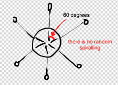
1. Thin filaments are composed of what 3 proteins? What protein does this ultimately produce? 2. "thin filament" is synonymous with the term ________ 3. T/F. the hinge region of the myosin molecule can bend, meaning the neck region is flexible 4. the myosin molecule has ________ heads, with each head containing a(n) ________ 5. the angle between adjacent myosin heads is ________ |
1. tropomyosin, troponin, G-Actin subunits; F-Actin is ultimately produced 2. F-Actin 3. True 4. 2, actin binding-site 5. 60 degrees |
|
|
What do myosin heads look like in cross-section? |
|
|
|
1. there are ________ myosin heads at the M-Line; this is because this region is deficient of ________ 2. the myosin heads ________ around the thick filaments; they are not randomly arranged 3. endoplasmic reticulum in muscle cells is referred to as ________ 4. the myofibrils are surrounded by ________ organelles 5. The holes of the sarcoplasmic reticulum have the effect of increasing the ________ |
1. ZERO, thin filaments 2. spiral 3. sarcoplasmic reticulum (SR) 4. sarcoplasmic reticulum 5. surface area |
|
|
1. most of the calcium in the muscle cell is located in the ________ and very little in the ________ 2. the concentration gradient of calcium in sarcoplasmic reticulum favors its movement ________ 3. the triad is a structure that includes the ________ and the ________ 4. the thick part of the SR are the ________ 5. skeletal muscle cells have multiple nuclei because of ________ |
1. sarcoplasmic reticulum, sarcoplasm 2. out of the cell 3. T-Tubule and the SR 4. 2 pieces of terminal cisterna 5. the long length of muscle cells; this requires multiple nuclei to control each region |
|
|
1. THE FUNCTION OF SARCOPLASMIC RETICULUM IS ________ 2. IF MUSCLES RUN OUT OF CALCIUM, THE NEXT SOURCE OF CALCIUM IS ________ 3. MANY MYOFILAMENTS MAKE UP A ________, WHICH IN LARGE NUMBERS MAKE UP A ________ 4. ________ FILAMENTS HAVE DIRECT ATTACHMENT TO THE Z-DISC; ________ FILAMENTS HAVE INDIRECT ATTACHMENT TO Z-DISCS 5. THE DISTANCE BETWEEN ADJACENT Z-DISCS ________ DURING MUSCLE CONTRACTION |
1. CALCIUM ION STORAGE 2. BONE 3. MYOFIBRIL, MYOFIBER 4. ACTIN (THIN); MYOSIN (THICK) 5. SHORTEN |
|
|
1. ________ FILAMENTS ARE THE ONES THAT SLIDE DURING MUSCLE CONTRACTION 2. THE SLIDING FILAMENT THEORY PROPOSES THAT WHEN MUSCLE CONTRACTS THE ________ SLIDES ALONG PARALLEL TO STATIONARY ________ AND AS THE ACTIN SLIDES IT PULLS THE ________ WITH IT, THUS BRINGING THEM CLOSER TOGETHER, SHORTENING THE SARCOMERE, AND SHORTENING THE WHOLE MUSCLE 3. THE H-ZONE IS DEFINED BY ________ FILAMENTS; THE A-BAND IS DEFINED BY THE ________ FILAMENTS 4. ________ CAUSES THE NECKS OF MYOSIN HEADS TO FLEX |
1. ACTIN (THIN) 2. ACTIN, MYOSIN, Z-DISCS 3. THIN FILAMENTS; THICK FILAMENTS 4. ATP |
|
|
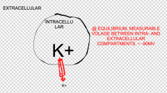
1. BESIDES POTASSIUM, ________ IS ALSO TRAPPED INSIDE THE NERVE CELL. 2. THE POTASSIUM CONCENTRATION IS ________ INTRACELLULARLY, AND ________ EXTRACELLULARLY 3. IS POTASSIUM DISTRIBUTED EVENLY THROUGHOUT THE BODY? |
1. ORGANIC ANIONS 2. GREATER, LESS 3. NO |