![]()
![]()
![]()
Use LEFT and RIGHT arrow keys to navigate between flashcards;
Use UP and DOWN arrow keys to flip the card;
H to show hint;
A reads text to speech;
43 Cards in this Set
- Front
- Back
- 3rd side (hint)
|
Why some animals do not need a transport system |
Get nutrients from the water in which they live - Diffusion Food reaches cells by pouches in gut, oxygen absorbed and waster excreted by means of diffusion |
|
|
|
Why animals do need a transport system |
Cells too far away from cells that obtain food/are in direct contact with environment Diffusion not good enough Special transport system necessary |
|
|
|
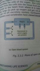
Open blood system |
Blood pumped through blood vessels Reach interconnected bloodfilled spaces/sinuses - haemocoels Organs receive the O2 and food via diffusion from blood they are bathed in CO2 and waste shed into haemocoels via diffusion Blood enters heart through small holes Small animals, molluscs, arthropods etc |
|
|
|
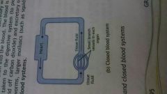
Closed blood system |
heart -> large blood vessels -> smaller blood vessels capillaries reach organs food + blood diffuses into tissue fluid co2 + waste tissue fluid -> blood\ heart -> digestive system (food) -> lungs (o2) -> excretory organs (get rid of waste) earthworms, large molluscs, vertabrates
|
|
|
|
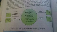
why a transport system is needed in humans |
-cells far away -food far, o2 far -waste needs to be taken away -circulatory and lymphatic system |
|
|
|
The Heart - Structure |
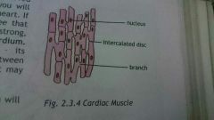
-hollow -closed fist -walls = thick muscles (cardiac muscles) -middle of chest cavity/thorax -apex slightly to left -held by blood vessels,, protected by ribs + sternum -fat on surface -covered by strong double walled sac (pericardium) - |

|
|
|
pericardium |
the strong, double walled sac that covers the heart -if cut open you will see -coronary arteries + veins -pulmonary artery + aorta -vena cavae -pulmonary veins |
|
|
|
Coronary arteries + veins |
-small blood vessels -run on surface of heart -to and from heart muscles |
|
|
|
Pulmonary artery + aorta |
-large arteries -away from heart -pulmonary = deoxygenated blood to lungs -aorta = oxygenated blood to body |
|
|
|
superior and inferior vena cava |
-carry deoxygenated blood to right side of the heart |
|
|
|
pulmonary veins |
-oxygenated blood from lungs to left side of heart |
|
|
|
Internal structure of heart - chambers
|
- 4 chambers - upper chambers = small, thin-walled (Atria) - lower chambers = larger, thick muscular walls (ventricles) - septum = muscle that separates left side of heart from right
|
chambers |
|
|
internal structure of heart - valves |
1. tricuspid - between right atrium and right ventricle - 3 flaps - prevent blood flowing back into atrium when ventricle contracts 2. bicuspid - aka mitral valve - between left atrium and ventricle - prevent blood flowing back into atrium whenventricle contracts 3. semi-lunar valves - moon-shaped - beginning of pulmonary artery and aorta - prevent blood flwoing back into ventricles when they relax |
valves |
|
|
the cardiac cycle |
- contraction and relaxation of heart muscles - contraction = systole - relaxation = diastole - heart beat has 3 stages - atrial systole, ventricular systole, general diastole |

* each heart beat takes about 0,8 seconds |
|
|
Cardiac cycle: atrial systole |
-SA node sends electrical impulses to muscle fibres in atria -atria contract -tri and bi cuspid valves open - blood flows into relaxed ventricles -0,1 seconds |
SA node = sino-atrial node, small patch of tissue in the right atrium * atria in systole, ventricles in diastole |
|
|
Cardiac cycle: ventricular systole |
-0,3 seconds -ventricles contract -tri and bi cuspid valves close (preventing blood from flowing back into atria) |
*ventricles in systole, atria in diastole |
|
|
cardiac cycle: general diastole |
- both atria and ventricles relax -semi-lunar valaves close -blood enters atria through vena cavae + pulmonary veins then flows into ventricles |
*valves close when blood is pushed back against them |
|
|
SA node |
Sino-atrial node; Pacemaker; Speed up or slow down heart beat; |
|
|
|
Blood vessels |

Arteries; Capillaries; Veins; |
|
|
|
Walls of arteries and veins |
1. Outer fibrous connective tissue layer 2. Middle smooth muscle layer (thicker in arteries) 3. Inner endothelium layer (capillaries are made of one layer of these only) |
|
|
|
4 differences between arteries and veins |
Arteries carry blood away from heart, veins to heart; A = thick walled (middle smooth muscle layer thick), V = thin walled (middle smooth muscle layer is thin); A= blood under pressure V = blood not under pressure; A = no valves except by heart V = semi lunar valves to prevent blood back flow |
|
|
|
Arteries (branching off) |
Arteries -> arterioles -> Capillaries |
|
|
|
Capillaries - structure and function |
Single layer of endothelial cells. Thin layer allows O2 and nutrients to diffuse from blood -> tissues, CO2 and waste diffuse from tissues -> blood. Join to form venules |
|
|
|
Capillaries to veins |
Waste + CO2 diffuse from tissues to capillaries -> capillaries join to form venules -> venules join to form veins |
|
|
|
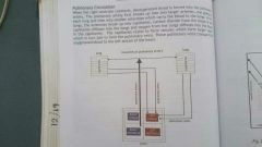
Blood circulation (double circulation) |
Two main routes of blood flow - pulmonary and systemic, (Pulmonary = lungs, systemic= various systems within body) Pulmonary - blood flows from heart to lungs to heart Systemic - heart -> body -> heart |
|
|
|
Pulmonary circulation |
Right ventricle contracts, deoxygenated blood.forced into pulmonary artery. Pulmonary artery breaks into two arteries which break up into arterioles, one to each lung. Arterioles break up into capillaries in lung. Oxygen diffuses into blood. Capillaries rejoin -> venules -> pulmonary veins. Veins carry oxygenated blood to left atrium. |
|
|
|
Systemic circulation |
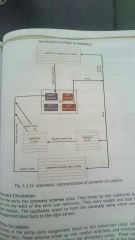
Left ventricle contracts, oxygenated blood -> aorta which branches all through body -> in tissues arteries break up into arterioles then capillaries. O2 and food diffuses into cells. CO2 diffuse into capillaries, blood = deoxygenated. Capillaries -> venules -> veins -> superior and inferior vena cavae -> right atrium.
|
|
|
|
Coronary circulation |
Specialised part of systemic circulation. Two coronary arteries form from aorta, they break into capillaries, pass into walls of atria and ventricles. Carry O2 and food to heart muscles. Capillaries rejoin -> coronary veins, carry blood to right atrium. |
|
|
|
Portal circulation |
Specialised part of systemic circulation. Branches of aorta carry blood to alimentary canal (stomach intestines etc) arteries -> arterioles ->Capillaries which absorb digested food in alimentary canal. Capillaries -> venules -> veins -> one large vein called hepatic portal vein which carries blood to liver. From liver blood is carried away by several hepatic veins to inferior vena cava -> right atrium |
|
|
|
Blood tissues - characteristics |
Salty, slightly sticky, reddish, pH little over 7 (slightly alkaline |
|
|
|
Composition of blood - plasma |

55% plasma, 45% blood corpuscles Plasma = 90% water, 10% dissolved substances (digested food, salts, gases, waste, plasma proteins, hormones, enzymes, antibodies) Fibrinogen is an eg of plasma protein. Helps in blood clotting.
|
|
|
|
Composition of blood - blood corpuscles |

3 types of corpuscles. 1. Red blood corpuscles / erythrocytes - disk shaped, no nuclei, contain haemoglobin (makes blood red) 2. White blood corpuscles / leukocytes - irregularly shaped, with nuclei 3. Blood platelets - cell fragments no nuclei |
|
|
|
Functions of blood tissue - plasma |
Transport system for --> digested food substances eg glucose and amino acids; excretory products eg urea and uric acid; plasma proteins eg fibrinogen ; salts ; gases eg O2 and CO2 ; hormones and enzymes. |
|
|
|
Functions of blood tissue - erythrocytes |
Transport O2. O2 dissolves in haemoglobin and forms oxyhaemoglobin before being transported. |
|
|
|
Functions of blood tissue - leukocytes |
Protect the body. Engulf microbes, form barrier against microbes, produce antibodies to destroy microbes. |
|
|
|
Functions of blood tissue - Blood platelets |
Clotting blood |
|
|
|
Functions of the blood system |
Absorbs digested food from alimentary canal and transports it throughout the body.. Absorbs O2 in lungs and transports it.. Transports excretory waste to excretory organs (kidneys, lungs, sweat glands) |
|
|
|
Relationship between the blood system and lymphatic system |

Arteries branch off and eventually form capillaries. Capillaries are under great pressure and are thin walled so some blood plasma exits the capillary and surrounds the tissues. Tissue fluid. Some enters capillaries again, but the rest enters lymph capillaries and is now called lymph. |
|
|
|
Main lymph vessels |
Lymph capillaries join to form larger lymph vessels. They unite to form left thoracic duct (draws lymph from intestines, legs and left side of body) or right lymphatic duct (upper body and fight side of the body)... Left thoracic duct ->> left sub-clavian vein (located under clavicle) Right lymphatic duct ->> right sub-clavian vein.... They join at vena cavae, so lymph is back in blood. |
|
|
|
Functions of the lymphatic system |
Return tissue fluid, transport digested fats from villi of small intestines, at nodes of lymph vessels, antibodies and lymphocytes are produced which protect the body against toxins and microbes. |
|
|
|
Diseases of the heart and circulatory system - heart disease and heart attack |

Arteries become narrow due to fatty deposits. Blood clots form at fatty deposit - coronary thrombosis / blood clot form in another part of body and gets stuck at fatty deposit - coronary embolism (both are coronary artery diseases, oxygen to heart reduced or stopped completely, heart muscles die = heart attack) |
|
|
|
Factors linked to heart disease |
Heredity, age, gender (males more likely than females as female sex hormones offer some protection), smoking, cholesterol, exercise (or lack thereof), hypertension (high blood pressure), stress, obesity, diabetes. |
|
|
|
Treatments of heart disease |
Drug treatment, Angioplasty (blockages broken up by laser beam given off tip of tube inserted through blood vessel), Spent insertion (small usually metal sometime fabric tube which prevents artery from becoming narrow and blocked or bursting, some are coated with meds), Open heart surgery (operation, cut heart open, replace or repair parts eg valve replacement), Pacemaker implantation (natural Pacemaker SA node causes heart to eat irregularly, artificial one insert under clavicle, connected to heart via electrodes), Heart transplant, By-pass surgery (operation, bypass portion of artery that is blocked by smaller vein from arm or leg) |
|