![]()
![]()
![]()
Use LEFT and RIGHT arrow keys to navigate between flashcards;
Use UP and DOWN arrow keys to flip the card;
H to show hint;
A reads text to speech;
28 Cards in this Set
- Front
- Back
|
Why do animals need transport systems?
|
All animals need a supply of oxygen and nutrients to survive.
They also need to remove waste products so they do not build up and become toxic Once an animal has several layers of cells any nutrients or oxygen diffusing in will be used up by the outer cells Animals need a lot of energy from food so they can move around. releasing energy from food via respiration requires oxygen quickly |
|
|
What is single circulatory system?
|
A single circulatory system for fish has blood flowing from the heart to the gills and then onto the body before returning to the heart.
|
|
|
What is a double circulatory system?
|
Mammals have a circulation that involves two separate circuits . One circuit carries blood to the lungs. this is know as the pulmonary circuit and the other circuit carries the oxygen and nutrients around the body to the tissues. this is the systemic circulation
|
|
|
What does open circulatory system mean?
|
The blood is not always held within the blood vessels. instead the blood fluid circulates through the body cavity so the tissues and cells of the animal are bathed directly in blood
some animals use the movement of the muscles to circulate the blood. Insects have a tube that lies under the dorsal fin. blood enters the heart though ostia. the heart then pumps blood towards the head and blood goes into the body cavity |
|
|
What does closed circulation system mean?
|
The blood always stays entirely inside vessels. a separate fluid called tissue fluid bathes
in a fish this goes heart-arteries-gills-veins-body tissues-veins-heart |
|
|
|
|
|
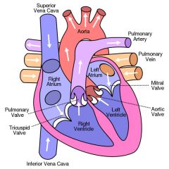
What is the atria like structurally and what does it do?
|
The muscle is very thin. This is because they do not need to create much pressure.
they push blood into the ventricles |
|
|
What is the right ventricle like structurally and what does it do?
|
The walls are a lot thicker than the atria. This lets the ventricles pump blood out of the heart
|
|
|
What is the left ventricle like structurally and what does it do?
|
The walls can be two or three times thicker than that on the right ventricle.
the blood from the left ventricle is pumped through the aorta and needs sufficient pressure to overcome the resistance of the circulation system |
|
|
What is the first phase of the cardiac cycle and what happens in it?
|
Diastole- Both the atria and the ventricles are relaxing, the internal volume increases and blood flows into the heart. blood flows into the atria then through the atrioventricular valves and into the ventricles
|
|
|
What is the second stage of the cardiac cycle and what happens in it?
|
Atrial systole- The atria contract. this small increase in pressure pushes blood into the ventricles. this stretches the walls of the ventricles and ensures they are full of blood. when the ventricles are full they begin to contract. blood fills the atrioventricular valve causing it to snap shut.
|
|
|
What is the third stage of the cardiac cycle and what happens in it?
|
Ventricular systole- all four heart valves are closed. The walls of the ventricles contract. this raises the pressure very quickly. The contraction starts in the apen of the heart so pushes blood back up towards the arteries. The semilunar valves open and blood is pushed out the heart. the ventricles then relax
|
|
|
How is heart action coordinated?
|
At the top of the right atrium is the sinoatrial node. this small patch of tissue generates electrical activity. It emits a wave of excitation at regular intervals.
This wave spreads over the walls of both atria. It travels along the membranes of the muscle tissue. This causes the cardiac muscles to contract at the base of the atria there is a disk of tissue that cannot conduct the wave. at the top of the septum is the atrioventricular node. this is the only route through the non-conducting tissue. this delays the wave and allows time for the atria to finish contracting before the ventricles start to contract. After this the wave is carried down the purkyne tissue. At the base of the septum the wave spreads over the walls of the ventricles. this causes the muscles to contract. this means it contracts from the base upwards |
|
|
How do you interpret electrocardio diagrams?
|
P = excitation of the atria
QRS = excitation of the ventricles T = diastole |
|
|
How can you tell from a electrocardio diagram is a heart is malfunctioning?
|
Elevation of the ST section indicates a heart attack
small and unclear P wave indicates atrial fibrillation deep S waves indicates abnormal ventricular hypertrophy |
|
|
What is the structure of an artery?
|
From outside to inside
Collagen fibres, smooth muscles, elastic fibres, endothelium, lumen |
|
|
What is the structure of a vein?
|
from outside to inside
Collagen fibres, smooth muscle, elastic fibres, endothelium, lumen |
|
|
What is the structure of a capillary?
|
From outside to inside
Endothelium, lumen |
|
|
What is the function of an artery?
|
Carries blood away from the heart. the blood it at high pressure so the artery must be able to withstand it
|
|
|
What is the function of a vein?
|
Carries blood back into the heat. The blood is at low pressure and the walls do not need to be thick
|
|
|
What is the function of a capillary?
|
They have very thin walls to allow exchange of materials between the blood and the cells of tissues via the tissue fluid.
|
|
|
Features of blood
|
Cells are erythrocytes, leucocytes and platelets
proteins are hormones and plasma proteins fats are sometimes transported by lipoproteins There are more amino acids there is more oxygen there is little carbon dioxide |
|
|
Features of tissue fluid
|
Cells are sometimes phagocytic white blood cells
proteins are sometimes hormones and proteins secreted by body cells it has no fats it has less glucose it has less amino acids it has less oxygen it has more carbon dioxide |
|
|
Features of lymph
|
Cells are lymphocytes
it has some proteins it has more fats than blood it has less glucose it has less amino acids it has less oxygen it has more carbon dioxide |
|
|
How is tissue fluid formed from plasma?
|
At the end of the capillaries leading from the arterioles are under high pressure due to the contraction of the heart materials, known as hydrostatic pressure. This will push the blood out of the capillaries
Fluid that leaves the blood consists of plasma with dissolved nutrients and oxygen. apart from the stuff too large to fit through the gaps. The fluid that leaves the capillary is know as tissue fluid |
|
|
What is the role of haemoglobin in terms of carrying oxygen and carbon dioxide?
|
Haemoglobin has four subunits that each consists of a polypeptide and a haem group. The haem group consists of an iron ion and this has an affinity for oxygen. They take the the oxygen out of the solution to maintain a steep diffusion gradient
At high oxygen tension the haemoglobin dissociates and release the oxygen as well as carbon dioxide |
|
|
What is the significance of the dissociation curves of adult oxyhaemoglobin at different carbon dioxide levels? (the bohr effect)
|
This shows an s-shaped curve.
At low oxygen tension does not readily take up oxygen molecules as the haem that attracts the oxygen are in the centre of the haemoglobin. as the tension increases, the diffusion gradient increases and one diffuses into the haemoglobin molecule and changes the shape slightly allowing more oxygen into the haemoglobin. once it gets three molecules it becomes more difficult for the fourth one to diffuses into it so the graph curves off. |
|
|
What is the significance of fetal haemoglobin having a different affinity than adult haemoglobin for oxygen? |
Fetal haemoglobin must be able to pick up oxygen from the placenta. The fetal haemoglobin must absorb oxygen from the fluid in the mother's blood.
This reduces oxygen tension within the blood fluid which makes the maternal haemoglobin release oxygen. |