Reading...
![]()
Play button
![]()
Play button
![]()
Use LEFT and RIGHT arrow keys to navigate between flashcards;
Use UP and DOWN arrow keys to flip the card;
H to show hint;
A reads text to speech;
83 Cards in this Set
- Front
- Back

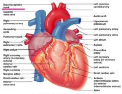
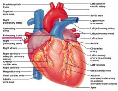
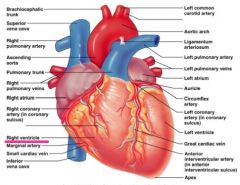
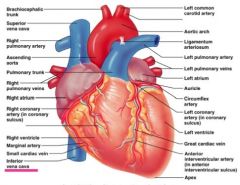
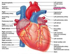
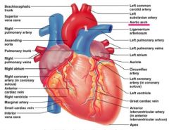
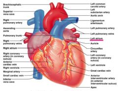
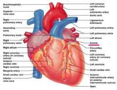
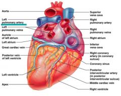
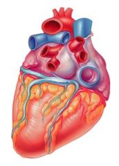
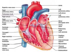
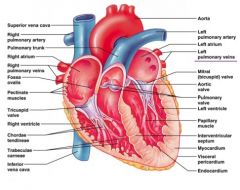
Locate the Brachiocephalic trunk
|
|
|

Locate the Superior vena cava
|
|
|

Locate the Right pulmonary artery
|
|
|

Locate the Ascending aorta
|
|
|

Locate the Pulmonary trunk
|
|
|

Locate the Right pulmonary veins
|
|
|

Locate the Right atrium
|
|
|

Locate the Right coronary artery
|
|
|

Locate the Right ventricle
|
|
|
Locate the Inferior vena cava
|
|
|

Locate the Left common carotid artery
|

|
|

Locate the Left subclavian artery
|
|
|

Locate the Aortic arch
|
|
|

Locate the Ligamentum arteriosum
|
|
|

Locate the Left pulmonary artery
|
|
|

Locate the Left atrium
|
|
|

Locate the Auricle
|
|
|

Locate the Left coronary artery
|
|
|

Locate the Left ventricle
|
|
|

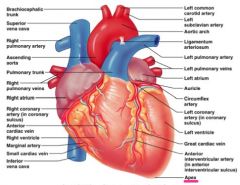
Locate the apex
|
|
|

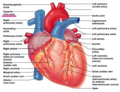
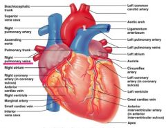
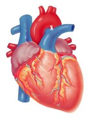
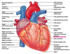
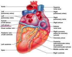
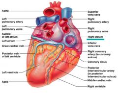
Locate the aorta
|

|
|

Locate the left pulmonary artery
|
|
|

Locate the Auricle of the left atrium
|

|
|

Locate the left atrium
|
|
|

Locate the left ventricle
|

|
|

Locate the apex
|

|
|

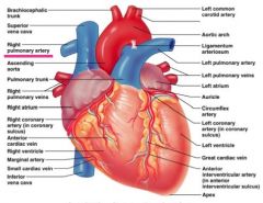
Locate the superior vena cava
|
|
|

Locate the right pulmonary artery
|

|
|

Locate the right pulmonary veins
|
|
|
Locate the Right atrium
|
|
|

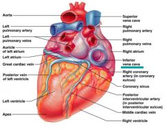
Locate the Inferior vena cava
|
|
|

Locate the Right coronary artery
|
|
|

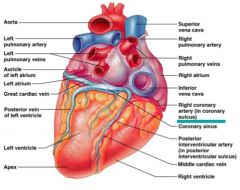
Locate the Coronary Sinus
|

|
|

Locate the Right ventricle
|

|
|

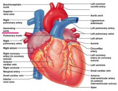
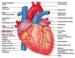
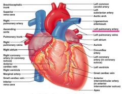
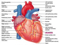
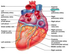
Locate the superior vena cava
|

|
|

Locate the Pulmonary trunk
|
|
|

Locate the right atrium
|

|
|

Locate the right pulmonary veins
|

|
|

Locate the fossa ovalis
|

|
|

Locate the Tricuspid valve
|

|
|

Locate the right ventricle
|

|
|

Locate the chordae tendineae
|

|
|

Locate the Inferior vena cava
|

|
|
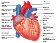
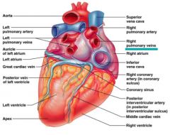
Locate the aorta
|

|
|

Locate the left pulmonary artery
|

|
|

Locate the left atrium
|

|
|

Locate the left pulmonary veins
|
|
|

Locate the mitral (bicuspid) valve
|

|
|

Locate the aortic valve
|

|
|

Locate the pulmonary valve
|

|
|

Locate the left ventricle
|

|
|

Locate the papillary muscle
|

|
|

Locate the Interventricular septum
|

|
|
|
Aortic stenosis - Systolic or diastolic?
List the cause and characteristic sounds |
systolic
Cause: turbulent blood flow through the narrowed aortic valve as blood is ejected from the left ventricle Characteristic Sound: WOOSH |
|
|
Mitral valve prolapse - Systolic or diastolic?
List the cause and characteristic sounds |
systolic
Cause: blood regulates back from left ventricle into left atrium during systole. Characteristic Sound: LUB-WOOSH-DUP |
|
|
Aortic regurgitation (aortic insufficiency) - Systolic or diastolic?
List the cause and characteristic sounds |
diastolic
Cause: aortic valve doesn't close completely, blood flows back from the aorta into the left ventricle during diastole Characteristic Sound: LUB-DUP-WOOSH |
|
|
What are AV (Atrioventricular Valves)?
|
• prevent back flow into atria when ventricles contract
• chordae tendineae anchor AV valves to papillary muscles (prevent prolapse) |
|
|
What are SL (Semilunar Valves)?
|
• prevent back flow of blog into the ventricles
|
|
|
Pathway of blood
|
Right atrium → Right AV valve → right ventricle → pulmonary valve → pulmonary arteries → lungs → pulmonary veins → left atrium → left AV valve → left ventricle → Aortic SL valve → aorta → systemic circulation
|
|
|
What is an EMG?
|
electromyogram
an external recording of the electrical activity of muscle Gives information on the speed of electrical conduction through the cardiac conduction system used to diagnose abnormalities and disease of the heart, measures electrical activity of muscles at rest and during contraction |
|
|
When stimulated by a motor neuron, a skeletal muscle fiber forms _______ that activate contraction of the fiber.
|
action potentials
|
|
|
The EMG records the combined action potential signals produced by _____ of activated muscle fibers
|
several
|
|
|
Define motor unit
|
motor neuron & all muscle fibers it supplies (4 to 1000’s)
• when motor neuron fires (action potential) all muscle fibers it innervates contract |
|
|
Define motor unit recruitment
|
an increase in # of active motor units
• ↑ motor unit recruiment = ↑ force of contraction |
|
|
Define antagonistic muscles
|
muscles that work in opposing directions
|
|
|
Define fatigue
|
muscle is physiologically unable to contract
|
|
|
Increasing the force of muscle contraction is accomplished by _____ recruitment of motor units, which increases the number of muscle fibers activated in the muscle.
|
increasing
|
|
|
The more muscle fibers that are stimulated to produce action potentials, the _____ the EMG signal will be
|
stronger
|
|
|
Explain how your results from Part A demonstrate recruitment of motor units.
|
Part A demonstrates recruitment of a motor unit because the amount of action potentials increases with the increase in the amount of weight being lifted. The body produces more action potentials when more energy effort is used.
|
|
|
Explain how your results from Part B demonstrate fatigue of the muscle
|
Part B shows that as the muscle fatigues more and more motor units got used.
|
|
|
What is meant by the term antagonistic muscles?
|
Antagonistic muscles are opposing muscle pairs
|
|
|
Summarize your results from Part C and explain briefly what the results demonstrate about the control of antagonistic muscles by the CNS.
|
In Part C the control of antagonistic skeletal muscles are complete opposites while in flexion the biceps produce a larger amount of ATP. The triceps keeps a small constant amount of ATP. During extension, the triceps had a very large amount of ATP being produced while the biceps had a moderate amount at the beginning but subsided.
|
|
|
Define sphygmomanometer
|
used to measure arterial blood
• consists of an inflatable cuff connected to a pressure gauge |
|
|
Define blood pressure
|
force exerted by blood on the walls of the blood vessels
• expressed in millimeters of mercury (mm Hg) • must be regulated to meet requirements for blood flow to the body organs. • driving force for blood flow |
|
|
Define Korotkoff sounds
|
sequence of sounds produced as the cuff pressure is decreased from systolic to diastolic pressure
• divided into 5 phases: 1. brief loud tapping 2. longer, softer murmurs 3. loud thumping sounds 4. soft muffled sounds 5. silence |
|
|
Define mean arterial pressure
|
the average blood pressure in the aorta and large arteries.
• The MAP is related to the cardiac output (CO) and total peripheral resistance (TPR) according to the formula: MAP = CO x TRP |
|
|
Define pulse pressure
|
the difference between the systolic and diastolic pressure (systolic P - diastolic P)
|
|
|
Define systolic pressure
|
pressure exerted on the arterial walls during ventricular contraction
|
|
|
Define diastolic pressure
|
lowest level of arterial pressure during a ventricular cycle
|
|
|
High and Low blood pressure levels
|
Systolic Pressure under 120
Diastolic Pressure under 80 |
|
|
Hypertension
|
chronically elevated arterial blood pressure, the heart works harder to pump blood around the body
|
|
|
Equation to calculate MAP
|
Diastolic Pressure + (Pulse Pressure/3)
|
|
|
How to take your blood pressure:
|
Subject must sit on the table with their left or right arm resting on the table at the same level with their heart. Put the cuff of the sphygmomanometer around the arm just above the elbow. Palpate the pulse in the brachial artery and close the screw valve. Inflate the cuff while listening to the brachial artery. Inflate the cuff about 20mmHg above the point where the pulse disappears. Place the diaphragm of the stethoscope over the brachial artery open screw valve slightly to slowly release pressure rate of 3 mmHg per second. Watch the pressure gauge while listening to the artery. Record systolic pressure as the point where you hear the 1st Korotkoff sound. Diastolic pressure is the point where the sound disappears.
|