![]()
![]()
![]()
Use LEFT and RIGHT arrow keys to navigate between flashcards;
Use UP and DOWN arrow keys to flip the card;
H to show hint;
A reads text to speech;
23 Cards in this Set
- Front
- Back
|
1.1)* Describe why animals have a heart and circulatory system. |
Animals have a heart and circulatory system because diffusion is too slow to move materials (e.g. oxygen and food) throughout the body fast enough and in sufficient quantities. A heart and circulatory system allows materials to be moved around quickly and in sufficient amounts. |
|
|
1.2)** Define the terms 'mass flow' and 'diffusion'. |
Mass Flow: the transport of substances in bulk from one part of an organism to another, rapidly.
Diffusion: for smaller organisms. Movement of molecules or particles from a higher concentration to lower concentration. |
|
|
1.3)** Describe the features of a mass flow system and explain why certain organisms need mass flow. |
System of vessels to carry substances. Moving substances fast enough to supply needs of the organism/ways of maintaining a concentration gradient so substances move quickly from one place to another. Suitable transport medium.
Certain Organisms need mass flow because diffusion is too slow for their needs and they require a way of moving substances rapidly from one place to another. |
|
|
1.4)** Describe the difference between an open circulatory system and a closed circulatory system. |
In an open circulatory system, the blood leaves the blood vessel and pumps out into cavities(small animals and insects), whereas in a closed circulatory system the blood remains in the blood vessel(larger animals). |
|
|
1.5)** Identify the key features of single circulatory systems and doubles circulatory systems and explain their features. |
In a single circulatory system blood only flows through the heart once for a complete circuit(found in fish). In a double circulatory systems the blood flows through the heart twice for a full circuit(human heart). |
|
|
1.6)** List the differences between pulmonary circulation and systemic circulation. |
Pulmonary circulation is a part of cardiovascular system responsible for carrying deox blood from heart to lungs and back to heart for it to transfer oxygenated blood to the rest of the body. Whereas systemic circulation is a part of the cardiovascular system which is responsible for carrying oxygenated blood away from heart, to body and return deoc blood back to heart |
|
|
1.7)** List the components of blood and describe their functions. |
Red blood cells: transport oxygen to body cells and deliver CO2 to lungs. White blood cells: cells of immune system, protect against infectious disease and foreign invaders. Platelets: make blood clots. Plasma: transport nutrients, hormones+proteins around body, also removes waste products. |
|
|
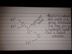
1.8)*** Describe with the aid of a diagram, the structure of a water molecule illustrating hydrogen bonding. |
|
|
|
1.9) Describe how the polar nature of water makes it a useful solvent. |
Water is a polar molecule. This means it has a slightly negative and a slightly positive charge. This means it can bond by a hydrogen bond to another water molecule(or any other polar molecule). This is useful because when many water molecules join together it causes its useful features(high boiling point, liquid at room temperature. |
|
|
1.10)*** List the structures of arteries, veins and capillaries and relate these to their functions. |
Arteries: Flow away from heart, oxygenated blood(except pulmonary artery), high blood pressure, small lumen, thick walls, lots of collagen, elastic fibres+smooth muscle, no valves(except aorta+pulmonary artery. Veins: Towards heart, deox blood(except pulmonary vein), low blood pressure, large lumen, less thick walls, some collagen, elastic fibres+smooth muscle, valves present. Capillaries: Blood away from heart>vein, oxygenated blood, higher blood pressure than vein, small lumen, very thin walls, endothelium only, no valves present. |
|
|
1.11)*** Label and annotate diagrams of the heart both internal and external. |
Add Picture |
|
|
1.12) Explain why the walls of the ventricles are thicker that the walls of the atria. |
Walls of the ventricles are thicker that walls of the atria because they need to be stronger to allow them to have enough power to pump blood around the body, the atria only squeeze blood returning from lungs into ventricles. |
|
|
1.13)** Explain the reason why the left ventricle is thicker that the right ventricle in terms of pulmonary and systemic circulation. |
The right ventricle pumps blood into the pulmonary circulation into ling and the left ventricle pumps blood into the systemic circulation through the aorta. The left ventricle is thicker that the right because the left needs to pump blood to most of the body, whereas the right ventricle only needs to fill the lungs. |
|
|
1.14)*** Recall the three main stages of the cardiac cycle. |
Atria Contract(atrial systole) Ventricles Contract(ventricular systole) Ventricles Relax(ventricular diastole).151 |
|
|
1.15)*** Describe in detail the sequence of events of atrial systole, ventricular systole and diastole in terms of blood pressure, flow and valves opening and closing. |
1) Atrial systole: Blood pushed into ventricles from atria because AV valves are open. 2) Ventricular Systole: Ventricles contract much more forcefully than atria(120mmHg LV, 40mmHg RV) contraction closes AV valves. Valve tendons prevent flaps from pushing up into atria. Papillary muscles contract to assist as well. 3)Ventricular Diastole: pressure inside falls rapidly, but semilunar valves prevent blood from returning into ventricles from arteries. These slam shut causing 2nd heart sound. Blood flows from atria into ventricles as ventricular blood pressure falls. Therefore blood pressure rises, pushes AV valves open- atria then contracts to complete the filling of ventricles. |
|
|
1.16)** Describe and explain what causes the valves to open and close. |
When the ventricles relax the atria pressure exceeds the ventricle pressure and the AV valves are pushed open. When the ventricles contract, ventricular pressure exceeds the atrial pressure causing the AV valves to shut. |
|
|
1.17) Interpret pressure diagrams of the cardiac cycle and to identify points at which valves open/close and to calculate heart rate. |
? |
|
|
1.18)** Define what is meant by the terms myocardial infarction, stroke, angina, aneurysm and thrombosis. |
Myocardial Infarction: death to areas of the cardiac muscle Stroke: sudden death of brain cells due to lack of oxygen when blood flow is impaired by blockage or rupture of an artery to the brain. Angina: pain in the chest due to partial blockage of an artery. Aneurysm: blood filled balloon like bulge in wall of blood vessel. Thrombosis: formation of a blood blot inside a blood vessel. |
|
|
1.19) Define what an atheroma is and explain how an atheroma can turn into a plaque. |
An atheroma is a build up of fatty material, calcium salts and fibrous tissue are stimulated to build up in the region of the atheroma producing a plaque. |
|
|
1.20) Explain how a plaque leads to narrowing of the arteries and thrombosis. |
Plaque leads to narrowing of the arteries and thrombosis by bulging into the lumen of the artery causing reduction in elasticity. Plaque can become very large and tear the endothelium of the artery, underlying cells come into contact with blood, triggers formation of blood clot(thrombosis). |
|
|
1.21) *** List the sequence of events that leads to atherosclerosis. |
Inflammatory responce--atheroma--plaque--reduction in elasticity(atherosclerosis)--thrombosis--blood pressure rises. |
|
|
1.22) Explain how atherosclerosis can lead to increasing blood pressure. |
Atherosclerosis blood pressure because the narrower your arteries, the harder your heart has to pump blood through them, this further increases the blood pressure of the blood flow, causing blood pressure to rise even more. |
|
|
1.23)*** List the sequence of events that leads to the formation of a thrombus(blood clot). |
Platelets come into contact with damaged cells--platelets change shape(flattened disc to spheres with long projections-- cell surface changes--stick to exposed collagen in artery wall--form platelet plug--release substances to activate more platelets-- triggers series of chemical reactions--blood clot. Stage 1- Conversion of prothrombin into thrombin: Thromboplastin(an enzyme, released by the platelets) and Ca2+(in the plasma) catalyse the conversion of promthrombin(inactive enzyme- a plasma protein) to thrombin(active enzyme) Stage 2- Conversion of soluble fibrinogen into soluble fibrin by thrombin: Thrombin converts fibrinogen(soluble plasma protein) into loose fibrin threads(insoluble). TAKES ROUGHLY 20S
|