• Shuffle
    Toggle On
    Toggle Off
  • Alphabetize
    Toggle On
    Toggle Off
  • Front First
    Toggle On
    Toggle Off
  • Both Sides
    Toggle On
    Toggle Off
  • Read
    Toggle On
    Toggle Off
Reading...
Front

Card Range To Study

through

image

Play button

image

Play button

image

Progress

1/71

Click to flip

Use LEFT and RIGHT arrow keys to navigate between flashcards;

Use UP and DOWN arrow keys to flip the card;

H to show hint;

A reads text to speech;

71 Cards in this Set

  • Front
  • Back

fourprimary types of tissues

1.Connective tissue
2.Muscle tissue
3.Epithelial tissue
4.Nervous tissue

ConnectiveTissue functions

1. Forms metabolic and structural connections between other tissues
2. Forms a protective sheath around organs andhelps to insulate the body
3. Acts as a reserve for energy
4. Provides the frame that supports the body
5. Composes the transport medium that transportssubstances from one region of the body to another
6. Plays a role in the healing process and thecontrol of invading microorganisms

Ground substance

1. The medium through which cells exchangenutrients and waste with the blood stream
2. Is amorphous (without shape) andhomogeneous (even consistency) material called GAG’s
3. Ranges in texture: from a liquid or a gelor a calcified solid
4. Protects the more delicate cells that itenvelopes
5. Is an effective obstacle for invadingmicroorganisms

ConnectiveTissue Components

•Extracellularmatrix
•Cells

Extracellular matrix composed of

–Extracellular fibers
–Ground substance

Extracellular fibers consist of

Collagenousfibers


Reticularfibers


Elasticfibers

Collagenous fibres


–Strong,thick strands of collagen (a fibrous protein)
–Organizedinto bundles of long, parallel fibrils
–Variabledensity and arrangement of fibers
–Foundin tendons and ligaments

Reticularfibers

–thin,delicate, branched networks of collagen


–Providesupport for highly cellular organs: endocrine glands, lymph nodes, spleen, bone marrow, and liver


–Alsofound around: blood vessels, nerves,muscle fibers and capillaries

Elasticfibers

–branchednetworks composed of: primarily proteinelastin


–Composedof coiled bundles of microfibrils


–Occurin tissues commonly subjected to stretching ie: vocal cords, lungs, skin, and walls of bloodvessels

CellTypes

FixedCells


TransientCells

Fixed cells involved in

the production & maintenance of the matrix

fixed cell types


and involvement

–“blasts”:
Fibroblasts(make fibers)
chondroblasts(make cartilage)


osteoblasts (make bone)

– Adipocytes (fat cells)
– Reticular cells (maintain reticular fibersscle fibers and capillaries

Transient cells

involved in the repair and protection oftissues

transient cell types

–Leucocytes:WBC’s
–Mast cells (involved withthe immune system)
-Macrophages (phagocytose foreign material)

Looseconnective tissue
types

–Areolar –Adipose –Reticular

•Dense connective tissue:types

–Denseregular –Denseirregular –Elastic

AreolarConnective Tissue

•Looseconnective tissue
•Fibersand cells suspended in a thick, translucentgroundsubstance •Predominantcell is the –Manufacturesthe elastic, reticular, andcollagenousfibers
•Location:–Surrounds every organ
–Formsthe SQ layer that connects skin to muscle
-Envelopesblood vessels, nerves and lymph nodes
–Presentin all mucous membranes

AdiposeTissue

•Areolartissuein which adipocytespredominate •Highlyvascular
•Function:acts as energy storehouse and a thermal insulator
•Location:–inthe SQ layer
–Mammaryglands
–surroundingorgans

ReticularConnective Tissue

•Networkofthin reticular fibers.
•Containsloosely arranged fibersandmany fibroblasts suspendedin asupportive groundsubstance
•Function:forms the framework of organs such as the spleen and lymph nodes

DenseRegular Connective Tissue

•Composedof tightly packed, parallelcollagen fibers
•Relativelyavascular
•Location:
–Makesup tendons and ligaments(tendons connect muscle to bone-ligaments attach bone to bone)
– can be found in sheets called fascia thatcovers muscles

DenseIrregular Connective Tissue

•Composedprimarily of collagen fibersarrangedin thick bundles
•Fibersare interwoven to form a singlesheet •Location:
–in the dermis of the skin and in the fibrous covering of many organs
–formsthe tough capsule of joints

ElasticConnective Tissue

•Primarilycomposed of elastic fibers
•Fibersmay be arranged parallel or in interwoven patterns with fibroblasts and collagenous fibers interspersed
•Location:
–Inthe spaces between the vertebrae
–Inareas of the body that require stretching: walls of arteries, stomach ,bronchi, bladder

SpecializedConnective Tissues

Cartilage
Bone
Blood

Cartilage types

–Hyalinecartilage
–Elasticcartilage
–Fibrocartilage

Cartilage


•Location:Injoints and the ear, nose and vocal chords
•Function: forms a framework on which boneis formed
•Structure: no innervation (no nerves);avascular (no blood vessels)
•Cells:
–Chondrocytes:live in the hollowed out pockets in the matrix called lacunae
•Matrix:
–Groundsubstance:gel of inorganic molecules called GAG’s
–Fibers:collagen fibers are the most common found in the matrix. But elastic fibers arealso present in varying amounts

HyalineCartilage

•Most common type of cartilage foundinthe body
•Composed of closely packed collagen
•Location:
–Inthe joints at the ends of long bones
–Growthplates of long bones
– tracheal rings
– connects the ribs to the sternum
– composes mostly of embryonic skeletons

ElasticCartilage

•Containselastic fibers in densebranchingbundles
•Flexible:can withstand repeatedbending •Location:
–Inthe epiglottis of the larynx
–Inthe pinnae of ears of animals

Fibrocartilage

•Usually found merged with hyaline cartilageanddense connective tissue
•Contains thick bundles of collagen fiberswithfewer chondrocytes thanhyalinecartilage
•Location:
–Inbetween vertebrae of the spine
–Betweenbones in the pelvic girdle
–In thestifle joint

Bone

•Matrix is a combination of organic collagenfibers and inorganiccalciumsalts
•Well vascularized
•Haversian canal contains bothvascular and nerve supply
•Canaliculi: channels within the matrix support thepassage of blood vessels into deeper portions of tissue


•Cells: –Osteoblasts:manufacture the fibers that are part of the matrix-Lacunae and canaliculi are created as the osteoblasts manufacture the bonymatrix–––Osteocytesreside in the lacunae, maintain bone structure–––Osteoclastswill break down bone when necessary

Blood

•Matrix:
–groundsubstance:plasma
–fibrouscomponent: protein
•Cells:
–Erythrocytes:RBC’s
–Leucocytes: WBC’s
–Thrombocytes:platelets

MucousMembranes aka mucosae

•Location:line organs with connections to the outside environment (mouth, intestines,nasal passages etc)
•Usuallycomposed of eitherstratifiedsquamous epitheliumor simplecolumnar epithelium covering a layer of areolarC.T. •Submucosa:connective tissue layer that connects the mucosa to underlying structuresshcards/create


•Maycontain goblet cells or multicellular glands-can produce large quantities of mucous (goblet)-mucus consists primarily of water, electrolytes and protein mucin
•Somemucosae also can absorb:(eg theepithelial layer in the intestine-nutrients)

SerousMembranes (serosae)

•Location: -line the walls and cover organsof the body cavities:
pleuralines the thorax cavity,peritoneumlinesthe abdominopelviccavity
•Consist of a continuous sheet doubledover on itself to form two layers: --Parietallayer lines the portion of the membrane that lines the cavity wall -Viscerallayer covers the portion of the membrane that covers the outer surface oforgans

CutaneousMembrane
Aka: integument of skin

•Outerlayer of stratifiedkeratinized squamous epithelium: epidermis
•Innerlayer of denseirregular C. T. :dermis
•Thedermis containscollagenous, reticular, andelasticfibers which enable skin to be both strong & elastic

SynovialMembranes

•Location: line the cavities of joints
•Composedof:loose connective tissue and adipose tissue covered by a layer of collagen fibers and fibroblasts
•Function:manufacture the synovial fluid that fills the joint spaces

MuscleTissue
•Composedof actinand myosin fibers(complex proteins) which act together to cause contraction
•Threetypes of muscle tissue:
–skeletal
–smooth
–cardiac

SkeletalMuscle

•Longcellsthat contain hundreds of nucleiand mitochondria
•Usuallycontrolled through conscious efforts (voluntary muscle) •Skeletalmuscle cells are striated(a result of the arrangement of actin and myosin within the cell)
•Skeletalmuscles are parallel bundles of cells that are often attached to bone bytendons
•Thecollagenfibers that surround the cells merge with the collagen fibers in tendons.

SmoothMuscle

•Composedof small, spindle-shaped cells that lack striations (non-striated)
•Musclecontractions cannot be consciously controlled (involuntarymuscle)
•Location: Wallsof hollow organs-gitract, in exocrine glands, and along the respiratory tract
•Function: –Peristalsis:movement of material through the gi tract–Constrictionof blood vessels –Emptyingof urinarybladder

CardiacMuscle

•Location:walls of the heart
•Containsspecialized pacemaker cells that supply signal for heart to contract at regularintervals
•Entirelyinvoluntaryand striated
•Cardiacmuscle cells connected to one another via intercalateddisks
•Cardiacmuscle cells are multinucleate in animals and the cells are branched

EpithelialTissues

•Sheetsof cells that cover and line other tissues•Protectunderlying tissues and may act to filter biochemical substances
•Mayabsorb, secrete, or excrete biochemical substances
•Mayplay a role in the reception of sensory input

Characteristicsof Epithelia

•Eachepithelial cell has an apical surface and a basal surface
–Apicalsurface facesthe lumen or outside of the organ
–Basalsurface facesthe basal lamina and blood vessels
•Lateralsurfaces are connected to neighboring cells by junctional complexes.
•Epithelialcellsare avascular
•Mostepithelialcells are innervated(havenerve supply).

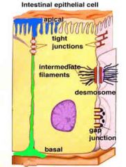
CellularAttachments


•Threemajor types of intercellularjunctions:


1. tight junctions
2. Desmosomes
3. Gap junctions

TightJunctions

•Formedby the fusion of the outermost layers of the plasma membranes of adjoiningcells
•Location:found in tissues in which there can be no leaks (urinary bladder, digestivetract)

Desmosomes

•Mechanical coupling formed by:filaments that interlock with one another
•Location: Found in tissues that undergorepeated episodes of tension and stretching (skin, heart uterus)
•Function: add strength

GapJunctions

•Tubularchannel proteins (connexons)that extend from the cytoplasm of one cell to the cytoplasm of another
•Function:allow exchange and passage of ions and mutrients
•Location:found in the epithelial cells, the heart, and smooth muscle tissue

BasementMembrane
aka basal lamina
function

•Function:
– meshwork of fibers that cements theepithelial cell to the underlying connective tissue; varies in thickness
– helps prevent the cell from being torn off byintraluminal pressures
– acts as partial barrier between theepithelial cell and the underlying connective tissue

SurfaceSpecialization


•Surfacesof epithelial cellsvary dependingon where they are located and what role they play in the function of the tissue


4 types


–Smooth:where no specialization is needed
–Microvilli(brush border):increase absorptive capability
–Cilia:move material past the cell surface


–Keratin:waterproofing and strength

tight junctions
desmosomes
gap junctions

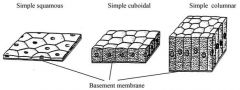
Classificationof Epithelial Tissue

•Numberof layers of cells:
– simple (one layer)
– stratified (2 or more layers)
•Shapeof the cells:
– squamous
– cuboidal
– columnar
•Presenceof surface specializations:
– cilia


– keratin

SimpleSquamous Epithelium

•Structure:fragile and thin
•Location:found lining the surfaces involved in the passage of either gas or liquid
–liningair sacs in the lung
– lining the lumen of blood vessels
•Flatand smooth(never ciliated or keratinized)

SimpleCuboidal Epithelium

•Structure:
–Singlelayer of cube shaped cells
–Round,dark staining central nuclei aligned in a single row
•Location:occurs in areas of the body where secretion and absorption take like
-lining kidney tubules
-ducts of small glands

SimpleColumnar Epithelium

•Structure:
–elongated and closely packed together
–Nucleialigned in a single row at the base of the cell near the basement membrane
-often ciliated
•Location:found in many excretory ducts as well as in the digestive tract.

StratifiedSquamous Epithelium

•Structure:multilayer of flat cells
•Function:protect underlying tissues
•Location:inareas of the body subject to mechanical and chemical stresses
–Maybe keratinized: surface skin-epithelium
–Maybe non-keratinized: lining oral cavity,vagina

StratifiedCuboidal Epithelium

•Structure:usually two layers of cuboidal cells •Function:protects underlying tissues


•Location:found primarily along the large excretory ducts (salivary & mammary glands)

StratifiedColumnar Epithelium

•Structure:several layers of cuboidal cells with a columnar layer on top
•Function:in secretion and protection
•Location:uncommon- found only in select parts of the respiratory, digestive,reproductive systems and along someexcretory ducts

PseudostratifiedColumnar Epithelium

•Structure:single layer of columnar cells that appear to be stratified because the cellnuclei are found at different levels across the length of the tissue
•Function:are often ciliated –move material pat the cell surface
•Location:found in respiratory tract and in portions of the male reproductive tract

TransitionalEpithelium

•Structure: Stratified epithelium with a basal layerofcuboidal or columnar cellsand asuperficiallayerof cuboidalorsquamous cells

•Location/function: found in areas of thebody required to expand and contract as part of their normal function (liningthe urinary tract)

GlandularEpithelium
Groupsof cells that manufacture and discharge a secretion

Classification of glands

1.Presenceor absence of ducts
2.Numberof cells that compose them
3.Shapeof the secreting ducts
4.Complexityof the glandular structure
5.Typeof secretion they produce
6.Mannerin which the secretion is stored and discharged

EndocrineGlands

•Partof a complex, biochemical network known as the endocrine system
•Structure:glands that do not have ducts or tubules and whose secretions are distributedthrough the body
•Function:produce and secret hormones into the bloodstream or lymphatic system
•Examples:thyroid gland, adrenal gland, pituitary gland etc

ExocrineGlands

structure

dischargesecretions via ducts directly into local area- the secretions eventually leavethe body

multicellular exocrine glands

•most exocrine glands
–salivaryglands
–sweatglands
–mammaryglands

unicellular exocrine glands

•Gobletcells
–secretemucus
–location: found along columnar cells of the respiratorytract and digestive tract and conjunctiva of the eye

exocrine glands ducts

Simple: main duct is unbranched

Compound: main duct is branched

exocrine glands

Shape of secretory portions
Tubular: secretory cells form a long channel of even width

Alveolar (or acinar): secretory unit forms a rounded sac

Tubuloalveolar, or tubuloacinar: secretory units possess both tubular and alveolar qualities
ExocrineGlands

Type of secretion produced:

•Seroussecretions
–watery
–contain a high concentration of enzymes
•Mucoussecretions
thick, viscous

–Composedof glycoproteins.
•Mixedexocrine glands contain both mucous and serous components.

NervousTissue

•Location:found in the brain, spinal cord and peripheral nerves
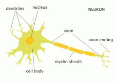
•Composedof two general cell types:
–Neurons(nerve cells)
–Neuroglia(supporting cells)

Neurons
Neurons
•Longest cells in the body; threeprimaryparts:
–Perikaryon(soma): the cell body; contains thenucleus
–Dendrites: short cytoplasmc extentions(from the soma); receive inpulses
–Axons: long, single extension; conducts impulsesaway from the cell body
Neuroglial cells
–Supportthe neurons

Typesof Neurons

•Sensoryneurons: nerve cells that carry information(nerve impulses) toward the CNS •Motorneurons: nerve cells that carry information away from the CNS
•Associationneurons: nerve cells that compose the CNS ( brain and spinal cord)
Typesof Nerves

•Sensorynerves: bundles of sensory neurons that carry information(nerve impulses)toward the CNS
•Motornerves: bundles of motor neurons that carry information away from the CNS
•Mixednerves: bundle of both types of neurons that carry information both toward andaway from the CNS