Reading...
![]()
Play button
![]()
Play button
![]()
Use LEFT and RIGHT arrow keys to navigate between flashcards;
Use UP and DOWN arrow keys to flip the card;
H to show hint;
A reads text to speech;
65 Cards in this Set
- Front
- Back
|
Tight Junctions
|
Formed by the fusion of the outermost layers of the plasma membranes of adjoining cells near apical surface.
Found in tissues in which there can be no leaks (urinary bladder, digestive tract). |
|
|
Desmosones
|
Mechanical coupling formed by filaments that interlock with one another
Tonofilaments extend from the plaque into the cytoplasm. Found in places of extreme stretching (skin, heart, uterus). |
|
|
Gap Junctions
|
Tubular channel proteins (connexons) that extend from the cytoplasm of one cell to another.
Allows exchange and passage of ions and nutrients. Found in intestinal epithelial cells, the heart, and smooth muscle tissue. |
|
|
Basil Lamina
|
Mesh work of fibers that cements the epithelial cell to the underlying connective tissue aka Basement Membrane.
Helps prevent cell from being torn off by intraluminal pressures. |
|
|
Microvilli
|
Increase surface area of intestine cells for absorption.
|
|
|
Cillia
|
Tiny hairlike projections from cells in respiratory tract.
Movement moves protective mucus up and out of airway. |
|
|
Epithelial Tissue
|
Composed of sheets of cells that cover and line other tissues, various sub-types.
May absorb secrete or excrete biochemical substances. -usually linings of cavities or coverings of glands, internal and external surfaces Are Polar: Apical surface faces the lumen or outside of the organ, Basal surface faces the basal lamina and blood vessels. Are avascular. Are innervated. |
|
|
Connective Tissue
|
Supports, protects and holds organs and tissues in place, provides a transport systems for nutrients and waste, stores energy reserves as fat, and contributes to healing.
Components: ground substance, extracellular fibers, cells. |
|
|
Muscle Tissue
|
Composed of actin and myosin fibers. Responsible for organized movement within the body.
Three types: Skeletal, Smooth, and Cardiac. |
|
|
Nervous Tissue
|
Found in the brain, spinal cord, and peripheral nerves.
Composed of two types: Neurons; longest cells in the body, generate and conducts nerve impulses, Neuroglia; support the neurons. |
|
|
Simple Cuboidal Epithelium
|
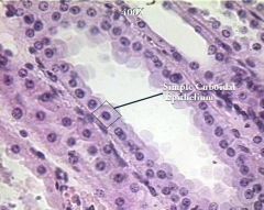
Found in ducts associated with hollow tubes, areas of secretion and absorption.
shape: usually simple found exp: exocrine glands, salivary glands, pancreas, sweat glands |
|
|
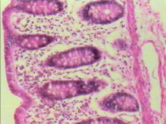
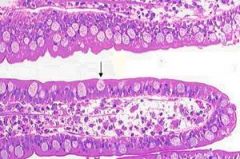
Simple Columnar Epithelium
|
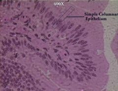
The cells absorb nutrients (from the small and large intestine. Have microvilli on top to aid in absorption and increase surface area
shape: tall, narrow cells simple found: small intestine, large intestine and digestive glands |
|
|
Pseudostratified Columnar Epithelium
|
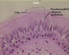
Has active cilia on top of the cells which aid in moving mucus up into the throat.
Works with goblet cells (produce mucus)- mucociliary transport system shape: appears stratified but simple found: respiratory tract |
|
|
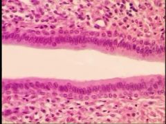
Simple Squamous Epithelium
|
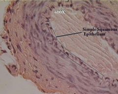
Delicate and thin, found lining surfaces involved in the passage of either gas or liquid areas of diffusion (alveoli), osmosis (capillaries) and filtration (kidneys).
shape: flat hexagonal, simple found: paratial pleura, visceral pleura, paratieal peritoneum, visceral peritoneum |
|
|
Stratified Squamous Epithelium
|
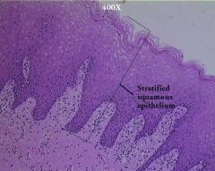
Designed for protection- very thick
shape: stratified found: skin and were skin has contact with internal structures (ear, eye, vaginal canal, rectum, mucous membranes) also regulate body temp., sensory/afferent info to the brain, excretion, vitamin D synthesis |
|
|
Stratified Cuboidial Epithelium
|
Protects underlying tissue, usually two layers of cells.
found: along large excretory ducts, salivary glands. |
|
|
Transitional Epithelium
|
Has ability to stretch, found in areas of the body required to expand and contract as part of their normal function.
A basal layer of cubodial or columnar cells and a superficial layer of cuboidial or squamous cells. shape: stratified found: only in ureters, uterus, urinary bladder, urethra. |
|
|
Glandular Epithelium
|
Synthesizes enzymes, Has ability to manufacture and discharge a secretion.
found: makes up all glands, endocrine and exocrine -acini- the termination of a duct lined by glandular epithelium |
|
|
Stratified Columnar Epithelium
|
Function in secretion and protection.
found: only in select parts of the respiratory, digestive, reproductive systems and along some excretory ducts. |
|
|
Blood Tissue
|
Specialized connective tissue that circulates.
Matrix: Ground substance, plasma. Fibrous components, protein. Cells: Erythrocytes, Leukocytes, Thrombocytes. Carries nutrients to tissues (oxygen) and waist away (CO2). |
|
|
Endocrine Gland
|
Glands that do not have ducts or tubules and whose secretions are distributed throughout the body.
Produce and secrete hormones into the bloodstream or the lymphatic system. Part of a complex, biochemical network. |
|
|
Exocrine Gland
|
Discharge secretions via ducts directly into local areas (except for goblet cells).
Unicellular or multicellular. |
|
|
Goblet Cell
|
Unicellular exocrine gland, ductless and composed of modified columnar epithelial cell.
Secrete the protein mucin. found: among columnar cells of the respiratory and digestive tracts and the conjunctiva of the eye. |
|
|
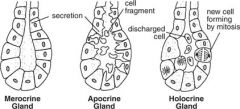
Multicellular Exocrine Glands
|
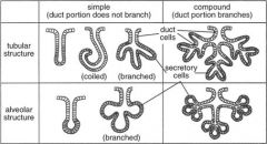
Composed of a secretory unit and duct surrounded by connective tissue rich in blood and nerve fibers.
Serous secretions are watery and contain high concentrations of enzymes. Mucous secretions are thick and viscous composed of glycoproteins. Merocrine- Exocytosis to secrete (unscented sweat). Apocrine- Partial cell sacrifice (smelly sweat). Holocrine- Total cell sacrifice (sebaceous oily). |
|
|
Ground Substance of connective tissue
|
Medium through which cells exchange nutrients and waste.
Acts to protect the more delicate cells it envelops and serves as an obstacle for invading microorganisms. Amorphous, homogeneous material ranging in textures from a liquid or gel to a calcified solid. |
|
|
Collagenous Fibers
|
Extracellular fiber component of connective tissue.
Strong, thick strands of collagen, organized into bundles of long, parallel fibrils composed of bundled microfibrils. Variable density and arrangement of fibers. found: tendons and ligaments also known as white fibers. |
|
|
Reticular Fibers
|
Extracellular fiber component of connective tissue.
Thin, delicate, branched networks of collagen provide support for highly cellular organs. found: endocrine glands, lymph nodes, spleen, bone marrow, liver, also around blood vessels, nerves, muscle fibers, and capillaries. |
|
|
Elastic Fibers
|
Extracellular fiber component of connective tissue.
Branched networks composed primarily of the protein elastin. Composed of coiled bundles of microfibrils. found: in tissues commonly subjected to stretching, vocal cords, lungs, skin, and walls of blood vessels. |
|
|
Cells of connective tissue
|
Fixed cells: involved in production and maintenance of the matrix (fibroblasts, chondroblasts, osteoblasts, reticular cells).
Transient (Wandering) cells: involved in repair and protection of tissues (leukocytes, mast cells [histamine], macrophages). |
|
|
Areolar Connective Tissue
|
Loose connective tissue proper.
Fibers and cells suspended in a thick translucent ground substance. Predominant cell is fibroblast: manufactures the elastic, reticular, and collagenous fibers. Surrounds every organ, forms the SQ layer that connects the skin to muscle, envelopes blood vessels, nerves, and lymph nodes; present in all mucous membranes. |
|
|
Adipose Connective Tissue
|
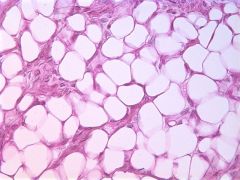
Loose connective tissue proper.
Areolar tissue in which adipocytes predominates, highly vascular. Acts as an energy storehouse and thermal insulator. Brown fat: thermal regulation White fat: stores energy |
|
|
Reticular Connective Tissue
|
Loose connective tissue proper.
Network of thin reticular fibers, loosley arranged and many fibroblasts suspended in a supportive ground substance. Forms the stroma (framework for all the organs). |
|
|
Dense Regular Connective Tissue
|
Composed of tightly packed, parallel collagen fibers, relatively avascular.
Makes up the tendons and ligaments. Can be found in fascial sheets that cover muscles. |
|
|
Dense Irregular Connective Tissue
|
Composed primarily of collagen fibers arranged in thick bundles, fibers are interwoven to form a single sheet.
Found in the dermis of the skin and in the fibrous coverings of many organs, forms the tough capsule of joints. |
|
|
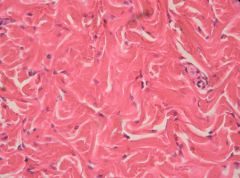
Cartilage
|
Specialized connective tissue found in joints and in the ear, nose and vocal cords.
Forms a framework on which bone is formed. No innervation, avascular. Cells: chondrocytes, live in hollowed-out pockets in the matrix called lacunae. Matrix: ground substance, gel of chondroitin sulfate, hyaluronic acid, and chondronectin. Collagen fibers are most commonly found in the matrix, but elastic fibers are also present in varying amounts. |
|
|
Hyaline Cartilage
|
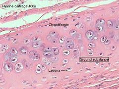
Most commonly type of cartilage found in the body, composed of closely packed collagen.
Found in joints at the ends of long bones, growth plates of long bones, tracheal rings, and connections of the ribs to the sternum. Composes most of the embryonic skeleton. Enclosed within a perichondruim. Most rigid cartilage. |
|
|
Elastic Cartilage
|
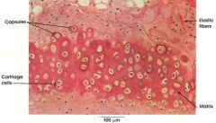
Contains elastic fibers in dense branching bundles, flexible can withstand repeated bending.
Found in the epiglottis of the larynx and in pinnae of ears of animals. |
|
|
Fibrocartilage
|
Usually found merged with hyaline cartilage and dense connective tissue.
Contains thick bundles of collagen fibers with fewer chondrocytes than hyaline cartilage. Lacks a perichondrium Found in spaces between vertebrae of the spine, between bones in the pelvic girdle, and in the knee joint. |
|
|
Elastic Connective Tissue
|
Primarily composed of elastic fibers, may be arranged parallel or in interwoven patterns with fibroblasts and collagenous fibers interspersed.
Found in spaces between vertebrae and in areas of the body that require stretching, walls of arteries, stomach, bronchi, bladder, ect. |
|
|
Bone Tissue
|
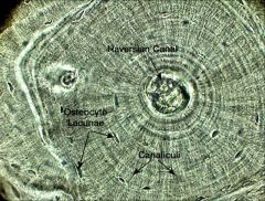
Specialized connective tissue.
Matrix is a combination of organic collagen fibers and inorganic calcium salts, well vascularized. Haversian Canal: contains both a vascular and a nerve supply. Canaliculi: channels within the matrix support passage of blood vessels into deeper tissues. Osteoblasts manufacture the fibers that are part of the matrix (lacunae and canaliculi are created as the osteoblasts create bony matrix). Osteocytes reside in lacunae (cellular extensions pass through the canaliculi). Osteoclasts take up old, damaged bone tissue to be recycled. |
|
|
Fixed Cells
|
Not able to move.
Exp. osteoblasts |
|
|
Wandering/Transient Cells
|
Able to move through tissues.
Exp. Mast Cells, Leukocytes |
|
|
Diapedesis
|
The outward passage of leukocytes cells through intact vessel walls. The leukocytes secrete proteases that degrade the basement membrane, allowing them to escape the blood vessel.
|
|
|
Mucous Membranes
|
Mucosae, line organs with connections to the outside environment (mouth, intestines, nasal passages, etc.)
Usually composed of either stratified squamous or simple columnar epithelium covering a layer of loose connective tissue. Submucosa: connective tissue layer that connects the mucosa to underlying structures. May contain goblet cells or multicellular glands which can produce large quantities of mucus. Some mucosae also can absorb nutrients (exp. the epithelial layer in the intestine). |
|
|
Mucus
|
Consists primarily of water, electrolytes, and the protein mucin.
|
|
|
Serous Membranes
|
Serosae, line walls and cover organs of body cavities (exp. thorax and abdominopelvic cavities.
Consists of a continuous sheet doubled over on itself to form two layers. |
|
|
Parietal Layer
|
The portion of the serous membrane that lines the cavity wall.
|
|
|
Visceral Layer
|
The portion of the serous membrane that cover the outer surface of the organ.
|
|
|
Mesenteries
|
Areas of the abdominopelvic cavity where visceral layers of serosa merge.
|
|
|
Cutaneous Membrane
|
Integument or skin.
Composed of an outer keratinized stratified squamous epithelium or epidermis. Epidermis is attached to an underlying layer of dense irregular connective tissue called the dermis. |
|
|
Dermis
|
Layer of dense irregular connective tissue on which the epidermis is attached.
Contains collagenous, reticular, and elastic fibers which enable skin to be both strong and elastic. |
|
|
Synovial Membranes
|
Lines the cavities of joints.
Composed of loose connective tissue and adipose tissue covered by a layer of collagen fibers and fibroblasts. Manufacture the synovial fluid that fills the joint spaces. |
|
|
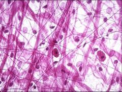
Skeletal Muscle
|
Large cells that contain hundreds of nuclei and mitochondria.
Voluntary muscle. Skeletal muscles are striated, cells are bundles of fibers held together by loose connective tissue. The collagen fibers that surround the cells merge with the collagen fibers in tendons (which connect to bone). |
|
|
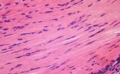
Smooth Muscle
|
Composed of small, spindle shaped cells that lack striations.
Involuntary muscle. Found in the walls of hollow organs, in exocrine glands, and along the respiratory tract. Responsible for peristalsis in gastrointestinal tract, constriction of blood vessels, and emptying of urinary bladder. |
|
|
Cardiac Muscle
|
Found only in the heart. Contains specialized pacemaker cells that supply signal for the heart to contract at regular intervals.
Entirely involuntary and striated. Cardiac muscle cells connect to one another via intercalated discs. Does not regenerate. |
|
|
Perikaryon
|
Nerve cell body, contains the nucleus.
|
|
|
Dentrites
|
Short cytoplasmic extensions of a nerve cell, receives impulses.
|
|
|
Axons
|
Long, single extension of a nerve cell, conducts nerve impulses away from the cell body.
|
|
|
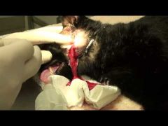
Inflammation
|
Body's initial response to injuries to limit further damage and eliminate any harmful agents.
Steps: Vasodilation, Swelling, Clot formation, Phagocytosis, Capillaries return to normal size, blood flow and fluid leakage into the affected area abate. |
|
|
Tissue Repair
|
Formation and organization of granulation tissue, regeneration of lost tissue, epithelialization, and formation of scar tissue.
|
|
|
Granulation Tissue
|
Tissue that forms beneath the overlying blood clot or scab.
Composed of a layer of collagen fibers infiltrated with capillaries (that have branched off existing capillaries in deeper layers of damaged tissue: capillary migration). Eventually is replaced by fiberous scar tissue. |
|
|
Epithelialization
|
The proliferation of new epithelium into an area devoid of it but that naturally is covered by it.
|
|
|
First Intention Wound Healing
|
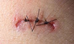
Edges of wound held in close apposition. Skin forms a primary union without formation of granulation tissue or significant scarring.
|
|
|
Second Intention Wound Healing
|
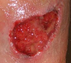
Edges of wound seperated from each other. Granulation tissue forms to close gap; scarring results.
|
|
|
Third Intention Wound Healing
|
Contaminated wound left open until contamination is reduced and inflammation subsides; later closed by first intention, also called delayed primary closure.
|